Achalasia cardia (achalasia of the esophagus) is a disease characterized by the lack of opening of the lower esophageal sphincter in response to the passage of food or liquid through the esophagus, due to a violation of the conduction of nerve impulses to the muscles of the esophagus and cardia. It is necessary to immediately clarify the correct name of the disease, because people call both achalasia of the cardia and achalasia of the esophagus. Previously, some doctors believed that it was correct to say achalasia of the esophagus, but the international classification of diseases of the 10th revision put an end to these disputes; it indicated achalasia of the cardia, which means this is the correct name for this disease. Achalasia affects equally often both men and women, more often only at the age of 30 - 45 years. This disease occurs in 3 - 20% of all diseases of the esophagus. Previously, it was believed that achalasia occurs in 1 person per 100,000 population, but with improved diagnosis and accumulation of data, reports began to appear that it occurs with a frequency of 5 and some authors believe that even 10 people per 100,000 population
Cause of achalasia cardia
It must be said right away that the exact cause of achalasia cardia is not known. There are various theories about the origin of this disease. The genetic theory is based on the fact that patients with esophageal achalasia have changes in the same gene loci, in addition, the risk of developing achalasia in children with Down syndrome is 200 times higher than in the population. Infectious hypothesis - based on the fact that when infected Trypanosoma cruzi, which causes Chagas disease, also causes dysphagia, which is similar in mechanism to cardial achalasia. In addition, the possibility of developing achalasia against the background of infection with the Herpes zoster virus and measles virus is being considered. Autoimmune theory is based on the fact that autoimmune diseases often occur in patients with esophageal achalasia. Thus, in patients with achalasia cardia, the risk of developing such diseases as increases: type 1 diabetes mellitus by 5.4 times, hypothyroidism by 8.5 times, Sjogren's syndrome by 37 times, systemic lupus erythematosus by 43 times, uveitis by 259 times . But none of these theories has received convincing confirmation to date, so it cannot be said that the cause of this disease is known.
Doctor's comment
If the symptoms characteristic of achalasia cardia cause physical and psychological suffering, you have the opportunity to get rid of the disease quickly and painlessly, at the same time giving up many medications that bring only temporary relief. There are several ways to treat this disease; we will choose the one that will be effective in your case. The low-traumatic operation is performed using endoscopic equipment, which allows all manipulations to be performed as delicately as possible. Moreover, the improved technique allows surgical intervention without the risk of further relapses. Do not wait for complications to develop and do not waste time on ineffective conservative therapy; timely treatment gives every chance of a quick recovery!
Head of the surgical service at SwissClinic Konstantin Viktorovich Puchkov
Classification of achalasia cardia
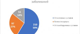
The classification of achalasia cardia is important, since depending on the degree of the disease, a decision is made on the choice of one or another treatment tactic. There are 4 stages in total:
Stage I (early) - transient disturbances in the passage of food, the diameter and peristalsis of the esophagus are not changed outside of an attack; Stage II - persistent disruption of the passage of food, peristalsis of the esophagus is enhanced, the esophagus is not significantly dilated; Stage III – a stricture of the cardiac part of the esophagus is revealed, the latter is significantly dilated, peristalsis is sluggish and superficial. There is fluid in the esophagus on an empty stomach; Stage IV – stage of complications with ulcerations and erosions of the esophagus. The latter is expanded to 10 cm, S-shaped, peristalsis is either completely absent or multidirectional, superficial.
What contributes to the disease achalasia
Experts do not name the specific cause that causes the development of the disease. It is associated with infections, compression of the esophagus, inflammation, tumors of malignant origin, infiltrative lesions, etc. In childhood, esophageal achalasia is usually detected after five years of age.
The first manifestations of the disease go unnoticed, so it is diagnosed late. If a child has dysphagia and starts vomiting after eating, you need to sound the alarm. These are common symptoms characteristic of achalasia.
Clinical picture of achalasia cardia
Most often, it will not be difficult for an experienced gastroenterologist or surgeon to suspect achalasia of the esophagus based on the patient’s complaints. Most often, patients complain of dysphagia, difficulty or complete impossibility of food passing through the esophagus. So-called paradoxical dysphagia often occurs. when solid food passes more easily than liquid food. Over time, dysphagia progresses and in particularly severe cases, patients lose the ability to eat through the mouth, which leads them to exhaustion. A common symptom of achalasia cardia is the so-called wet pillow symptom, when the patient experiences reflux of the contents of the dilated esophagus into the mouth, and if the person is sleeping, the contents flow out onto the pillow, this can be either sputum or food debris. Often the pain intensifies against the background of stressful situations, or when the patient is nervous, which often leads patients with achalasia cardia to see a psychiatrist instead of a surgeon.
Often this disease is accompanied by quite intense pain. At the beginning of the disease, the pain is spastic, paroxysmal, and as the disease progresses, the pain becomes less intense, but almost constant.
Another common symptom of achalasia cardia is regurgitation, which is the reflux of the contents of the dilated esophagus into the mouth, the so-called esophageal vomiting. This condition actually resembles vomiting, but unlike it, the patient does not experience nausea, and it occurs most often when bending over or lying down.
A common symptom of esophageal achalasia is also a cough, and the development of bronchitis, and sometimes pneumonia. The problem is that during regurgitation, aggressive and infected contents enter the respiratory tract, which causes the above complications.
Advantages of treatment at the Swiss University Hospital
- The clinic is equipped with the latest generation equipment, which allows the operation to be performed without damaging nearby structures, the muscularis mucosa is dissected without damaging the mucosa, intraoperative complications are also excluded, etc.
- The treatment method is selected for each patient individually, taking into account his state of health. Great importance is given to preserving the natural anatomical location of the esophagus, diaphragm and stomach, which avoids the occurrence of reflux esophagitis and sphincter insufficiency in the future.
- When treating patients, the clinic uses a technique that makes it unnecessary to leave a transgastric tube after surgery, which makes the postoperative course easier. Thanks to the original method used, the risk of further relapse is minimal; in more than 97% of patients, good results were achieved, confirmed by fibroesophagogastroduodenoscopy, X-ray examination and esophagomanometry.
- In our clinic, patients with several diseases requiring surgical treatment can undergo simultaneous surgery. Performing several surgical interventions during one anesthesia reduces the burden on the body, hospitalization time, and recovery, compared to conventional surgery, takes much less time.
Diagnosis of achalasia cardia
Diagnosis of achalasia cardia is most often not difficult. FGDS (gastroscopy) - it is necessary to begin the examination with this procedure. When performing an FGDS, a narrowing of the esophagus is detected at the point where it enters the stomach; endoscopically, the device passes into the stomach with force. With achalasia grades 3 and 4, the esophagus is dilated, contains food debris on an empty stomach, and ulcers and erosions of the esophagus are often detected.
Barium X-ray of the esophagus - allows you to evaluate the speed of passage of contrast through the esophagus in patients with esophageal achalasia. The transverse size of the esophagus, peristalsis, and the presence of bends are also assessed. It is on the basis of radiography that the degree of achalasia of the cardia is determined.
CT examination - performed to assess the presence or absence of inflammation in the surrounding tissues, the presence of bronchitis or pneumonia. Motility of the esophagus - is considered the “gold standard” for diagnosing achalasia cardia, however, the equipment for its implementation is extremely expensive, and its use is quite limited. In this regard, several clinics, mainly research ones, have such equipment.
Treatment
At the initial stage, when problems with swallowing are minor, conservative therapy can be used; the drugs used are aimed at eliminating spasm and alleviating the patient’s condition. Endoscopic bougienage may also be performed to widen the narrowed area in the esophagus. To obtain the result, several procedures will be required, but the effect of dilatation is short-lived.
A very effective way to treat esophageal achalasia is esophagomyotomy - a surgical procedure during which part of the wall of the esophagus is dissected, but only to the submucosal layer - without opening the lumen, thus restoring normal swallowing. If there are contraindications to surgical treatment, as well as in elderly patients with recurrent disease who have previously undergone surgery, endoscopic dilatation is indicated.
If there are contraindications to surgery and endoscopic cardiodilation, endoscopic administration of an intrasphincteric injection of botulinum toxin A is possible. The procedure is safe and suitable even for patients with severe diseases of the cardiovascular and respiratory systems. But the effect lasts only 2-3 months, so repeated procedures may be required.
In our clinic, for each patient, a technique is selected that will be most effective in his or her case. If in the area of narrowing there are no traces of cardiodilation performed in the past, and there is also no depletion of the muscular wall of the esophagus, surgery is recommended; its timely implementation leads to recovery. Surgical intervention is performed by laparoscopy and even with a stretched esophageal wall leads to good results.
In this case, the operation itself is performed using video endoscopic equipment, which allows the surgeon to perform all manipulations with maximum precision, excluding damage to structures located in this area (vessels, nerves, fascial spaces). Our specialists have improved the technique, so patients can count not only on a shorter recovery period, but we have managed to minimize the incidence of intraoperative complications and relapses. Today this figure does not exceed 0.2%.
Common symptoms
The main symptom of achalasia is dysphagia, which occurs in almost all patients. The time between the appearance of the first symptoms and a visit to the doctor can range from one to ten years. The second most common symptom is congestion in the esophagus. Manifested by coughing and attacks of suffocation at night.
Heartburn and chest pain can also be signs of achalasia. Localized mainly behind the sternum, squeezing or squeezing pain is felt in the lower jaw, neck and back. Achalasia is often mistaken for gastroesophageal reflux or another disease accompanied by heartburn. If it does not occur from food and does not go away after taking antacids, there is a possibility that the problem is related to esophageal achalasia.
What are the complications?
With achalasia, irreversible changes occur throughout the body. Among the most common complications associated with esophageal disease are:
- squamous cell carcinoma;
- bezoars;
- volumetric formations on the neck;
- exfoliating submucosal layer;
- distal diverticulum;
- phlebeurysm;
- lung disease;
- purulent pericarditis;
- pneumopericardium, etc.
With prolonged progression of the disease, the esophagus expands and its walls become thinner, which becomes the cause of complications. In most cases (85%) those suffering from achalasia experience noticeable weight loss.
Achalasia
(from the Greek - lack of relaxation)
cardia
is a disease of the esophagus of unknown etiology, the pathogenesis of which is based on inflammation and gradual degeneration of the neurons of the intermuscular plexus, leading to the loss of normal peristaltic activity of the smooth muscle part of the thoracic esophagus, impaired relaxation of the lower esophageal sphincter (LES) and dysphagia [1].
The gold standard for diagnosing achalasia is high-resolution esophageal manometry. According to the Chicago classification of disorders of motor function of the esophagus [2], characteristic manometric signs of achalasia cardia are an increase in the total relaxation pressure (IRP - integrated relaxation pressure) of more than 15 mm Hg. and the absence of normal peristaltic contractions in the thoracic esophagus.
The use of high-resolution manometry made it possible to establish that impaired peristalsis on the part of the smooth muscle portion of the esophagus in patients with achalasia cardia can be represented by three main types (Table 1)
Table 1. Types of achalasia cardia according to the Chicago classification v. 3.0 [2] [2].
Achalasia type I:
complete absence of contractions in the thoracic esophagus, intrabolus pressure is not increased (Fig. 1).
Rice.
1. Cardiac achalasia type I. a — fluoroscopy of the esophagus: saccular expansion of the esophagus, narrowing of the terminal section in the form of a bird’s beak; b — manometry of the esophagus: increased level of total relaxation pressure (IRP), absence of contractions of the thoracic esophagus. Achalasia type II:
absence of peristaltic contractions along with preservation of the tone of the esophageal walls, causing a total increase in intrabolus pressure in at least 20% of swallows (Fig. 2).
Rice.
2. Cardiac achalasia type II. a — fluoroscopy of the esophagus: slight dilation of the esophagus, narrowing of the terminal section in the form of a bird’s beak, retention of the contrast agent in the lumen of the esophagus; b — manometry of the esophagus: increased level of total relaxation pressure (IRP), absence of peristaltic contractions of the thoracic esophagus, total increase in the level of intrabolus pressure. Achalasia type III:
spastic non-peristaltic contractions of the smooth muscle of the esophagus in at least 20% of swallows (Fig. 3).
Rice.
3. Cardiac achalasia type III. a — fluoroscopy of the esophagus: spastic contractions of the thoracic esophagus; b — manometry of the esophagus: increased level of total relaxation pressure (IRP), spastic contractions of the thoracic esophagus. In this case, the term “achalasia” reflects the form of disturbances in the motor function of the esophagus, and not the nosological form “Achalasia of the cardial part of the esophagus” (ICD code K22.0).
There is a hypothesis that the described types of achalasia cardia are not three different forms, but successive stages in the development of this disease [3]. It is believed that at the initial stage of the disease (occurring with the gradual death of inhibitory motor neurons), changes in motility are observed according to the type of spastic achalasia (type III), then, as excitatory neurons die and the contractile function of the esophagus is inhibited, a picture of cardia achalasia type II is observed. With the total death of the motor neurons of the intermuscular plexus, changes in motility occur, described as type I achalasia, accompanied by a significant dilation of the esophagus and its S-shaped deformation [3].
Interestingly, some patients with characteristic symptoms of achalasia cardia may experience normal peristaltic contractions of the esophagus, despite a significant increase in the total relaxation pressure of the LES (IRP>15 mm Hg) (Fig. 4).
Rice. 4. Esophageal manometry: obstruction of the esophagogastric junction. This disorder of esophageal motility refers, according to the Chicago classification, to “obstruction of the esophageal-gastric junction” (EGJ outflow obstruction) [2].
Patients with obstruction of the esophagogastric junction require additional clarifying studies: computed tomography (CT) of the chest and abdominal organs, endosonography (endoultrasound). During the examination, the manometric conclusion “obstruction of the esophagogastric junction” will most likely be transformed into a diagnosis of “achalasia cardia” or organic obstruction of the esophagogastric junction zone will be identified (stricture or tumor of the esophagus, eosinophilic esophagitis, sliding axial or paraesophageal diaphragmatic hernia, etc. .) [2, 4]. It must be remembered that previous bariatric surgery or fundoplication may also cause the manometric pattern of esophagogastric junction obstruction [5, 6].
A series of studies conducted among patients with a manometric diagnosis of “obstruction of the esophagogastric junction” showed that in 20-40% of cases, spontaneous resolution of the obstruction occurs. However, 12-40% of patients with such a conclusion were subsequently treated for achalasia cardia [7-10].
The most recent revision of the Chicago Classification does not yet include esophageal motility disorders that do not fully meet the manometric criteria for achalasia. For example, in recent years, more and more studies have appeared that prove the possibility of the existence of a separate type of achalasia cardia - with a normal total relaxation pressure of the LES. In this case, we are talking about a subgroup of patients with characteristic clinical symptoms of achalasia cardia, who have no peristaltic contractions of the esophagus during manometry; during fluoroscopy, a long-term retention of barium in the lumen of the esophagus is observed, but the IRP level does not reach 15 mm Hg. [eleven]. According to available publications, low total relaxation pressure can be observed in patients with advanced stages of the disease (with achalasia type I) with low basal pressure in the LES (Fig. 5).
Rice. 5. Achalasia of the cardia with a normal level of total relaxation pressure. a — fluoroscopy of the esophagus: saccular expansion of the esophagus, S-shaped deformation of the esophagus; b — manometry of the esophagus: normal IRP value, absence of peristaltic contractions of the thoracic esophagus (in accordance with the Chicago classification v. 3.0: absence of contractility).
In addition to cardial achalasia, patients with dysphagia (in the absence of organic causes) may experience changes in esophageal motility, which according to the Chicago classification belong to the group of “significant disturbances of peristalsis” [2].
Significant disturbances of peristalsis never occur in healthy individuals, occur with obvious clinical symptoms and include the following nosological types:
distal esophagospasm
— in the thoracic esophagus (in 20% of swallows or more) spastic contractions are observed with normal average values of the IRP level (Fig. 6);
Rice.
6. Esophageal manometry: distal esophagospasm. hypercontractile esophagus
— in the thoracic esophagus (in 20% of swallows or more) extremely powerful contractions are recorded (distal contractile integral DCI >8000 mm Hg × cm × s) with normal average values of the IRP level (Fig. 7);
Rice.
7. Esophageal manometry: hypercontractile esophagus. lack of contractility
— in 100% of swallows there are no contractions of the esophagus (failed, DCI <100 mm Hg × cm × s), the average values of the IRP level are within the normal range, which does not allow establishing the diagnosis of “achalasia cardia” (Fig. 8).
Rice.
8. Lack of contractility in a patient with systemic scleroderma; the tone of the lower esophageal sphincter is not determined. In hypercontractile esophagus, contractions are non-peristaltic, extremely intense, multi-peak, and are accompanied by severe retrosternal pain and dysphagia [4].
According to modern data, the most effective methods for treating hypercontractile esophagus and distal esophagospasm are surgical interventions used to treat achalasia cardia, and primarily oral endoscopic myotomy [12], which allows these disorders of the motor function of the esophagus to be classified as a special group of diseases (Table 2) ,
Table 2. Differential diagnosis of “achalasia syndromes” [12, 13] Note. DCI—distal contractile integral; endoUS - endosonography; CT - computed tomography; MRI - magnetic resonance imaging. Note. DCI - distal contractile integral; endosonography - endosonography; CT - computed tomography; MRI - magnetic resonance imaging. united by the term “achalasia syndromes” [12, 13].
Lack of contractility is a conclusion of a manometric study that requires clarification. Lack of contractility is often observed as a result of degenerative processes in the wall of the esophagus in patients with systemic scleroderma (see Fig. 8). We can also talk about achalasia cardia with a low level of pressure in the LES (achalasia cardia with a normal level of IRP). The diagnosis of achalasia in the absence of contractility can be confirmed by barium fluoroscopy (with achalasia cardia, there is a long retention of contrast material in the lumen of the esophagus) or using provocative tests during a manometric study (a test with multiple rapid swallows will demonstrate an increase in the level of intrabolus pressure).
To date, pathogenetic treatment of achalasia cardia, which can stop the degeneration of neurons in the myenteric plexus of the esophagus, has not been developed. The main goals of treatment for achalasia cardia and “achalasia syndromes” are the resolution of obstruction from the LES, reducing the severity of dysphagia, achieved surgically.
The comparative effectiveness and safety of the three most common invasive treatments for achalasia cardia—balloon pneumocardial dilation (BPCD), oral endoscopic myotomy (POEM), and laparoscopic Heller myotomy (LMG)—has been intensively studied in prospective randomized trials, but no definitive conclusion has yet been made.
BPCD has remained the most common treatment for achalasia cardia for many years. This method involves inserting a cylindrical balloon (30, 35 or 40 mm in diameter) into the lumen of the esophagus, installing it (under x-ray control) along the LES and manually pumping air into the lumen of the balloon [14]. The effectiveness of pneumocardiodilation in the treatment of achalasia cardia, estimated in randomized controlled trials, is 62–90% [1]. It is important that the positive effect of therapy persists for several years after BPCD. On average, 30% of patients experience recurrence of symptoms 4-6 years after the procedure, necessitating repeat dilatation. The success of BPCD largely depends on the patient's age (the best results are observed in people over 45 years of age), gender (female), the degree of dilatation of the esophagus (the larger the diameter of the esophagus, the worse the treatment results) and the type of achalasia cardia (the best results in type II) [ 15-18].
An alternative to BPKD is LMG, in which the circular muscle fibers of the LES are surgically separated. Since LMG is often complicated by the development of reflux esophagitis, it is often combined with fundoplication. The operations of choice in this case are anterior fundoplication according to DOR (180°) or fundoplication according to Toupet (270°). Nissen fundoplication is not recommended for achalasia cardia, since it can lead to the development of postoperative dysphagia in 15% of patients [19-21].
A significant number of studies (Table 3)
Table 3. Randomized controlled trials of the effectiveness of laparoscopic Heller myotomy and balloon pneumocardiodilation in the treatment of achalasia cardia [1] Note. BPCD - balloon pneumocardiodilation; LMG - laparoscopic myotomy according to Heller. Note. BPKD - pneumatic balloon dilation; LMG - laparoscopic Heller myotomy. devoted to comparing the value of LMG and BPCD in the treatment of achalasia cardia [1, 19-28]; the largest of them is the European multicenter randomized controlled trial.
The conclusion of these studies was the conclusion that the effectiveness (about 90%) of BPCD (especially when carrying out several procedures) and LMG is comparable; the differences did not reach statistical significance [22, 23]. The incidence of complications (esophageal perforation) of these methods when performed by an experienced physician and using modern technologies does not exceed 1% [24].
POEM is a relatively new method of endoscopic treatment of achalasia cardia, which includes the creation of a submucosal tunnel in the esophagus with dissection of the circular muscle layer in the thoracic esophagus and in the area of the LES. In recent years, more and more data have confirmed the high effectiveness and advantages of POEM over other methods of treating achalasia cardia.
The obvious advantages of POEM are the minimally invasive nature of the intervention, short recovery time, and the absence of the need for intubation anesthesia. From a technical point of view, endoscopic myotomy makes it possible to regulate the length of the dissection of the circular layer of muscles, focusing on the data of endosonography and manometry of the esophagus, up to the upper esophageal sphincter. The endoscopic procedure, unlike LMG, does not have the risk of damage to the vagus nerve [13].
The effectiveness of POEM in the treatment of achalasia cardia has been assessed in a large number of uncontrolled clinical studies and is more than 90% (Table 4)
Table 4. Studies assessing the effectiveness and safety of POEM [13] Note. GERD is gastroesophageal reflux disease. Note. GERD - gastroesophageal reflux disease. [29-41].
According to the founder of the method, Professor H. Inoue [29], who published the results of the largest study (500 people) based on the results of POEM, this treatment method allows normalizing pressure in the LES and relieving clinical symptoms. Similar results were obtained in patients who underwent POEM after BPCD or LMG [42, 43]. Severe complications of the procedure include bleeding, esophageal perforation, and pneumothorax.
A multicenter comparative study evaluating complications and side effects of endoscopic myotomy showed that among 1826 cases of POEM, complications (mild, moderate and severe) were recorded in 6.4; 1.7 and 0.5% of patients, respectively. The most common side effect of POEM was reflux esophagitis, noted in 58% of patients [39].
The issue of postoperative esophagitis was also studied in a systematic review and meta-analysis comparing the outcomes of POEM (1958 patients) and LMG (5834 patients). It has been shown that in the short term (average follow-up duration was 16 months), POEM is more effective than LMG in relieving dysphagia, but at the same time is associated with a higher risk of developing postoperative erosive esophagitis [44].
Interestingly, a significant proportion of patients after POEM may not experience clinical symptoms of gastroesophageal reflux disease (GERD), despite the development of esophagitis. A study by Y. Werner et al. [45] showed that erosive esophagitis was detected in 31% of patients with a good clinical effect of POEM and no complaints during 29 months of follow-up after surgery, and Barrett’s esophagus was detected in 2 patients. This observation indicates the need for closer endoscopic monitoring of patients after POEM [39, 45]. A group of authors (T. Rösch et al., [46]) even published a work in 2021 with the self-explanatory title “Will reflux destroy the POEM technique?”
In 2021, F. Ponds et al. [30] published the results of a large randomized clinical trial that compared the effectiveness of POEM and BPCD in patients with achalasia cardia. After POEM or BPCD procedures, observation of 126 patients continued for 2 years. Treatment was considered successful if the Eckardt score was ≤3, there were no serious complications and there was no need for re-treatment. The results of endoscopic POEM were successful in 92% of patients, while BPCD led to a permanent cure in only 54% of patients. At the same time, postoperative reflux esophagitis was diagnosed in 41% of patients after POEM and only in 7% after BPCD [30].
Thus, despite a significant number of works showing higher efficiency of POEM compared to BPKD and LMG (Table 5)
Table 5. Comparison of the effectiveness of oral endoscopic myotomy with other methods of treating achalasia cardia Note. POEM—peroral endoscopic myotomy; BPCD - balloon pneumocardiodilation; LMG - laparoscopic myotomy according to Heller. Note. POEM - peroral endoscopic myotomy; BPKD - pneumatic balloon dilation; LMG - laparoscopic Heller myotomy. [30–35], additional studies are needed to evaluate the long-term risks and benefits of POEM, the optimal length of myotomy, and the likelihood of developing reflux esophagitis after treatment.
Before undergoing POEM, patients should be informed about the possible need for long-term and even lifelong use of proton pump inhibitors (PPIs) to avoid the development of erosive esophagitis and to relieve symptoms of GERD. Given the technical complexity of the procedure, POEM should be performed in expert centers by an experienced surgeon [47,48].
It should be noted that the effectiveness indicators discussed above for certain methods of treating achalasia cardia were obtained in studies conducted without taking into account the type of achalasia. However, today no specialist has any doubt that the prognosis of the effectiveness of therapy and the choice of treatment method for achalasia cardia depend not only on the stage of the disease, but also on the data obtained during manometry.
Thus, according to the results of several new studies, when using methods such as BPCD or LMG, the best treatment results are observed in type II achalasia cardia (treatment is effective in 96-100% of patients) [49-51]. The effectiveness of treatment of patients with achalasia cardia type I reaches 81% (results depend on the degree of dilatation of the esophagus) (Table 6)
Table 6. Treatment effectiveness depending on the type of achalasia [12] Note. LMG—laparoscopic myotomy according to Heller; BPCD - balloon pneumocardiodilation. Note. LMG - laparoscopic Heller myotomy; BPKD - pneumatic balloon dilation. [16, 49-51]. Achalasia cardia type III is the most difficult to treat with traditional methods (treatment is effective only in an average of 66% of patients) [49].
A distinctive feature of type III achalasia cardia is the presence of spastic contractions in the thoracic esophagus, therefore treatment methods (BPCD, LMG) that primarily affect the LES area are often ineffective. In this case, the use of POEM is more justified, since, as mentioned above, this technique allows you to “calibrate” the length of the myotomy depending on the length of the spastic segment.
The effectiveness of POEM in the treatment of patients with achalasia cardia type III, according to a meta-analysis by M. Khan et al. [52], is 92% (95% CI; 84-96%) with a myotomy length of 17.2 cm (95% CI; 13-19.7 cm). In addition, POEM is a highly effective technique for the treatment of other spastic diseases of the esophagus (Table 7):
Table 7. The effectiveness of oral endoscopic myotomy in the treatment of spastic diseases of the esophagus [52] distal esophagospasm - 88% (95% CI; 61-97%) and hypercontractile esophagus - 72% (95% CI; 55-83%) [52]. However, it should be noted that the data accumulated in the mentioned meta-analysis were obtained from short-term studies without a control group [12].
Interestingly, there are studies showing extremely encouraging results from the use of POEM not only for type III achalasia, but also in the treatment of advanced stages of achalasia with sigmoid deformity and dilatation of the esophagus (Fig. 9)
Rice. 9. X-ray of the esophagus in a patient with type I achalasia cardia. a - before treatment: the esophagus is dilated, curved; b - after treatment. (Data from A.M. Gasanov.) [53]. However, data are still limited, and there are other studies demonstrating an increased incidence of complications when performing POEM in patients with sigmoid deformity of the esophagus [54].
Of course, when choosing a treatment method for achalasia cardia, in addition to the type of esophageal motility disorders, it is necessary to take into account other factors: the degree of dilatation of the esophagus and its sigmoid deformation, the presence of a diaphragmatic hernia, large supradiaphragmatic diverticula, and also to exclude possible organic causes of obstruction from the LES [13].
Taking into account the data accumulated to date, several expert consensus documents were published in 2021 [12, 13], regulating the choice of the optimal method of treating achalasia depending on the type of disorders of the motor function of the esophagus. The main provisions of these expert documents are presented in table. 8,
Table 8. Recommended treatments for achalasia depending on the type according to the Chicago classification [12, 13] Note. BPCD - balloon pneumocardiodilation; LMG—laparoscopic myotomy according to Heller; POEM—peroral endoscopic myotomy; GERD is gastroesophageal reflux disease. Note. BPKD - pneumatic balloon dilation; LMG - laparoscopic Heller myotomy; POEM - peroral endoscopic myotomy; GERD - gastroesophageal reflux disease. 9
.
Table 9. Recommended methods of treatment for “achalasia syndromes” [12, 13] Note. BPCD - balloon pneumocardiodilation; LMG—laparoscopic myotomy according to Heller; POEM—peroral endoscopic myotomy; GERD is gastroesophageal reflux disease. Note. BPKD - pneumatic balloon dilation; LMG - laparoscopic Heller myotomy; POEM - peroral endoscopic myotomy; GERD - gastroesophageal reflux disease.
Speaking about the treatment of achalasia cardia, one cannot fail to mention drug methods, such as the use of botulinum toxin [55-58], calcium channel blockers and nitrates [59], and phosphodiesterase inhibitors [60]. However, despite some relief of symptoms during therapy with these drugs, in most patients with achalasia the disease progresses: dilation of the esophagus, stasis of food in the lumen of the organ [12].
The use of calcium channel blockers and nitrates leads to a decrease in pressure in the LES in 47-64% of patients and some relief of dysphagia [59]. Unfortunately, these drugs have a significant range of side effects (headache, orthostatic hypotension, edema), which makes them unsuitable for long-term use. As a rule, they are prescribed to patients who cannot undergo surgical or endoscopic treatment: nifedipine 10-30 mg orally 30-45 minutes before meals, isosorbide dinitrate 5-10 mg orally 15 minutes before meals.
5-phosphodiesterase inhibitors (sildenafil) reduce pressure in the LES and the severity of spastic contractions of the distal esophagus by enhancing the effect of nitric oxide (by blocking the enzyme that breaks down cyclic guanosine monophosphate). The possibility of using sildenafil for the treatment of achalasia and spastic disorders of esophageal motility has been described [60]. However, clinical studies proving the effectiveness and safety of long-term use of this drug are insufficient.
Injections of botulinum toxin into the LES area block the release of acetylcholine from nerve fiber endings. According to P. Pasricha et al. [55], the use of botulinum toxin leads to relief of dysphagia in 66% of patients with achalasia cardia within 6 months. The temporary effect of the drug is due to the regeneration of nerve fibers; it lasts no more than 1 year [56, 59, 60]. Repeated injections of botulinum toxin complicate subsequent surgical and endoscopic interventions, therefore the use of this method is not recommended as 1st line therapy. Botulinum toxin can be used to relieve symptoms in patients with an unclear diagnosis, as well as to treat patients with syndromes that do not fully satisfy the diagnosis of “achalasia” (see Table 9)
[1, 12, 13].
Possibility of restoring peristalsis of the thoracic esophagus after myotomy
One of the key criteria for establishing the diagnosis of “achalasia” when performing high-resolution manometry is (along with impaired relaxation of the LES) the absence of normal peristaltic contractions of the thoracic esophagus. The question of whether it is possible to restore normal peristaltic function of the esophagus after resolution of obstruction from the LES is of concern to both researchers and patients. Indeed, there are studies showing the restoration (in some cases) of normal esophageal motility after LMG [12]. Most likely, this is possible only in patients with the initial stages of the disease, with limited death of neurons located in the LES region. It is believed that in this case, the absence of peristalsis in the thoracic esophagus is secondary and is a consequence of LES obstruction and resolves after myotomy of the LES [61]. This phenomenon was clearly demonstrated in an experiment on cats: when a ligature was applied to the area of the LES, contractions in the thoracic esophagus disappeared. Removal of the ligature led to complete restoration of peristalsis [62, 63]. In humans, similar disturbances in esophageal motility may occur after bariatric gastric band surgery. Loosening or removal of the band leads to normalization of motor function of the esophagus [64, 65].
Interestingly, work involving morphological studies has shown that in patients with spastic achalasia (when contractile activity is preserved in the body of the esophagus), the number of neurons in the intermuscular nerve plexus is within normal limits, only their inflammation is noted, while in classical achalasia cardia is observed death of ganglia and their replacement with fibrous tissue [66]. Thus, the researchers concluded that achalasia is a disease that affects the myenteric nerve plexus and occurs through a series of successive stages - from inflammation to aganglionosis and fibrosis. This hypothesis was supported by a study published in 2016, in which patients with achalasia type I showed more neuronal death than those with achalasia type II. In this regard, there was an assumption that the manometric types of achalasia cardia represent successive stages of one process, among which type I achalasia is the most advanced [67].
Within the framework of this concept, it becomes clear that hope for restoration of esophageal peristalsis is possible only in patients with the early stages of the disease, in which there is inflammation of the intermuscular nerve ganglia. The absence of peristaltic contractions of the esophagus after myotomy indicates neuronal death, aganglionosis and fibrosis of the myenteric plexus [12].
In a study using the FLIP (functional impedance planimetry) technique, recovery of peristalsis after myotomy was recorded in 10 of 10 patients with type III achalasia, in 8 of 26 patients with type II achalasia, and in 5 of 15 patients with type I achalasia [68].
Despite the fact that achalasia cardia as a separate disease was described several centuries ago, its etiology is still unknown, and there is no pathogenetic treatment. The goals of treatment for achalasia are to relieve symptoms and slow the progression of the disease. Today, there are several alternative methods of surgical and endoscopic treatment of achalasia cardia, aimed primarily at resolving the obstruction of the lower esophageal sphincter. The effectiveness of these methods is generally comparable, and therefore there is no clear understanding among attending physicians of their advantages and possible complications, indications for the use of a particular method.
However, over time and the appearance of data from an increasing number of clinical studies, it becomes obvious that in order to select an adequate treatment method and achieve its optimal result, it is necessary to take into account several aspects (duration of the disease history, degree of dilatation of the esophagus, etc.), among which the key is the type of motor dysfunction esophagus according to the Chicago classification.
According to the data accumulated to date, the methods of choice in the treatment of type I and II achalasia are oral endoscopic myotomy, laparoscopic Heller myotomy and balloon pneumocardiodilation. For the treatment of type III achalasia and other spastic syndromes (hypercontractile esophagus, distal esophagospasm), oral endoscopic myotomy is recognized as 1st-line therapy.
Oral endoscopic myotomy should be performed in expert centers by experienced specialists, since the operation requires certain skill and technical skills and is dangerous with serious complications (perforation of the esophagus). It must be remembered that most methods of invasive treatment of achalasia (especially oral endoscopic myotomy) may be complicated by the development of postoperative reflux esophagitis, and therefore patients should be warned about the possible need for lifelong use of proton pump inhibitors and should be monitored with periodic endoscopic examinations .
The authors declare no conflict of interest.
The authors declare no conflict of interest.
Information about authors
Kaibysheva V.O. - https://orcid.org/0000-0003-0114-3700; e-mail
Nikonov E.L. — https://orcid.org/0000-0002-5231-711X
Plakhov R.V. — https://orcid.org/0000-0001-7197-0985
Fedorov E.D. — https://orcid.org/0000-0003-2671-1655
Shapovalyants S.G. — https://orcid.org/0000-0003-0629-3871
Corresponding author
: Kaibysheva V.O. — email
Kaibysheva V.O., Nikonov E.L., Plakhov R.V., Fedorov E.D., Shapovalyants S.G. Comparative effectiveness of modern methods of treating achalasia cardia. Evidence-based gastroenterology
. 2019;8(4-5):44-60. https://doi.org/10.17116/dokgastro2019804-05144