NEMATODOSE
ASCARIDASIS
Ascariasis is a human helminthiasis caused by the round helminth Ascaris lumbricoides
. It is characterized by an allergic syndrome in the early phase of the disease and dysfunction of the gastrointestinal tract in the late phase.
Choice of antimicrobials
Drugs of choice:
pyrantel, 10 mg/kg once.
Alternative drugs:
levamisole, mebendazole, albendazole.
ANKYLOSTOMIDOSIS
Hookworm disease is a helminthiasis caused by parasitism of nematodes of the Ancylostomatidae
:
Ancylostoma duodenale
and
Necator americanus
. It is characterized by dysfunction of the gastrointestinal tract and the development of iron deficiency anemia.
Choice of antimicrobials
Drugs of choice:
pyrantel.
Alternative drugs:
levamisole, mebendazole, albendazole.
If there are signs of anemia, specific treatment is supplemented with the administration of iron supplements and, if necessary, folic acid.
TRICHOCEPHALESIS
Trichocephalosis is a human helminthiasis caused by the whipworm - Trichocephalus trichiuris
. It is characterized by a chronic course with a predominant dysfunction of the gastrointestinal tract.
Choice of antimicrobials
Drugs of choice:
mebendazole.
Alternative drugs:
albendazole.
ENTEROBIOSIS
Enterobiasis (oxyurosis) - human helminthiasis caused by pinworms - Enterobius (Oxyuris) vermicularis
. Characterized by intestinal disorders and perianal itching.
Choice of antimicrobials
Drugs of choice:
pyrantel.
Alternative drugs:
mebendazole, albendazole.
Typically, the selected drug is prescribed in two doses with an interval of 7-10 days, since a single dose may not have the desired effect due to frequent reinfestations and superinvasions. Indispensable conditions for successful deworming for enterobiasis are the simultaneous treatment of all family members (team) and strict adherence to a hygienic regime to eliminate the possibility of re-infection.
STRONGYLOIDOSIS
Strongyloidiasis is a chronic helminthiasis with predominant damage to the gastrointestinal tract and general allergic manifestations. The causative agent is the intestinal eel ( Strongyloides stercoralis
).
Choice of antimicrobials
Drugs of choice:
albendazole, course 3 days.
Alternative drugs:
ivermectin once.
TRICHINOSIS
Trichinosis is a helminthiasis caused by parasitism of nematodes of the genus Trichinella
. It is characterized by an acute course, fever, muscle pain, edema, hyperzosinophilia and other allergic manifestations.
Treatment of trichinosis is largely individualized and includes specific (etiotropic) and pathogenetic therapy.
Choice of antimicrobials
Etiotropic therapy is aimed at destroying intestinal Trichinella, stopping the production of larvae (larvae), disrupting the encapsulation process and increasing the death of muscular Trichinella.
In Russia and abroad, mebendazole is mainly used for these purposes, but there is no uniform treatment regimen for these drugs.
In Russia, it is customary to prescribe the drug at a dose of 0.1 g every 8 hours for 7-14 days; abroad, higher doses are used - in the first 3 days, 0.2-0.4 g every 8 hours, then 0.4-0.5 g every 8 hours for the next 10 days. In recent years, albendazole has been used at a dose of 10 mg/kg/day in 3 divided doses for 7-14 days. It is most effective to carry out etiotropic therapy in the incubation period, when it is possible to prevent clinical manifestations, or in the first days of the disease, when intestinal Trichinella is still present. During the muscular stage of the disease and encapsulation, the effectiveness of etiotropic therapy is significantly reduced and its use during this period can contribute to an exacerbation of the disease.
Symptomatic therapy includes the prescription of antihistamines, NSAIDs, etc. In case of severe invasion with neurological disorders, myocarditis, pulmonary failure, glucocorticoids are used: prednisolone orally at a dose of 20-80 mg/day for 5-7 days, followed by a dose reduction and discontinuation of the drug after 1 -1.5 weeks Due to the fact that glucocorticoids can prolong the period and amount of larvoproduction in the intestine, it is recommended to use anthelmintic drugs (mebendazole or albendazole) during the entire period of glucocorticoid use and several days after their discontinuation.
TOXOCAROSIS
Toxocariasis is a disease that develops as a result of infestation of humans by the larvae of dog roundworms - Toxocara canis
(less commonly parasites of cats -
T.cati
). The clinical symptoms of toxocariasis are polymorphic and determined by the localization of the larvae: asthmatic condition, fever, urticarial skin rashes, blurred vision, hepatomegaly, epileptiform symptoms, eosinophilia.
Choice of antimicrobials
Drugs of choice:
albendazole 10 mg/kg/day for 10-20 days.
Alternative drugs:
diethylcarbamazine 3 mg/kg/day for 3 weeks.
FILARIATOSIS
Filariasis is a group of tropical helminthiasis caused by nematodes of the family Filariidae
, characterized by transmissible transmission, extremely slow development and a long course. The most common are lymphatic filariasis (wuchereriosis and brugiosis), onchocerciasis and loiasis.
LYMPHATIC FILARIATOSIS
Lymphatic filariasis includes wuchereriosis and brugiosis, the causative agents of which are the filamentous round helminths Wuchereria bancroffti
and
Brugia malayi
. These helminthiases are characterized by predominant damage to the lymphatic system.
Choice of antimicrobials
Drug of choice:
Diethylcarbamazine. Course - 12-14 days. To determine individual tolerance and prevent severe adverse reactions in the first days of treatment, the drug is used in fractional doses in gradually increasing doses, starting with 1/3 of the daily dose on the first day, and from the 3-4th day - the full dose. If after 3-6 months microfilariae are again detected in the peripheral blood, repeat courses of treatment are carried out.
Alternative drugs:
ivermectin as monotherapy or in combination with diethylcarbamazine or albendazole.
If complications develop (lymphadenitis, lymphangitis, etc.), specific treatment should be combined with the use of antibacterial therapy and surgery. In the obstructive stage of the disease with the development of elephantiasis, the effectiveness of conservative treatment is low.
LOAD
Loiasis is a filariasis characterized by swelling of soft tissues, damage to the conjunctiva, serous membranes and genital organs. The causative agent is a nematode of the species Loa loa
.
Choice of antimicrobials
Drugs of choice:
Diethylcarbamazine, course - 2-3 weeks. Start treatment with a small dose of the drug and gradually increase it to reach the therapeutic dose over 3-5 days.
When treating loiasis, diethylcarbamazine often causes severe allergic reactions due to the death of helminths, and in case of intensive infestation, even the development of meningoencephalitis. Therefore, therapy is recommended to be carried out in a hospital setting with the mandatory prescription of antihistamines. For intensive invasion, glucocorticoids are prescribed. For microfilaremia of more than 1000 per 20 mm3, exchange transfusion of blood is recommended before prescribing diethylcarbamazine in order to reduce the intensity of invasion.
ONCHOCERCASIS
Onchocerciasis (“river blindness”) is a filariasis caused by the thread nematode Onchocerca volvulus
, characterized by damage to the skin, subcutaneous tissue, lymph nodes and eyes, up to the development of blindness.
Choice of antimicrobials
Drugs of choice:
ivermectin once. As the most effective and safe drug, it is widely used for mass chemotherapy in areas of onchocerciasis. This prevents severe eye damage and blindness, but in some patients the reproductive capacity of the filariae is restored. Treatment regimens and intervals for using ivermectin are being worked out. It has been established that for an effective effect on adult helminths, large doses of the drug are needed - a total of up to 0.8 mg/kg per course. Onchocerciasis nodes are removed surgically.
Alternative drugs:
Diethylcarbamazine, initially used in small doses, which are gradually increased to standard doses and treatment is continued for 10 days. The drug has an effect only on helminth larvae - microfilariae, so it is combined with the use of suramin. Instead of diethylcarbamazine, ivermectin can be used once at the beginning and at the end of the course of treatment with suramin.
As a result of the massive death of microfilariae during chemotherapy, severe adverse reactions are possible, including optic neuritis, up to blindness, therefore, antihistamines are used simultaneously, and, if indicated, glucocorticoids.
Symptoms
Different helminthiases are characterized by different periods of manifestation of the disease. With ascariasis, signs of the disease are visible on the 2-3rd day, with most other helminthiases - after 2-3 weeks.
Characteristic signs of infection:
- fever;
- swelling;
- recurrent itchy skin rashes;
- enlarged lymph nodes;
- increase in leukocytes in the blood;
- myalgia;
- arthralgia;
- symptoms of lung damage;
- an increase in the size of the spleen and liver;
- stomach ache;
- dyspeptic disorders;
- Symptoms of damage to the nervous system may vary in severity.
The chronic course of the disease is usually asymptomatic . Signs of the disease may appear in the presence of large-sized helminths: broad tapeworm, roundworms, taeniaids, etc.
ECHINOCOCCOSIS
Echinococcosis is a chronic helminthiasis caused by the parasitism of larvae of tapeworms of the family Taeniidae
—
Echinococcus granulosus
and
E. multilocularis
.
The main treatment method for patients with echinococcosis remains surgical.
Conservative therapy for patients with echinococcosis is indicated for multiple lesions of the liver, lungs and other organs, in which surgical intervention is associated with a high risk to life and is technically impossible. It is also used as an anti-relapse treatment for rupture of echinococcal cysts (in history or as a result of damage during surgical interventions).
Choice of antimicrobials
Chemotherapy regimens for echinococcosis have not been fully developed. For these purposes, albendazole is currently used predominantly, and mebendazole is used less frequently.
Drugs of choice:
albendazole 10-20 mg/kg/day. The duration of the continuous cycle is from 3 weeks to several months; number of cycles - from 1 to 20 or more. The intervals between cycles are 21-28 days or treatment is carried out continuously for several years. The effectiveness of treatment with albendazole for hydatid echinococcosis of the liver and lungs ranges from 41% to 72%; relapses occur in an average of 25% of patients. The effectiveness of conservative therapy for alveolar echinococcosis is lower, and the relapse rate is higher. Conservative and surgical treatment of echinococcosis complement each other and require a strict individual approach.
Alternative drugs:
mebendazole In the first 3 days, use 0.5 g every 12 hours, in the next 3 days, 0.5 g every 8 hours, then 25-30 mg/kg/day in 3-4 doses for 15-24 months (from taking into account portability).
During therapy, complications may arise associated with a decrease in vital activity and death of the parasite: suppuration of cysts, spontaneous ruptures, the appearance of cavities of decay of alvolar echinococcus with subsequent abscess formation. A high incidence of complications is observed in the treatment of pulmonary echinococcosis, especially with cysts larger than 6 cm in diameter. In these cases, the development of lung abscesses and pleural empyema is observed. These complications quite often require surgical treatment.
Table. Preparations for the treatment of human helminthiasis
| 0.2-0.4 g once 0.4 g every 24 hours for 3 days 0.4-0.8 g every 12-24 hours for 3-5 days 0.4 g every 24 hours for 1- 3 days 10 mg/kg/day in 3 divided doses for 7-10 days 10 mg/kg/day in 3 divided doses for 10-20 days 0.2 g once, see text | Not applicable up to 2 years | ||
| Levamisole | Ascariasis Ankylostomiasis | 0.15 g once 0.15 g twice with an interval of 7-10 days | 2.5 mg/kg once 2.5 mg/kg twice with an interval of 7-10 days |
| Mebendazole | Ascariasis Ankylostomiasis Trichocephalosis Trichinosis Enterobiasis Echinococcosis | 0.1 g every 24 hours for 3 days 0.1 g every 12 hours for 3 days 0.1 g every 12 hours for 3-6 days 0.3-0.6 g/day in 3 divided doses for 7-14 days 0.1 g once see text | Single doses: up to 3 years - 0.025 g, 3-6 years - 0.05 g, over 7 years - 0.1 g. Prescription regimens as for adults |
| Pyrantela pamoate | Ascariasis Ankylostomiasis Enterobiasis | 10 mg/kg once 10 mg/kg every 24 hours for 3 days 10 mg/kg twice with an interval of 1 week | Single doses: 6 months-2 years - 62.5 mg; 2-6 years - 0.125 g; 6-12 years - 0.25 g; 12-15 years - 0.375 g Prescription regimens as for adults |
| Praziquantel | Opisthorchiasis Clonorchiasis Paragonimiasis Schistosomiasis Hymenolepiasis Diphyllobothriasis Teniarynchosis Teniosis | 75 mg/kg/day in 3 divided doses for 1 day 40-75 mg/kg/day in 2-3 divided doses, 1 day 20-25 mg/kg twice with an interval of 10 days 20-25 mg/kg once | Not applicable up to 4 years |
| Niclosamide | Hymenolepidosis Diphyllobothriasis Taeniarinhoz Teniosis | 2.0-3.0 g/day (see text) | up to 2 years - 0.5 g/day 3-5 years - 1.0 g/day 5-12 years - 1.5 g/day |
| Ivermectin | Strongyloidiasis Onchocerciasis | 0.2 mg/kg once 0.15 mg/kg once | Not applicable up to 5 years |
| Diethylcarbamazine | Filariasis | 6 mg/kg/day in 3 divided doses for 10-28 days (see text) | Not applicable under 6 years of age |
CESTODOSES
Teniarinhoz
Taeniarhynchosis is a helminthiasis caused by the bovine tapeworm ( Taeniarhynchus saginatus
). It is characterized primarily by dysfunction of the gastrointestinal tract and spontaneous passage of helminth segments from the intestine.
Choice of antimicrobials
Drugs of choice:
praziquantel 25 mg/kg once.
Alternative drugs:
niclosamide - 2.0 g in the evening before bed and 1.0 g in the morning on an empty stomach. After 5 hours, take a laxative.
TENIOSIS, CYSTICERCOSIS
Taeniasis is a helminthiasis caused by parasitism of the pork tapeworm ( Taenia solium
). Cysticercosis is an invasion in humans that occurs when the larval stage of the same helminth is parasitized; the most severe form is neurocysticercosis.
Choice of antimicrobials
Intestinal taeniasis
Drugs of choice:
niclosamide 2.0 g at night. It is recommended to drink 1.0-2.0 g of sodium bicarbonate 15 minutes before taking it. The drug is highly effective, causing the death of the scolex and immature joints.
Alternative drugs:
praziquantel 25 mg/kg once.
Cerebral cysticercosis
Drugs of choice:
praziquantel - 50 mg/kg/day in 3 doses for 14 days or more; albendazole - 15 mg/kg/day in 3 doses for 10 days. It is recommended to carry out 3 cycles of treatment with an interval of 2-3 weeks. Along with anthelmintic drugs, patients with cysticercosis are prescribed glucocorticoids.
DYPHYLLOBOTRIOSIS
Diphyllobothriasis is a helminthiasis caused by tapeworms of the genus Diphyllobothrium
.
The main pathogen is the broad tapeworm ( Diphyllobotrium latum
). It is characterized by a chronic course with a predominant dysfunction of the gastrointestinal tract and the possibility of developing megaloblastic anemia.
Choice of antimicrobials
Drugs of choice:
prazivantel 25 mg/kg once. For severe anemia, vitamin B12 is prescribed before deworming.
HYMENOLEPIDISIS
Hymenolepiasis is a helminthiasis of humans and small rodents caused by tapeworms of the genus Hymenolepis
. It is characterized by a predominant disruption of the digestive organs.
Choice of antimicrobials
Drugs of choice:
praziquantel, two doses of 2 mg/kg with an interval of 10 days.
Treatment
In the acute period, the goal of treatment is detoxification . According to indications, hormonal drugs may be prescribed, but only in case of severe cases of certain helminthiases or to prevent allergic complications.
Today, medicine has highly effective anthelmintic drugs to combat most helminthiases. A necessary condition for successfully getting rid of some helminths is the simultaneous treatment of all family members (team) and strict adherence to a hygiene regime to prevent re-infection. Treatment is not limited to the prescription of anthelmintic drugs: therapeutic measures must be taken in combination and be aimed not only at the destruction of helminths, but also at eliminating the consequences of their vital activity. At SM-Clinic, therapy includes taking antihistamines to prevent allergies. If necessary, after taking an anthelmintic drug, patients are prescribed a course of enterosorbents and probiotic drugs.
Therapist-gastroenterologist, hepatologist, Ph.D. Ilchishina T.A.
Helminthic nematodes are diseases caused by parasitic roundworms (helminths) and their larvae in the human body.
Hookworm infections include two types of helminthiases: hookworm disease, caused by Ancylostoma duodenale, and necatoriasis, caused by Necator americanus. Both helminths parasitize in the small intestine, mainly in the duodenum, sucking with hooks (hookworm) or cutting blades (necator). Infection occurs when larvae penetrate human skin or when they are ingested with contaminated vegetables, fruits, or water. Within 7-10 days, the larvae migrate through the lymphatic and blood vessels, then, after 1-1.5 months. After turning into sexually mature individuals in the small intestine, they begin to lay eggs. Their lifespan ranges from several months to 20 years.
During the period of larval migration, toxic-allergic reactions occur, small capillary ruptures and eosinophilic infiltrates appear in the lungs. Under the influence of hookworm secretions, hemolysis of red blood cells occurs, intestinal bleeding occurs, followed by the development of hypochromic anemia. When the larvae penetrate the skin, itching and burning of the skin (especially between the toes), swelling, and erythema are noted.
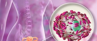
Ascariasis . The causative agent is the roundworm, which parasitizes the human small intestine. Infection occurs when invasive roundworm eggs are ingested from unwashed fruits, berries, vegetables, or water contaminated with sewage (Fig. 1.24).
Rice. 1.24. Ascariasis
A larva develops in the small intestine and migrates through the lymphatic and blood vessels within 7-10 days. Through the walls of blood vessels it penetrates into the lungs, then into the trachea, pharynx, is swallowed again and, when it re-enters the small intestine, develops into an adult helminth. The lifespan of roundworms is about a year.
In the migration stage, vascular damage occurs with the development of hemorrhages, eosinophilic infiltrates in various organs.
In the intestinal stage (two months from the onset of the disease), toxic-allergic and neuro-reflex manifestations of ascariasis are noted, the activity of the gastrointestinal tract is disrupted, nausea, unstable stools, pain in the epigastric region, dizziness, headaches, and increased nervous irritability occur.
Strongyloidiasis The causative agent is a nematode belonging to the genus Strongyloides stercoralis, a small filamentous parasite 2.2 mm long. It parasitizes mainly in the duodenum and upper parts of the small intestines, sometimes it can penetrate the ducts of the liver and pancreas. It enters the human body through the skin (when walking barefoot) and through the mouth (when consuming contaminated vegetables, fruits, water).
Strongyloides, unlike other helminths, can complete their life cycle in the human body without exiting into the external environment. Migrating with the bloodstream, the larvae damage the walls of blood vessels, leading to multiple hemorrhages in organs and ulcerations.
In addition to mechanical effects, they also cause toxic-allergic damage to organs and tissues.
Trichinosis . The causative agent of this type of helminthiasis is Trichinella. Human infection occurs by eating animal meat (domestic pigs, wild boars, badgers, etc.) infected with Trichinella. In the digestive tract, Trichinella are freed from capsules, quickly develop into adults, and after 3-4 days the females give birth to young Trichinella, which are carried throughout the body by the flow of blood and lymph, entering muscles rich in blood vessels (intercostal muscles, muscles of the diaphragm, tongue, eyes, pharynx ). Coiling into spirals, they are encapsulated and subsequently calcified, remaining viable for several decades. The intestinal stage lasts about 8 weeks.
Trichinella has a toxic-allergic effect on the body. By sensitizing the body, they lead to capillaropathy, edema, and muscle infiltrates. In addition, they cause trauma to the tissues in which they parasitize.
Trichostrongylosis . The causative agent is a small round helminth Trichostrongylidae. It parasitizes the small intestine of herbivores (cattle and small livestock). It is less common in humans. Infective larvae from animals enter the human intestine with contaminated water or food, where they develop into an adult parasite.
Trichuriasis is one of the most common helminthiases.
The causative agent of trichocephalosis is the whipworm (Trichocephalus trichiuris), which parasitizes the large intestine, mainly the cecum. Attached to the intestinal wall with a thin (hair-like) head end.
Human infection occurs when ripe whipworm eggs enter the mouth with unwashed vegetables, fruits and various food products, onto which they are carried by flies, dust or other means.
A larva develops in the intestines, turning into a mature helminth after a month.
The lifespan of a whipworm is about 5 years.
The whipworm, which fixes itself on the intestinal wall, injures the intestinal mucosa, causing inflammation, swelling of the mucous membrane, and reflex changes in other abdominal organs.
Being a hematophage, whipworm leads to the development of hypochromic anemia, sensitizes the body and has a toxic effect on vital organs and systems.
Enterobnoz . The disease is caused by pinworms, which parasitize the upper part of the large intestine. With intense infestation, pinworms can also be found in the lower part of the small intestine, in the appendix.
Eggs are laid not in the intestines, but in the perianal folds. They develop both in the external environment and on the human body, and therefore repeated self-infection is possible. From a pinworm egg that enters the human digestive tract, a larva is formed, which within 3 weeks turns into a mature pinworm.
Pinworms have a mechanical, toxic and allergenic effect on humans. Inflammatory processes occur in the intestinal tissues, since helminths promote the penetration of pathogenic microbes into the tissues. Inflammatory processes appear in the perianal area, urinary tract and genitals (especially in girls).
Opisthorchiasis . The causative agent is the cat fluke or Siberian fluke. It parasitizes the bile ducts of the liver, gall bladder and pancreatic ducts of humans, cats, dogs and some wild animals. It is characterized by focal distribution in the river basins of the Dnieper, Kama, Volga, Don, Donets, Northern Dvina, Pechora, Neman, Ob, Irtysh.
Infection occurs when eating raw thawed, frozen, lightly salted, insufficiently fried fish, mainly carp species (ide, chebak, dace, etc.). The cat fluke parasitizes the human body for a long time, often 20-40 years.
Opisthorchis suckers and spines covering their body injure the mucous membrane of the bile and pancreatic ducts, causing an inflammatory process. The accumulation of a large number of helminths impedes the outflow of bile and pancreatic juice.
Hyperplasia of the glandular epithelium in opisthorchiasis predisposes to the growth of tumors in the liver.
The toxic and neuro-reflex effects of the helminth can lead to dysfunction of a number of organs and systems (stomach, intestines, cardiovascular and nervous systems). In the initial period, severe allergization of the body is possible (eosinophilia in the blood).
Alveococcosis . The causative agent is the larval stage of alveococcus. The larvae, which are microscopic in size and resemble a bubble, most often parasitize the liver, sometimes in other organs in the form of large clusters. This type of helminthiasis, in addition to humans, affects rodents. The sexually mature stage of alveococcosis is observed in dogs, cats, foxes, arctic foxes, and wolves, from which humans become infected by drinking water from contaminated reservoirs or wild berries collected in the habitats of animals with alveococcosis.
The accumulation of alveococcal larvae in the liver, like tumor formations, infiltrates nearby organs and tissues, significantly impairs blood circulation, leading to degenerative changes and tissue atrophy. In addition, alveococcus larvae have a toxic and allergenic effect on the entire body.
Hymenolepidosis . The disease is caused by the dwarf tapeworm, which parasitizes the small intestine and passes through the human body as a larval and adult stage, i.e. man is its successively intermediate and final master. Human infection occurs by ingesting helminth eggs that come into contact with a patient or objects contaminated with his feces.
In the gastrointestinal tract, the helminth egg is freed from the shell, the oncosphere is attached to the intestinal villi with hooks and forms cysticercoids, which turn into sexually mature helminths. The development of the parasite ends within 3 weeks. In this case, inflammatory changes in the mucous membrane occur in the intestines, often with ulceration, and toxic-allergic reactions are also observed.
Rice. 1.25. Teniarinhoz. Pork tapeworm development cycle
Diphyllobothriasis . The causative agents of the disease are various types of tapeworms - wide, small, narrow, etc. The most common is the wide tapeworm, the length of which reaches 9 m, the life expectancy is tens of years. The definitive hosts are humans, dogs, cats, foxes, bears, the first intermediate hosts are Cyclops crustaceans, and the second hosts are freshwater fish (pike, burbot, ruffe, whitefish, ide). A person becomes infected by eating raw or half-raw fish or insufficiently processed caviar if they contain helminth larvae.
The tapeworm, attaching to the intestinal mucosa, injures it. Clusters of tapeworms can cause intestinal obstruction.
By absorbing vitamin B, the broad tapeworm leads to hypovitaminosis B12, a disruption of the biosynthesis of folic acid carried out by the intestinal microflora. This is the main reason for the development of anemia.
Teniarinhoz . The causative agent is bovine tapeworm. A person becomes infected by eating raw or insufficiently heat-treated cattle meat that contains bovine tapeworm larvae (Finns).
Infection is possible when tasting raw minced meat.
The development of taeniasis occurs with a change of hosts. The definitive host is humans, in whose small intestine the taeniids parasitize at the sexually mature stage; the intermediate host is cattle. In the small intestine of a person, a sexually mature helminth develops from swallowed finna within about 3 months, which has a toxic, mechanical and allergenic effect on the human body, injures the intestines, and inhibits the secretory function of the stomach.
Tennose . A disease caused by pork tapeworm (Fig. 1.25), which, unlike bovine tapeworm, can parasitize the human body in both the mature and larval stages. In the latter case, cysticercosis occurs. Infection with taeniasis occurs when eating raw or insufficiently heat-treated pork meat affected by Finns. An adult helminth develops in the small intestine from the finna.
Pork tapeworm has a mechanical, toxic-allergic and reflex effect on the human body. If oncospheres of a pork tapeworm enter the human stomach, which is possible during antiperistaltic movements of the intestine, if it contains sexually mature forms of pork tapeworm, as well as when consuming contaminated food products or from contaminated hands, cysticerci are carried by the bloodstream into various tissues and organs. Cysticerci can affect various parts of the brain, the organ of vision, the heart muscle, lungs, and diaphragm. The lifespan of cysticerci is calculated in years, sometimes decades.
Echinococcosis . The disease is caused by echinococcus in the larval stage. In the sexually mature stage, it parasitizes in the intestines of dogs, wolves, and jackals. The direct source of human infection with echinococcosis is dogs, which become ill by eating animal organs infected with echinococcosis. There may be helminth eggs on dogs' fur.
Echinococcus larvae have a mechanical, toxic-allergic effect on the human body, promote the development of immunological reactions, affect the liver, and less often the lungs, kidneys, and peritoneum. The bladder-shaped larva lives in the human body for many years. By squeezing tissue, blisters impair blood circulation and cause functional disorders. Suppuration and rupture of blisters with contamination of neighboring organs and anaphylactic shock are possible.