Osteomyelitis of the jaw
In surgical dentistry, osteomyelitis of the jaw is considered a common disease. Osteomyelitis of the jaw (ICD 10 code M.86) - (osteomyelitis from the Greek osteon bone + myelos bone marrow) is a severe inflammation affecting soft tissue elements, bone tissue and periosteum. It is accompanied by necrotic damage (decomposition) of the bone, which subsequently leads to its deformation. The development of the pathological process is provoked by bacteriological microorganisms that penetrate the jaw bone together with infected blood or in response to post-traumatic damage - fractures and wounds.
Osteomyelitis of the jaw (ICD code M.86) is a serious pathogenic process that, without timely treatment, can lead to complications. The risk group consists of male patients under 40 years of age. The primary and main thing is to identify the symptoms of the disease, thanks to a clear differential diagnosis. Thus, dental surgeons from the West Dental family clinic network in Yanino-1 and Vsevolozhsk will help diagnose and treat the problem at a reasonable price.
The causes and symptoms of the disease, which specialist to contact and how to treat - we will consider below in the article.
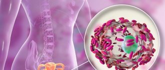
Vertebral infection
Not so long ago, this disease was considered incurable, since it was impossible to remove the infection from the skeletal system. Today, the process is successfully stopped through an operation during which infected tissue is removed, associated with intensive antibacterial drug action.
Spinal osteomyelitis is treated surgically
By the way. Osteomyelitis often occurs in children, affecting the bones in the legs and arms. Adults mainly suffer from an inflammatory process localized in the bones of the spine. In some cases, if an adult patient has diabetes, the process begins to develop from the lower extremities, if there are trophic ulcers on them.
Infection of the vertebrae cannot be considered a common cause of back pain, especially if the patient is young and, in principle, healthy. But cases of the disease are becoming more frequent, and osteomyelitis (the second name is spondylitis) is gradually becoming an urgent problem.
By the way. The most common route of infection into the vertebral segments is hematogenous, which is carried out through the bloodstream. In some episodes of infection, microbes appear in the vertebrae after various medical diagnostic procedures, such as urological cystoscopy.
Etiology of causes of osteomyelitis
To understand how bone tissue becomes infected, it is necessary to remember how the blood supply occurs in the spine.
Initially, the body of each vertebral segment is isolated from the rest by a cartilaginous intervertebral septum. But along the ridge there are arteries that branch from the neck and carry blood to all vertebral tissues, and, of course, through the vascular network, to the vertebrae. The lumbar zone receives nutrition with blood passing through the arterial canals of the lumbosacral zones. Each vertebra is entangled with venous vessels that connect to form veins.
Blood supply to the spinal cord
In this way, viruses can penetrate the vertebra through both veins and arteries. As soon as the pathogens find themselves in the bone tissue, the inflammatory stage is activated, during which it dies.
Accumulating obsolete microorganisms that are not removed in a timely manner form purulent accumulations in the spinal tissue. The outer vertebral membrane is destroyed, and the process spreads to other parts of the spinal column.
Who is at risk
Bone tissue is designed to be resistant to infection. Therefore, for osteomyelitis to begin, additional factors contributing to the development of the disease are necessary. These factors weaken bone tissue, increasing its vulnerability and reducing resistance to infection.
- Injuries accompanied by bone fractures, when infection can easily be introduced into the vertebra both by the bloodstream and by the tissue located next to the fracture.
- An operation to reposition bone fragments or a deep puncture was performed.
- Impaired blood circulation in the event that blood vessels are damaged or the blood flow is unstable. A deficiency of immune cells occurs, which reduces the ability to resist infection.
- Disease diabetes mellitus.
- Weakening and pathology of peripheral arteries, which may be associated with smoking.
- Infections.
- Deep carious dental lesions.
- Hereditary hemoglobinopathy expressed by sickle cell anemia.
- Placement of a dialysis or medical catheter.
- Intravenous drug use.
Causes of osteomyelitis
By the way. Despite the fact that catheters are absolutely necessary in various situations - dialysis, emptying the bladder, administering medications into veins for a long time - they carry perhaps the highest risk of unsterility and possible infection.
Greatest risk of infection during catheterization
Based on this, there are groups of patients who are most susceptible to osteomyelitis:
- elderly people;
- patients with drug addiction;
- people whose immune systems are weakened.
Drug-dependent individuals are most at risk of contracting osteomyelitis
Long-term use of corticosteroids for rheumatoid arthritis and other diseases, diabetes accompanied by insulin dependence, AIDS, people who have received organ transplants, suffer from malnutrition, and have cancer - all these patients have reduced immunity and an increased ability to resist infection at the bone-cellular level.
Causes
The main provocateur of the formation of osteomyelitis in the upper and lower jaw is considered to be pathogenic microflora - streptococci and anaerobes, present in the oral cavity of every person. They aggravate the infection, affecting the soft and hard components of the mouth. There are several ways to penetrate deeper into the pathological process:
- Missed dental caries, which destroyed the enamel with dentin and penetrated into the pulp chamber.
- Traumatization – crack, violation of the sealing material. The damage must be deep (fracture of the HF or LF) to provoke an infectious process (staphylococci).
- Chronic infection occurs in other parts of the body. Pathological microorganisms can reach the jaw through the lymph nodes or blood vessels (ENT organs).
In most patients, osteomyelitis is located on the lower jaw, since the lower units are more likely to be affected by caries or trauma. Also, people with immunodeficiency and blood diseases are at risk, because they have difficulty healing wound surfaces and the immune function of the body is weakened.
Other factors:
- smoking;
- alcohol;
- Diabetes type I and II;
- eating disorders;
- syphilitic infection;
- radiation and chemotherapy.
If you miss and do not treat acute osteomyelitis in the upper or lower jaw, then the process becomes chronic, which is more difficult to treat and leads to many complications.
Classification
According to the method of penetration of osteomyelitis infection on both jaws, a classification has been compiled:
- Hematogenous - pathogenic microorganisms move through the blood from other foci of infection. This feature is characteristic of secondary pathology of the upper oral part, which develops against the background of another disease (scarlet fever, diseases of the ENT organs). The bone elements of the oral apparatus are primarily affected, followed by the gingival tissue.
- Odontogenic – infection comes from a tooth, damaged pulp or root (pulpitis, periodontitis, cyst, alveolitis).
- Traumatic – advanced fractures and injuries of the maxillofacial area.
According to the symptoms, the process happens:
- spicy;
- subacute;
- chronic.
By distribution:
- local (with clear boundaries);
- vague (diffuse).
Features of symptoms
The main noticeable symptom is called pain, but in fact it is far from the first, it’s just that the symptoms of the disease are not obvious at the initial stage. In addition, pain also occurs with all other diseases of the spine, so even with the onset of pain, the infection goes unnoticed for a long time, for months. Patients begin to worry when general intoxication, a feeling of chills, a rise in temperature, increased sweating and weight loss are added to the pain, and the pain begins to manifest itself especially strongly at night.
The most obvious symptom of osteomyelitis is pain against the background of general intoxication and poor health
In accordance with the infecting ability of pathogens, three types of osteomyelitis developing in vertebral structures are distinguished.
- Spicy.
- Primary chronicle.
- Chronic.
Table. Symptoms of osteomyelitis by type.
| ● multi-stage sharp pain, increasing in amplitude at night; | ● mild episodic pain syndromes; | ● mild pain of unclear localization; |
| ● excessive sweating; | ● chills; | ● rises to 37°C, usually in the evening; |
| ● strong rise in temperature to 39°C and above; | ● mild intoxication; | ● pain may intensify at night; |
| ● convulsive conditions; | ● slight temporary increase in temperature; | ● feeling of stiffness of the spine; |
| ● hypotension; | ● increased sweating; | ● sweating; |
| ● swelling of soft paravertebral tissues. | ● general state of lethargy; | ● slight swelling and numbness of the spine. |
| ● vascular tension; | ||
| ● pain in the back muscles. |
By the way. Almost always, patients mistake the primary chronic and stable chronic form for manifestations of osteochondrosis or other vertebral and joint pathologies, or diseases of internal organs such as the lungs and heart.
During its development, osteomyelitis inflames not only the bone tissue, but also spreads to the entire blood supply system in the spine. This can lead to sepsis, disability and death.
Occasionally, the infection involves nerve structures in the inflammatory process by moving into the spinal canal, causing the formation of an epidural abscess. It puts pressure on the nerves, and if the source of infection is located in the neck or chest area, it can result in paraplegia (paralysis of either lower or upper limbs in pairs) and quadriplegia (simultaneous paralysis of all limbs).
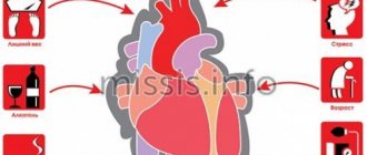
By the way. Victims of osteomyelitis most often become older men, especially those who experience frequent stress and excessive tension and have a large number of different diseases. This reduces immunity and makes it impossible to cope with bacteria that cause inflammation.
Victims of osteomyelitis are most often older men
When the disease takes a protracted chronic form, the number of purulent foci in the bone increases and their size also increases. New inflamed tissue may begin to grow, which comes into conflict with healthy tissue and infects it, expanding the area of infection. The bone marrow does not remain aloof from the inflammatory process, since the accumulating pus interferes with the normal functioning of the blood cells that provide it with nutrition.
Osteomyelitis of the upper and lower jaw: symptoms
The course of the disease process is slow, and the first sign is pain in the area of the affected unit.
Then joins:
- spread of pain to nearby segments;
- swelling and redness of the gum tissue;
- periodontal damage, which causes mobility of units;
- pain in the temporal region;
- numbness of the chin;
- difficulty eating;
- speech defects;
- unpleasant odor from the mouth;
- increase in l/u;
- swelling in the problem area.
Acute osteomyelitis on the lower jaw (ICD M.86) develops very rapidly in conjunction with an increase in temperature and chills. In the absence of outflow of pus, abscesses and phlegmons form, which requires immediate surgery. This pathology in a child may be confused with mumps (mumps).
With osteomyelitis on the lower jaw (ICD-10 code M.86), in addition to damage to the alveolar part and body, the inflammatory process can spread to its branch and processes. One important feature is complications from the soft tissue elements around the NP - many muscles and tissue spaces.
After the acute period (1-2 weeks), the subacute period begins, after the formation of a fistula for the discharge of pus. The condition improves, pain decreases, and the mobility of units increases—gastrointestinal diseases develop. The subacute form becomes chronic, which lasts several months. As a result, in adults the clinical history ends with the removal of necrotic tissue, with a favorable outcome.
Osteomyelitis is a disease characterized by the development of a purulent-inflammatory process in the bone caused by pathogenic microorganisms.
The main routes of infection into bone tissue are:
- hematogenous (with blood flow). Pathogenic microorganisms from any source of infection (for example, from carious teeth, inflamed tonsils) penetrate into the bone tissue through the bloodstream, leading to the development of osteomyelitis. In hematogenous osteomyelitis, the inflammatory process begins with the inner layers of the bone - the bone marrow and spongy bone are primarily affected. This type of osteomyelitis often occurs in childhood. This is due to increased blood supply to growing bones in children.
- direct infection of bone tissue. Direct infection of bone tissue can occur with open fractures, extensive, deep wounds. In some cases, pathogenic bacteria can penetrate this route during various orthopedic interventions.
- infection from nearby purulent foci (contact route). Pathogenic bacteria penetrate bone tissue from long-existing purulent-inflammatory foci in the surrounding soft tissues. An example is the development of osteomyelitis during felon (purulent inflammation of tissue on the finger).
The progression of the purulent-inflammatory process leads to the destruction of bone tissue. Its blood supply is disrupted, which further enhances necrotic processes in the bone. The transition of the inflammatory process to nearby tissues can lead to the formation of abscesses (circumscribed accumulations of pus), drainage of purulent contents to the outside, the development of arthritis (inflammation of the joints), and other complications.
Treatment of osteomyelitis requires the use of massive doses of antibacterial drugs. Surgical treatment is aimed at removing areas of dead bone tissue, purulent contents, and draining the inflammatory focus.
English synonyms
Osteomyelitis.
Symptoms
The main symptoms of osteomyelitis include:
- increase in body temperature
- severe pain in the area of the affected bone
- redness, swelling, increased temperature of the skin in the area of the purulent-inflammatory process in the bone
- decreased range of motion in joints adjacent to the area of inflammation
- formation of fistulas (channels connecting the purulent focus with the external environment) with purulent discharge
General information about the disease
Osteomyelitis is a purulent-inflammatory process in bone tissue caused by pyogenic microorganisms.
In children, the most common cause of osteomyelitis is the spread of infection through the hematogenous route (through the bloodstream) from some source of inflammation (for example, in the presence of boils, purulent tonsillitis).
A special feature of bone tissue in children is its abundant blood supply. In most cases, osteomyelitis develops in the growth areas of the bones (metaphyses). In this case, the long bones of the limbs are most often affected. Pathogenic microorganisms are carried through the bloodstream into these parts of the bone, which leads to the development of osteomyelitis.
With hematogenous osteomyelitis, the purulent-inflammatory process begins from the internal parts of the bone. In this case, the bone marrow is affected, then the spongy bone. Subsequently, the process moves to the compact substance (the outer dense layer of bone) and the periosteum.
Due to the peculiarity of the localization of the osteomyelitic process, children may subsequently experience a slowdown in bone development due to damage to the growth zones by the inflammatory process.
In adults, hematogenous osteomyelitis is observed in 20% of cases. However, this disease is more common in men.
With increasing age, the incidence of vertebral osteomyelitis in adults increases. The infection can penetrate the spine hematogenously from inflammatory foci in soft tissues, lungs, genitourinary tract, and carious teeth. Two adjacent vertebrae with an intervertebral disc are affected.
With this form of osteomyelitis, pain occurs in the area of the purulent-inflammatory process. The duration of the disease development can be up to 3 months. Destruction of the vertebrae and compression of the spinal cord can lead to motor and sensory disorders in these patients.
Osteomyelitis, which develops as a result of direct infection or spread of infection from nearby lesions into bone tissue, occurs more often in adults. Processes in which the immune status of the body decreases and the blood supply to bone tissue is disrupted predispose to the development of this disease. These include:
- Diabetes mellitus is a disease in which there is an increase in blood sugar levels. In this case, various types of metabolism are disrupted. People with diabetes often develop various purulent-inflammatory processes. This is aggravated by the fact that as a result of disorders of the nerve fibers (polyneuropathy), sensitivity is impaired. This leads to the fact that various wounds often go unnoticed by the patient, and inflammation quickly develops in them.
- Sickle cell anemia is a hereditary disease characterized by the formation of abnormal hemoglobin protein in red blood cells. This causes a decrease in the life expectancy of such red blood cells and their increased destruction. In this case, hypoxia develops (a decrease in the amount of oxygen supplied to the tissues) and thrombosis of various vessels.
As a result of the formation of blood clots in the vessels of the bones, the blood supply to this area of \u200b\u200bbone tissue is disrupted and it dies. The addition of infection can lead to the development of osteomyelitis.
- Human immunodeficiency virus (HIV) – with this disease, the cells of the immune system are destroyed, which makes the body vulnerable to various infections.
- long-term use of glucocorticoids. Glucocorticoids are hormones of the adrenal cortex. Long-term use of this group of drugs for various diseases reduces the body's immune status.
Acute osteomyelitis can become chronic. Complicating the treatment of this form of the disease is the disruption of the blood supply to the bone, which is observed during the course of the purulent-necrotic process. This makes it difficult for antibacterial drugs to penetrate and reduces the effectiveness of the local immune response in bone tissue. In such cases, the radical treatment method is wide resection (removal of the affected bone tissue); in some cases, amputation may be required.
Who is at risk?
Risk groups include:
- persons with severe bone tissue injuries, deep wounds
- persons who have undergone orthopedic surgery
- persons suffering from diabetes mellitus
- persons with foci of infection in various organs and tissues (for example, long-term non-healing ulcers on the skin)
- patients whose vital functions require special medical devices (for example, various catheters) - these medical devices facilitate the penetration of pathogenic microorganisms from the external environment into the human body
- injection drug addicts.
Diagnostics
Diagnosis of osteomyelitis is based on identifying the symptoms of the disease, performing radiography, computed tomography and magnetic resonance imaging to detect purulent-inflammatory foci and characteristic changes in bone tissue.
For further treatment, it is of great importance to conduct laboratory tests of purulent discharge, bone biopsy materials from the source of inflammation to identify the type of infectious agent and its sensitivity to antibacterial drugs. This makes it possible to select the most effective antibiotics against a given pathogenic microorganism.
Laboratory research:
- General blood analysis. This analysis allows you to determine the number of red blood cells, hemoglobin, leukocytes, and platelets in the blood. With osteomyelitis, as a result of a pronounced inflammatory process in the bone tissue, an increase in the level of leukocytes will be noted.
- Erythrocyte sedimentation rate (ESR). A nonspecific indicator of the occurrence of various pathological processes in the body, mainly of an inflammatory nature. ESR is one of the indicators that allows you to assess the activity of the inflammatory process and its dynamics. With osteomyelitis, the ESR will be increased.
- Leukocyte formula. The leukocyte formula is the percentage of different types of leukocytes, which can change as a result of pathological processes in the body. With osteomyelitis, a shift in the leukocyte count to the left may be observed, which indicates the presence of a pronounced purulent-necrotic process in the bone tissue.
- Culture of flora with determination of sensitivity to antibiotics (purulent discharge, aspiration material (carried out using a special needle and syringe) and open bone biopsy).
The resulting samples are placed on special nutrient media that promote the growth of pathogenic microorganisms. This allows you to determine the exact type of infectious agent. Then antibiotics are selected that most effectively suppress these pathogens.
- Blood culture for sterility with determination of sensitivity to antibiotics. The principle of the method is similar to the previous study. Normally, human blood is sterile. In osteomyelitis, the causative agent of infection is found less frequently in the blood than in material obtained from the area of bone tissue damage.
Research:
- Radiography. X-ray of the bone allows you to identify areas of bone destruction in the area of the purulent-necrotic process. The disadvantage of the method is its low information content in the early stages of the disease, since it takes several weeks for the formation of pathological changes visible on radiography for osteomyelitis.
- Computed tomography (CT), magnetic resonance imaging (MRI). Computed tomography is based on the ability of X-ray radiation to penetrate tissues of varying densities. The principle of magnetic resonance imaging is the action of a strong magnetic field on the tissues and organs being studied.
Both studies use computer processing of the received information, resulting in the formation of layer-by-layer, highly informative images of the internal structures of the body. Computed tomography and magnetic resonance imaging can detect changes in the bone and surrounding tissues in osteomyelitis.
Treatment
Treatment of osteomyelitis requires the use of conservative and surgical methods. Conservative therapy consists of using antibiotics that are most effective against pathogenic microorganisms that cause a purulent-inflammatory process in bone tissue.
Surgical treatment consists of evacuation of purulent contents, removal of areas of necrotic (dead) bone tissue, and drainage of the purulent-inflammatory focus.
Prevention
There is no specific prevention of osteomyelitis. Persons with an increased risk of developing purulent-inflammatory diseases (for example, patients suffering from diabetes mellitus) should pay special attention to even minor damage to the skin - treat them thoroughly with antiseptics, use sterile dressings.
Recommended tests
- General blood analysis
- Erythrocyte sedimentation rate (ESR)
- Leukocyte formula
- Culture of flora with determination of sensitivity to antibiotics
Blood culture for sterility with determination of sensitivity to antibiotics
Diagnostics
If you experience pain in the tooth area of unknown etiology, or changes in the gums, contact your dentist (he has a Leica M320 operating microscope in his arsenal), who will then refer you to a surgeon or orthopedist as necessary. At the initial stage of pathology, the doctor chooses to collect complaints and study them, visual and instrumental examination. The degree of mobility of the units and their pain, the state of the oral mucosa is determined.
It is important to distinguish the acute period of the disease from periostitis, cysts and periodontitis, so the experience of a specialist is very important.
This is a purulent infectious process, which means that general blood and urine tests will be of research significance. To type the pathogen, a tank is performed. sowing of pus.
In the chronic stage, bone changes are already noticeable, so X-rays (CT) are necessary, for which West Dental has a panoramic CBCT Pax-i3D. The image shows sequestration and the depth of the process.
Symptoms
The clinical picture depends on the stage
of Acute Pain, mobility of the causative tooth and those located nearby. Opening the mouth, difficulty swallowing, breathing with a putrid odor. Swelling and redness of the gums. Feverish state, lack of appetite, lethargy, headache. Inflammation of the lymph nodes. Subacute
The inflammatory process subsides, suppuration decreases, the mouth opens freely.
Teeth mobility is maintained. Improvement in general condition, decrease in temperature (slightly above 37°C). Regional lymph nodes are dense but painless. Chronic
Reduced tissue infiltration, subsidence of pain.
The jaw in the area of the inflamed lesion is thickened; upon incision, fistulas with purulent contents are revealed. CT scan reveals roughness and unevenness of dying bone tissue. Exacerbation
The clinical picture of the acute stage is repeated - general signs of intoxication suddenly appear. Local symptoms increase - swelling of the inflamed area, soreness and mobility of teeth, the formation of purulent fistulas.
Complications
If you see a doctor in time, make a diagnosis and choose the right treatment, the prognosis will be favorable.
If you ignore all the conditions, complications will arise:
- Meningitis
- Abscess of the brain and lung.
- Orbital phlegmon.
- Sinusitis.
- Vein thrombophlebitis.
- Sepsis.
- Mediastinitis.
Pathological data require immediate assistance to prevent death.
The chronic process, with its long course, affects the soft tissue and bone areas of the maxillofacial area, and is accompanied by:
- traumatization;
- changes in the TMJ;
- the formation of adhesions in the joints and scars on the masticatory muscles;
Such disorders limit chewing movements or lead to immobility.
Treatment
The effectiveness of therapy depends on the initial causes of the formation of the inflammatory process. Therefore, in osteomyelitis of the jaw, comprehensive treatment is important, not only by a dentist, but it is recommended to go for a consultation with doctors of medical specialties.
- Sanitation of the oral cavity with antiseptic solutions. Further spread of infection to surrounding structures is prevented. Also, necrotic soft tissue formations are eliminated.
- Anti-inflammatory drugs. Minimize intoxication of the body.
- Immobilize the fracture, and if there is a tooth in it, then remove it.
- Taking antibiotics, regardless of provoking factors.
- Intraosseous lavage may be necessary and will provide positive results. Minimizes the occurrence of complications and quickly prevents the growth of a harmful process.
- In case of a fistula, sequestrectomy is performed - necrotic areas of bone tissue are eliminated under anesthesia, taking into account the extent of the damage.
- If the teeth are mobile, they are splinted.
- After eliminating symptoms and carrying out all interventions, physical therapy, vitamins and immunomodulators are recommended.
Prevention of such pathology consists only of timely and regular visits to the dentist’s office and treatment of pathological processes in the oral cavity. Do not treat yourself at home, so as not to make it worse. Also, it is necessary to strengthen the body’s immune system, avoid injuries and not let the pathological process become chronic.
Prevention
Preventive measures are not only the key to preventing the development of osteomyelitis, but also a factor that reduces the risk of complications and shortens the recovery period if the disease cannot be avoided:
- Timely treatment of caries, even if it has no clinical manifestations.
- Maintaining normal immune status through regular physical activity, rational and nutritious nutrition.
- Sanitation of all chronic foci of infection in the body.
- In case of injury, in the postoperative period or after tooth extraction, compliance with all preventive medical prescriptions.
In conclusion, it should be noted that, despite all the achievements of modern medicine, osteomyelitis of the jaw in adults and children does not lose its relevance. Timely detection of its signs and adequate treatment increase the patient’s chances of a full recovery and maintaining a high quality of life.
»