Full text of the article:
Haemophilus influenzae infection includes a whole group of infectious diseases caused by Haemophilus influenzae. Haemophilus influenzae can come in different forms and over time tend to develop resistance to antibacterial drugs. The infection was first isolated by the German scientist Richard Pfeiffer at the end of the 19th century.
General information
Haemophilus influenzae infection (Hib infection) is caused by Haemophilus influenzae type b. It can cause acute infectious diseases - purulent meningitis, pneumonia (pneumonia), epiglottitis (inflammation of the epiglottis), arthritis (inflammation of the joints), as well as purulent damage to the whole body - sepsis. Hemophilus influenzae infection is characterized by predominant damage to the respiratory system, central nervous system and the development of purulent foci in various organs. The bacterium H. influenzae is localized in the nasopharynx, from where it can be transmitted to other people by airborne droplets. Only a very small number of those in whom the pathogen is localized in the nasopharynx develop a disease with clinical manifestations. However, carriers of H. influenzae in the nasopharynx are an important source of spread of the pathogen.
Consequences of hemophilus influenzae infection
Hemophilus influenzae infection can provoke the development of invasive (generalized) forms of diseases: secondary damage to the membranes of the brain (meningitis), joints (arthritis), lungs (pneumonia), pericardium (pericarditis), subcutaneous fatty tissue (cellulitis), epiglottis (epiglottitis).
Timely etiotropic treatment prevents the development of such complications:
- septicemia;
- respiratory failure;
- convulsions;
- disturbances of consciousness;
- neurological deficit;
- infectious-toxic shock.
Chance of getting sick
There are several risk groups for this disease. Firstly, children most often suffer from this infection. According to various studies, carriage of Haemophilus influenzae in children reaches 50%. Infants 6-12 months of age who are bottle-fed and do not receive small additional amounts of maternal antibodies in breast milk have a particularly high risk of developing the most severe forms of infection - pneumonia and meningitis. For this reason, artificial feeding is an additional indication for vaccination against Hib infection, starting from 3 months of age.
People of extreme ages (children under 2 years of age, the elderly) and people with low socioeconomic status are also susceptible to hemophilus influenzae infection. In addition, people with low socio-economic status, extremely weakened and suffering from alcoholism, patients with lymphogranulomatosis (Hodgkin's disease), sickle cell anemia are susceptible to the disease; persons who have undergone splenectomy (removal of the spleen).
The incidence increases in late winter and spring. In recent years, the incidence of morbidity in adults has increased markedly.
Introduction
Nongonococcal urethritis (NGU) is the most common disease of the lower urogenital tract in men [1]. Epidemiological data indicate a significant increase in the incidence of NGU over recent decades - from 50 million cases per year at the end of the last century to almost 90 million cases annually today [2]. Inflammation in the urethra can lead to urethrogenic spread of infection with the development of pathology in the area of the prostate-vesicular complex and scrotal organs, which contributes to a decrease in the reproductive potential of men [3, 4]. In addition, NGU in men is often associated with pelvic inflammatory diseases in their female sexual partners, and thereby affects women’s reproductive health [5–7].
The medical and social significance of NGU is due to the fact that the disease occurs in predominantly young men, which, in turn, increases the risk of infection and transmission of sexually transmitted infections (STIs), including HIV [8, 9].
Determining the reasons for the development of NGU in some cases presents certain difficulties. One of the most difficult stages in diagnosing the disease is to establish the etiological factor for the development of inflammation in the urethra, which directly affects the choice of adequate etiotropic treatment and helps to increase its effectiveness.
According to modern concepts, the generally accepted etiological agents of NGU are C. trachomatis, M. genitalium and T. vaginalis [10]. Less common causes of NGU are infections caused by herpes viruses types 1 and 2, Epstein-Barr virus [11] and adenoviruses [12, 13], as well as U. urealyticum in a highly concentrated bacterium at the locus [14, 15 ]. Among the possible causes of inflammation in the urethra in men, a number of aerobic bacteria are considered - Haemophilus spp. [16, 17], Neisseria meningitidis [18, 19], Moraxella catarrhalis [20, 21], Streptococcus pneumoniae [22, 23], protozoa - Giardia lamblia, Entamoeba histolytica [24, 25], but controlled studies regarding the association of these microorganisms with NGU, little research has been done.
According to experts, after excluding “recognized” pathogens related to STIs, in more than 35% of cases the etiological factor of inflammation in the urethra cannot be found [26]. In the literature, such urethritis is called pathogen-negative, or idiopathic [26, 27]. They can be caused by both non-infectious causes - trauma to the urethra, allergic reactions, metabolic disorders in the body, congestion (from Latin: tide, accumulation - for example, fluid) in the pelvic organs [28], and previously unidentified microorganisms or agents not currently recognized as etiological.
Research in recent years has shown the possibility of exchange of microflora between sexual partners [29, 30], when microorganisms found in the lower parts of the reproductive tract in women can be considered as possible etiological agents for the development of inflammation in the urethra in men.
One of the most common infectious diseases among women of reproductive age is bacterial vaginosis (BV). Its prevalence varies from 10–30% in developed countries to almost 70% in some regions of Africa [31–33]. BV is associated with an increased risk of perinatal complications, impaired reproductive health of women, and a negative impact on the quality of life of patients [34–36].
We conducted a longitudinal study that included examination of sexual partners and the formation of a control group to assess the clinical features of urethritis in men associated with BV in their sexual partners.
Symptoms
The duration of the incubation period is difficult to determine, since the disease is often a consequence of the transition of a latent infection to a manifest one. Both a local inflammatory process of the mucous membrane of the upper respiratory tract and diseases caused by hematogenous dissemination can develop.
Complications after an illness
Often, hemophilus influenzae infection occurs in children in the form of a common respiratory infection with corresponding symptoms. However, cases of more severe forms of infection are not uncommon. The most severe form of Haemophilus influenzae infection is purulent meningitis. According to some data in Russia, in children under 6 years of age, up to a third of all cases of purulent meningitis are caused by Haemophilus influenzae type b. Haemophilus influenzae meningitis is difficult to treat because its causative agent produces enzymes that make it resistant to antibiotics (about 20-30% of Haemophilus influenzae bacilli isolated from patients are not sensitive to many antibiotics). Therefore, treatment results are not always successful, and mortality in severe forms of the disease can reach 16–20%.
A third of patients who have had hemophilic meningitis develop irreversible neurological complications - seizures, delayed neuropsychic development, deafness, blindness, etc.
Pneumonia caused by Haemophilus influenzae type b occurs predominantly in children aged 2 to 8 years, and its course in 60% of cases also has various complications, including those affecting the heart and lungs.
Up to half of all purulent infections of the ear, throat, and nose are associated with hemophilus influenzae infection, in particular, recurrent purulent otitis (inflammation of the middle ear) and sinusitis (inflammation of the paranasal sinuses).
Hemophilus influenzae sepsis most often develops in children 6-12 months of age who are predisposed to this disease. It proceeds violently, often like lightning, with septic shock and rapid death of the patient.
Purulent arthritis is a consequence of hematogenous introduction of Haemophilus influenzae and is often accompanied by osteomyelitis.
What else is prescribed with this study?
Meningococcus, Haemophilus influenzae, streptococcus (Neisseria meningitidis, haemophilus influenzae, streptococcus pneumoniae, PCR) scraping, quality.
19.52.2. Scraping 2 days
610 ₽ Add to cart
Determination of pathogen sensitivity to antibacterial drugs (DDM)
01. 1 day
410 ₽ Add to cart
Determination of pathogen sensitivity to an expanded range of antibacterial drugs
02. 2 days
600 ₽ Add to cart
Determination of pathogen sensitivity to an expanded range of antibacterial drugs, with determination of the minimum inhibitory concentration (MIC, MIC)
13. 2 days
960 ₽ Add to cart
Sowing the discharge of the upper respiratory tract for microflora (nose, pharynx)
120.2. Scraping 4 days
650 ₽ Add to cart
Treatment
Haemophilus influenzae Hib infection is difficult to treat, since Haemophilus influenzae is record resistant to antibiotics. For this reason, even timely treatment with modern antibiotics is often ineffective. Penicillin antibiotics, erythromycin, chloramphenicol, tetracycline - the percentage of Haemophilus influenzae resistance to them is 80-100% (of the number of isolated bacterial samples). Without etiotropic therapy, some forms of hemophilus influenzae infection (meningitis, epiglottitis) almost always end in the death of the patient. It is necessary to begin treatment for hemophilus influenzae infection as early as possible.
Effectiveness of vaccination
In 2000, before the widespread introduction of Hib vaccine in resource-limited countries, Hib disease accounted for 8.13 million cases of serious illness in children aged 1–59 months (range, 7.33–13.2 million cases). ) and 371,000 deaths (fluctuation amplitude – 247,000=527,000). By 2008, when 136 WHO Member States introduced the Haemophilus influenzae vaccine, there were an estimated 203 000 deaths due to Hib infection in children under 60 months of age (range 136 000–281 000).
The effectiveness of Hib vaccines is 95-100%; the protective antibody titer lasts for at least 4 years.
Among vaccinated people in 10 European countries, Israel and Australia, Hib infection developed with a frequency of only 2 cases per 1 million; 18% of these children had problems, including prematurity; 33% had low immunoglobulin levels.
results
In 13 (7.8%) of 166 men initially included in the study, examination revealed microorganisms whose etiological role in the development of urethritis is considered proven (N. gonorrhoeae, C. trachomatis, M. genitalium, T. vaginalis, HSV 1, 2). These patients, as well as their sexual partners, were excluded from the study. Thus, 153 men and 161 women continued to participate in the study. Male study participants were divided into 2 clinical groups in accordance with the examination results:
— Group 1 — men with clinical and/or laboratory signs of urethritis (n=94);
— Group 2, control — men without clinical and/or laboratory signs of urethritis (n=59).
In order to trace a possible connection between the development of urethritis in men and bacteria associated with BV in women, we assessed the state of the microflora of women who were sexual partners of men in the study groups. Depending on the presence or absence of BV, the sexual partners of men who took part in the study were assigned the status of BV-positive or BV-negative (Table 1) .
Table 1. Distribution of patients into clinical groups depending on the presence of inflammation in the urethra and the BV status of their sexual partners
| Study Participants | Number of participants (%) |
| Group 1 (patients with urethritis) | 94 (100) |
| subgroup 1A (BV+) | 43 (46) |
| subgroup 1B (BV–) | 51 (54) |
| Group 2 (control) | 59 (100) |
| subgroup 2A (BV+) | 13 (22) |
| subgroup 2B (BV–) | 46 (78%) |
It was found that 46% of female sexual partners of men in group 1 (men with urethritis) had BV-positive status. Moreover, in group 2 (control), only 22% of sexual partners were diagnosed with BV.
Thus, within each clinical group, 2 subgroups were divided depending on the state of the microflora of sexual partners (presence or absence of BV).
Sociodemographic and behavioral characteristics of men
Among men with signs of inflammation in the urethra (group 1) and among men in the control group (group 2), there were no significant differences in the level of education, monthly income, as well as in the average age of the participants, approximately equal in both groups - 35.08 ± 8 .12 and 35.3±8.33 years (Table 2) . However, it was noted that patients with urethritis and BV-positive sexual partners were on average 3.6 years younger than patients with urethritis whose sexual partners had a negative BV status (the average age of patients was 33.1 ± 7.74 and 36.7±8.15, respectively). In addition, an analysis of the distribution of men with inflammation in the urethra by age category depending on the BV status of their sexual partners with a high level of confidence (p≤0.01) showed that 53.4% of patients with urethritis and a positive BV status of their sexual partners ( subgroup 1A) were at the age of greatest sexual activity (from 18 to 30 years). Only 25.4% of patients with negative BV status of sexual partners belonged to this age group (subgroup 1B). The peak of visits from patients in this group was noted in the age range of 31–45 years, while at the same time, among patients with urethritis over 45 years of age, BV-positive status of sexual partners was established more than 2 times less often than BV-negative status.
Table 2. Sociodemographic and behavioral characteristics of men
| Characteristic | Retreat patients | Control group (n=59) | pV | |
| subgroup 1A (BV+) (n=43) | subgroup 1B (BV–) (n=51) | |||
| Average age, years | 35,08±8,12 | 35,3±8,33 | ||
| 33,1±7,74 | 36,7±8,15 | |||
| Education (%): | ||||
| higher | 24 (55,8) | 30 (58,8) | 38 (64,4) | 0,66 |
| average | 19 (44,2) | 21 (41,2) | 21 (35,6) | |
| Level of monthly income, thousand rubles. (٪): | ||||
| <60 | 18 (41,9) | 17 (33,3) | 26 (44) | 0,49 |
| ≥60 | 25 (58,1) | 34 (66,7) | 33 (56) | |
| Circumcision (%): | ||||
| There is | 3 (7) | 14 (27,4) | 13 (22) | 0,04 |
| No | 40 (93) | 37 (72,6) | 46 (78) | |
| History of STI (٪): | ||||
| There is | 13 (30,2) | 20 (39,2) | 19 (32,2) | 0,62 |
| No | 30 (69,8) | 31 (60,8) | 40 (67,8) | |
| Number of sexual partners in the last 6 months (٪): | ||||
| <2 | 17 (39,5) | 27 (52,9) | 41 (69,5) | 0,01 |
| ≥2 | 26 (60,5) | 24 (47,1) | 18 (30,5) | 0,01 |
| Practice of unprotected sex during the last 1 month (٪): | ||||
| vaginal | 43 (100) | 51 (100) | 59 (100) | — |
| oral | 29 (67,4) | 29 (56,9) | 30 (50,8) | 0,25 |
| anal | 3 (7%) | 11 (21,5%) | 2 (3,4%) | 0,01 |
Sociodemographic and behavioral characteristics of male study participants are presented in Table. 2 .
There were no significant differences among men of different clinical groups in the presence of a history of STIs, however, a study of sexual history showed that men with a positive BV status of sexual partners were significantly more likely (p≤0.01) to have had 2 or more sexual partners in the last 6 months than in men with negative BV status of sexual partners and healthy men in the control group - 60.5, 47.1 and 30.5%, respectively. At the same time, the practice of unprotected anal intercourse with a high level of reliability was more often (p≤0.01) indicated by patients with urethritis and negative BV status of sexual partners (subgroup 1B).
Only 7% of men in subgroup 1A had no foreskin, while in 27.4% of patients in subgroup 1B and 22% of patients in group 2 it was preserved (p≤0.05).
Clinical manifestations of urethritis
Symptoms of urethritis as assessed by patients
An analysis of the clinical symptoms of urethritis in men in clinical groups is presented in Table. 3 .
Table 3. Comparative analysis of clinical symptoms of urethritis in men in clinical groups
| Symptom | Subgroup 1A BV+ (%) | Subgroup 1B BV– (%) | pV | ||
| Character of discharge from the urethra: | |||||
| transparent | 6 (13,9) | 12 (23,5) | 0,24 | ||
| cloudy | 9 (20,9) | 17 (33,3) | 0,18 | ||
| none | 28 (65,1) | 22 (43,1) | 0,03 | ||
| Number of allocations: | |||||
| meager | 4 (9,3) | 5 (9,8) | 0,94 | ||
| moderate | 10 (23,2) | 17 (39,5) | 0,28 | ||
| abundant | 1 (2,3) | 7 (13,7) | 0,05 | ||
| none | 28 (65,1) | 22 (43,1) | 0,03 | ||
| Subjective feelings: | |||||
| itching in the urethra: | |||||
| - weak | 5 (11,6) | 7 (13,7) | 0,76 | ||
| - moderate | 3 (6,9) | 4 (7,8) | 0,87 | ||
| - pronounced | 0 | 4 (7,8) | 0,06 | ||
| - absent | 35 (81,3) | 36 (70,5) | 0,23 | ||
| burning in the urethra: | |||||
| - weak | 3 (6,9) | 3 (5,8) | 0,83 | ||
| -moderate | 0 | 7 (13,7) | 0,01 | ||
| - expressed | 2 (4,6) | 7 (13,7) | 0,14 | ||
| - absent | 38 (88,3) | 34 (66,6) | 0,01 | ||
| Dysuria | 1 (2,3) | 4 (9,2) | 16 (31,3) | 25 (48,9) | 0,01 |
| Pain/discomfort in the lower abdomen/scrotum | 3 (6,9) | 9 (17,6) | |||
| No symptoms of urethritis | 25 (58,1) | 15 (29,4) | 0,01 | ||
As follows from the data given in table. 3 , patients with urethritis and positive BV status of sexual partners, compared with patients with negative BV status of partners, were significantly more likely (p≤0.05) not to notice discharge from the external opening of the urethra, whereas in patients with inflammation in the urethra , whose sexual partners were BV-negative, with a high degree of reliability, more often (p≤0.05) complained of heavy discharge from the urethra. Subjective signs of urethritis, such as itching and burning in the urethra, are less pronounced in patients of subgroup 1A; patients in this group reported complaints of dysuria and pain in the lower abdomen and scrotal organs significantly less frequently (p≤0.01) compared to patients in subgroup 1B (total 4 (9.2%) and 25 (48.9%) respectively). In general, the absence of any subjective signs of inflammation in the urethra was indicated with a high degree of confidence more often (p≤0.01) by patients with urethritis and a positive BV status of their sexual partners than by patients with a negative BV status of their partners.
Objective clinical signs of urethritis
Clinical signs of urethritis identified during examination of patients are presented in Table. 4 .
Table 4. Clinical signs of urethritis
| Signs of urethritis | Subgroup 1A BV+ (%) | Subgroup 1B BV– (%) | pV |
| Character of urethral discharge: | |||
| mucous membranes | 24 (55,8) | 17 (33,3) | 0,03 |
| mucopurulent | 14 (32,5) | 25 (49) | 0,11 |
| purulent | 4 (9,3) | 8 (15,6) | 0,36 |
| none | 1 (2,3) | 1 (1,9) | 0,9 |
| Intensity of urethral discharge: | |||
| meager | 25 (58,1) | 15 (29,4) | 0,01 |
| moderate | 16 (37,2) | 24 (47) | 0,34 |
| abundant | 1 (2,3) | 11 (21,5) | 0,01 |
| none | 1 (2,3) | 1 (1,9) | 0,9 |
| Hyperemia of the urethral sponges | 30 (69,7) | 41 (80,3) | 0,23 |
| Swelling of the urethral sponges | 9 (20,9) | 19 (37,2) | 0,09 |
Assessing the objective signs of urethritis presented in table. 4 , we can conclude that in patients of subgroup 1A significantly more often than in subgroup 1B (p≤0.05) discharge from the urethra was mucous in nature, while in patients of subgroup 1B mucopurulent and purulent discharge predominated. In addition, it was found that during a clinical examination of patients, scanty discharge from the urethra was observed with a high degree of reliability more often (p≤0.01) in subgroup 1A, and copious discharge was significantly more often (p≤0.01) observed in patients of subgroup 1B. Evaluation of objective signs of urethral inflammation also showed that hyperemia and swelling of the urethral sponges were less often observed in patients of subgroup 1A compared to patients of subgroup 1B (69.7 and 80.3%, respectively).
Laboratory signs of urethritis in examined men
Laboratory signs of urethritis identified during evaluation of microscopy results are presented in Table. 5 .
Table 5. The severity of urethritis depending on the BV status of sexual partners
| The severity of urethritis | Subgroup 1A BV+ (%) | Subgroup 1B BV– (%) | pV |
| Mild inflammation | 31 (72) | 22 (43,1) | 0,01 |
| Severe inflammation | 12 (28) | 29 (56,9) | 0,01 |
From the data in table. 5 shows that in patients with a positive BV status of their sexual partners, a high degree of severity of urethritis was noted significantly less often (p≤0.01) than in patients whose sexual partners had a negative BV status.
Vaccines
Currently, the only way to prevent this infection is vaccination. According to WHO recommendations, vaccination against Haemophilus influenzae is recommended for all children. The effectiveness of vaccination today is estimated at 95-100%. There have been numerous trials of polysaccharide conjugate Hib vaccines in Europe and North America. In particular, a clinical trial in the UK (1991-1993) showed an 87% reduction in the incidence of haemophilus influenzae meningitis. In Holland, during a similar study, a complete absence of cases of meningitis of hemophilic etiology was recorded within 2 years after the start of immunization.
Modern vaccines are chemically linked antigens of the Haemophilus influenzae capsule and tetanus toxoid, which is necessary for the main antigen to produce immunity in children under 18 months of age.
Recommended calendars. WHO recommends one of the following vaccination schedules against Hib infection:
- 3 doses as part of the primary vaccination course without a booster dose (3p+0);
- 2 doses as part of the primary course of vaccination and a booster dose (2p+1);
- 3 doses as part of the primary course of vaccination and a booster dose (3p+1).
In countries where the peak burden of severe disease from Haemophilus influenzae type b occurs in young infants, early administration of 3 doses of vaccine may be beneficial.
By Order of the Ministry of Health of the Russian Federation No. 125 of March 21, 2014, vaccination against hemophilus influenzae type b was introduced into the National Calendar of Preventive Vaccinations of the Russian Federation for children at risk.
More about vaccines
ANTIBACTERIAL THERAPY OF INFECTIONS IN OTORHINOLARYNGOLOGY
L.S. Strachunsky, E.I. Kamanin
"Russian Medical Journal", 1998; vol.6, no.11, pp.684-693
Epidemiology
In the available literature, we did not find modern data on the prevalence of ENT infections in Russia. To get an idea of their prevalence, we extrapolated data obtained in the United States and Western Europe [1]. Based on data from foreign colleagues, we can assume that every year in Russia 10 million people suffer from acute sinusitis that developed in a community setting.
According to foreign sources, acute otitis media (AOM) is the most common bacterial infection in children. To compare these data with domestic ones, we studied 400 randomly selected outpatient records of children in the first 5 years of life. It turned out that if, according to foreign data, 21-62% of children suffer from AOM in the first year of life, then in Russia - 3%, during the first 5 years of life in the USA and Western Europe, more than 90% of children suffer at least 1 AOM, and we have only 20%. All this indicates a low detection rate of AOM in children, since it is unlikely that there are any objective reasons that explain such huge differences.
Infections of the ENT organs are one of the leading indications for the prescription of antibiotics. In France, more than 3 million are prescribed annually [2], and in the USA there are about 30 million prescriptions for antibiotics for AOM [3].
Pathogens
The main bacterial causative agents of acute infections in otorhinolaryngology are pneumococcus ( Streptococcus pneumoniae
) and Haemophilus influenzae (
Haemophilus influenzae
).
Moraxella ( M. catarrhalis
), (group A b-hemolytic streptococcus (BHSA.
S.pyogenes
) is much less often isolated. The significance of viruses, which in special studies are isolated as the only pathogen in 6% of cases, is unclear. Recently, the first reports of the isolation of
Chlamydia pneumoniae
S.pneumoniae
and
H.influenzae
are of primary importance .
The main viral pathogens are rhinoviruses (25-40% of all viruses), coronaviruses, influenza and parainfluenza viruses. Less common are respiratory syncytial virus, adenoviruses, enteroviruses, reoviruses and picornaviruses. There is no specific therapy against viral infections of the upper respiratory tract. The exception is influenza type A, for which early administration of rimantadine is effective. Several studies have demonstrated the prophylactic effect of interferon (IFN) against rhinovirus infection. Leukocyte g-IFN was used intranasally for 4-5 days [4]. However, for infections caused by other viruses (influenza, parainfluenza, coronaviruses) or Mycoplasma pneumoniae
, the protective effect of IFN was not observed.
When symptoms of rhinovirus infection developed, the use of IFN had no effect on its course. In addition, with long-term use of IFN, irritation of the nasal mucosa (formation of dry crusts, bleeding) was observed. Due to the limited spectrum of activity, the tendency of viral infections to self-heal, and also taking into account the cost of therapy, the use of IFN has not become widespread [5]. The effectiveness of such popular drugs as oxolinic ointment, arbidol, and numerous immunomodulators has not been proven in randomized comparative trials. In general, we can agree with the statement of the prominent American infectious disease specialist J. Barlett, who recently wrote: “There are no antiviral drugs whose effectiveness in treating patients with upper respiratory tract infections has been established” [6]. Thus, the basis for the treatment of viral infections is symptomatic therapy and vis medicamentrics naturae
(healing power of nature - lat.).
We discuss the treatment of so-called acute respiratory infections or acute respiratory viral infections due to the fact that the unjustified prescription of antibiotics, especially for children, for these infections is one of the leading factors in the formation of antibiotic resistance.
Antibacterial therapy in otorhinolaryngology is based on empirical choice. However, it must be remembered that empirical therapy should be based on information obtained in prospective studies.
When planning empirical antibiotic therapy, the physician should ask himself four main questions:
- What is the most likely pathogen that could cause this disease?
- Which antibiotics have been proven effective in randomized clinical trials?
- What is the most likely sensitivity of the suspected pathogen to these antibiotics?
- What local data are available on its sensitivity?
The most difficult question is the last one, since most clinics and hospitals do not have reliable data on the sensitivity of true pathogens (and not contaminating microflora!) to modern antibiotics. Therefore, we will provide general information about the sensitivity of the most likely pathogens of infections of the ENT organs.
Pneumococcus.
Worldwide, the most significant problem in recent years is the rapid increase in resistance to penicillin in combination with resistance to macrolide antibiotics, tetracyclines, co-trimoxazole, oral cephalosporins and sometimes even to third-generation parenteral cephalosporins (cefotaxime, ceftriaxone). There are pneumococci with intermediate sensitivity and resistant. In the treatment of infections caused by strains with intermediate sensitivity, high doses of benzylpenicillin, III (cefotaxime, ceftriaxone) and IV (cefepime, cefpirome) generation cephalosporins, as well as clindamycin, are effective. Of the oral penicillins, the best is amoxicillin, and of the oral cephalosporins, cefuroxime axetil. If pneumococci are resistant to b-lactam antibiotics, vancomycin and rifampicin are used. Promising drugs are third generation quinolones (grepafloxacin, trovafloxacin, etc.), quinupristin/dalfopristin, oxazalidinones (linezolid).
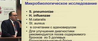
Haemophilus influenzae.
For many otorhinolaryngologists, this pathogen seems to be something exotic, since it is not isolated in most microbiological laboratories. And if this can be done, the results of determining sensitivity very often raise serious doubts. Thus, from domestic laboratories there are reports not just of resistance, but of high resistance to third-generation cephalosporins. This phenomenon has not been described in the world literature. The reason for the errors is the high demands of Haemophilus influenzae on agar and incubation conditions. Therefore, to obtain reliable data and their reproducibility, it is common practice throughout the world to use the recommendations of the US National Committee for Clinical Laboratory Standards (NCCLS). As shown in our laboratory, the domestic AGV medium cannot be used to determine the sensitivity of Haemophilus influenzae.
GABHS, or Streptococcus pyogenes
is the causative agent mainly only for tonsillopharyngitis. As in the case of other pathogens of respiratory infections, the isolation and determination of sensitivity of GABHS in domestic laboratories is carried out very rarely and the results obtained are often questionable. For example, GABHS strains resistant to penicillin or other b-lactam antibiotics have not yet been discovered anywhere in the world, although such results reach the attention of treating physicians and are published in the domestic literature. Using E-tests, we conducted a sensitivity study of GABHS strains isolated in 1994-1996. All GABHS were sensitive to penicillin, amoxicillin, cefaclor and cefuroxime. An alarming 12.6% resistance to erythromycin with cross-resistance to other macrolides (azithromycin, roxithromycin, dirithromycin). Resistance to tetracycline was 60%, which once again confirms the futility of using tetracyclines for tonsillopharyngitis.
Moraxella catarrhalis
(old name -
Branchamella catarrhalis
) is often described in the literature as one of the main causative agents of infections of the ENT organs. However, it seems to us that its importance is somewhat exaggerated. According to our data, even when conducting in-depth bacteriological studies, it is very rare and cannot “compete” with pneumococcus and Haemophilus influenzae. Thus, in the latest series of studies in 80 patients with acute sinusitis, it was isolated in only one case. Moraxella produces b-lactamase in almost 100% of cases and is therefore resistant to penicillin, ampicillin, and amoxicillin. At the same time, she is almost always sensitive to amoxicillin/clavulanate and oral cephalosporins of the third generation. Until recently, it was believed that Moraxella was also always sensitive to macrolides and co-trimoxazole. However, recent data from Spain shows an alarming increase in resistance. Thus, 42%, erythromycin 18%, azithromycin 3% of Moraxella strains isolated from respiratory infections were resistant to co-trimoxazole (E-102, J. Garcia ea A National, Multicenter, and Prospective Survey of Respiratory Bacteria Susceptibility to 12 Commonly Used Antimicrobials in Spain. ICAAC-96). Sensitivity to other macrolides was not determined. In general, the choice of antibiotics in the treatment of this infection does not pose a big problem.
There is no complete unanimity among specialists regarding the prescription of antibiotics for AOM, since in 60 out of 100 cases recovery occurs without the use of antibiotics. Antibiotics are actually needed in only one third of children with AOM, in whom eradication of the pathogen leads to a faster recovery, but identifying these children based on clinical data is difficult, if not impossible.
The tactics for managing children with AOM depends on factors such as the child’s age, the presence of concomitant and background diseases, ENT history, the socio-cultural level of the parents, and the availability of qualified medical care.
- Children under 2 years of age. According to most experts, antibiotics should be prescribed in all cases. If the child’s condition worsens after 24 hours, this may indicate the development of purulent complications that require immediate medical attention.
- In children over 2 years of age, in the absence of severe symptoms of intoxication, pain, or temperature above 38 0C during the day, symptomatic therapy can be limited. However, if symptoms persist or do not improve within 24 hours, antibiotics should be prescribed.
Reassessment of condition
carried out after 48-72 hours. If antibacterial therapy is effective, all the main symptoms of otitis media, except for exudation in the middle ear, should resolve. If this does not happen, then the prescribed treatment is ineffective. In this case, it is necessary to change the antibiotic, for example, instead of amoxicillin, prescribe amoxicillin/clavulanate or ceftriaxone intramuscularly. If indicated, tympanopuncture is performed with mandatory bacteriological examination of the obtained material. If the course of AOM is smooth, the child is examined on the 10-14th day, i.e. after completion of antibacterial therapy. Approximately half of the children still have effusion in the middle ear by this time, but this does not require continued antibiotic therapy.
As already mentioned, AOM is one of the main indications for prescribing antibiotics [7]. However, there is a constant reassessment of the effectiveness of antibiotics for this disease. Currently, three conditions for the effectiveness of antibiotics in AOM are quite clearly formulated: sensitivity of the pathogen to the antibiotic; the concentration of the antibiotic in the middle ear fluid and blood serum is higher than the MIC for this pathogen; maintaining serum concentrations above the MIC for 40-50% of the time between doses of the drug, which ensures 80-85% effectiveness [8]. If a decision is made to prescribe an antibiotic, oral amoxicillin is the drug of choice. Of all the available oral penicillins and cephalosporins, including drugs of the II-III generation, amoxicillin is the most active against penicillin-resistant pneumococci and, for example, is 4 times more effective than ampicillin. As a result, data on resistance to penicillin and ampicillin cannot be extended to amoxicillin. Its additional advantages are 2 times higher blood levels than ampicillin, a significantly lower frequency of unwanted side reactions from the gastrointestinal tract, and ease of administration. Amoxicillin is taken 3 times a day, regardless of the time of food intake, while ampicillin should be taken 4 times a day, 1 hour before or 2 hours after meals, since food reduces the bioavailability of this antibiotic by 2 times.
Exudative (effusion) otitis media
Antibacterial therapy, due to its low effectiveness, is carried out very selectively, especially in cases where hearing loss occurs. The prescription of an antibiotic should be preceded by an observation period of at least 3 months. Most often, amoxicillin is prescribed, the course duration is at least 10 days. If there is no effect, it is possible to conduct a second course of therapy with amoxicillin/clavulanate, the effectiveness of which has recently been proven in controlled clinical trials [9].
For chronic otitis media
Antibacterial therapy plays a secondary role compared to local sanitation and the use of ototopic drugs. It is advisable to use antibiotics if local therapy is ineffective, a clear picture of exacerbation develops and, what is extremely important, under the control of microbiological data, i.e. systemic therapy should be etiotropic [10].
Otitis externa
In localized forms of otitis externa, the main pathogen is Staphylococcus aureus. In some cases, erysipelas may occur involving the auricle and external auditory canal, caused by GABHS. Acute diffuse external otitis (“swimmer’s ear”) and malignant external otitis, which are almost always caused by gram-negative microflora and primarily Pseudomonas aeruginosa, should be distinguished from these forms. In chronic otitis externa, there is usually concomitant chronic otitis media. In such patients, it is necessary to exclude a fungal infection (aspergillus, candida).
Systemic antibacterial therapy must be combined with local treatment (hypertonic solutions, 2% acetic acid solution, 70-96% alcohol, ototopic antibacterial drugs). For staphylococcal otitis, oxacillin, amoxicillin/clavulanate, oral cephalosporins - generations, co-trimoxazole are prescribed; for streptococcal - phenoxymethylpenicillin and macrolides.
For a malignant form, a drug with antipseudomonal activity is prescribed. Considering the need for long courses of therapy (4-6 weeks), the prescription of stepwise therapy with ciprofloxacin is justified (initially intravenous 400 mg 2-3 times a day with a further transition to oral administration 500-750 mg 2 times a day).
Tonsillopharyngitis
When choosing antibiotics, the following must be taken into account: GABHS remains sensitive to penicillin and other b-lactam antibiotics: in some countries there is an increase in resistance to erythromycin and other macrolides; Russian data on the sensitivity of GABHS, obtained by internationally recognized methods, are very limited.
The goals of treatment for pharyngitis caused by GABHS are: reducing the severity and duration of symptoms; prevention of purulent complications, prevention of the spread of GABHS; reducing the risk of developing late complications - rheumatism and glomerulonephritis. Obviously, the last goal is the most important.
The leading drugs for the treatment of tonsillopharyngitis in children and adults are b-lactam antibiotics, and primarily phenoxymethylpenicillin, which is used in the usual daily dose, but not in 4 doses, as with other infections, but in 2-3 doses. Despite the complete sensitivity of pyogenic streptococcus to this antibiotic, the drug cannot be considered ideal for therapy, since phenoxymethylpenicillin must be taken 1 hour before or 2 hours after meals, the course of therapy is 10 days, cases of allergic reactions are not uncommon. In addition, eradication of the pathogen (the most important indicator of microbiological effectiveness) cannot be achieved in 11-16% of patients. One of the factors explaining incomplete eradication is the presence in the deep layers of the tonsils of so-called copathogens - microorganisms that are normal inhabitants of the oropharynx - which produce b-lactamases that destroy penicillin, ampicillin, amoxicillin. In this regard, two additional routes of therapy are possible: firstly, the use of antibiotics of other groups, primarily macrolides (erythromycin, spiramycin, roxithromycin, azithromycin, midecamycin), and in case of allergies to penicillins, macrolides or lincosamides are first-line drugs; secondly, the use of b-lactams that are resistant to the action of b-lactamases. These can be second generation cephalosporins (cefaclor, cefuroxime) or combinations of aminopenicillins with b-lactamase inhibitors sulbactam or clavulanate (ampicillin/sulbactam or amoxicillin/clavulanate). Inhibitor-protected aminopenicillins are more often used as second-line drugs in certain situations. Thus, indications for the use of amoxicillin/clavulanate are relapse of tonsillopharyngitis, the occurrence of tonsillopharyngitis during treatment with penicillin, macrolides.
Epiglottitis
Inflammation of the epiglottis and supraglottic structures is a rare but serious disease that most often occurs in children over 2 years of age. The most common pathogen is Haemophilus influenzae type B, less commonly - GABHS. Given the risk of asphyxia, patients are subject to urgent hospitalization. Parenteral antibiotic therapy should begin immediately after diagnosis. The drugs of choice are ampicillin/sulbactam, third generation cephalosporins (cefotaxime, ceftriaxone).
Acute sinusitis
In this article we will touch only on community-acquired acute sinusitis, in other words, acute sinusitis that developed in outpatients, the treatment of which can be carried out both in outpatient and inpatient settings. The importance of distinguishing this form of sinusitis from chronic sinusitis and nosocomial sinusitis in patients on mechanical ventilation is associated both with the high frequency of infection and with fundamental differences in etiology, pathogenesis and antibacterial therapy.
As the results of computed tomography show, in most cases of acute respiratory viral infection lasting more than 48 hours, an inflammatory reaction of the mucous membrane is noted. This is due to the fact that the paranasal sinuses are lined with mucous membrane, which is a continuation of the mucous membrane of the nasal cavity. Occlusion of the remaining opening due to hyperemia and edema leads to impaired sinus drainage and secondary infection. Thus, in most cases, acute sinusitis develops as a complication of an acute respiratory viral infection, which is observed in 2-5% of patients with ARVI [,]. Other predisposing factors are mechanical obstruction or allergic rhinitis. A common cause of maxillary sinusitis is odontogenic infection.
The most common causative agents of acute sinusitis are S.pneumoniae
and
H.influenzae
,
M.catarrhalis
, staphylococci, and non-spore-forming anaerobes are much less often isolated.
Such results are obtained all over the world with a correctly conducted microbiological study, which involves obtaining material by puncture of the sinuses, inoculating the material on appropriate media (blood, chocolate and anaerobic agars) and incubation at elevated CO2 levels and under anaerobic conditions. Our data obtained from the study of punctate maxillary sinuses in 85 patients show that bacterial pathogens were isolated in 71% of cases. The leading pathogen was S. pneumoniae
, which was isolated from 53% of patients, followed by
H. influenzae
and non-spore-forming anaerobes [13].
The goal of antibacterial therapy for acute sinusitis is to suppress symptoms and shorten the duration of the disease, destroy pathogens, which reduces damage to the mucosa, leading to chronicity of the disease. Adequate antibiotic therapy also prevents the development of intracranial and orbital complications.
Traditional drugs for the treatment of acute sinusitis in adults were oral antibiotics: amoxicillin (in Russia - ampicillin), doxycycline, co-trimoxazole. However, as noted above, such a choice in modern conditions may not always lead to good results. This is due both to the increasing resistance of microflora to these drugs, and in the case of co-trimoxazole with insufficient safety. At the same time, one should remember such an obvious advantage of traditional drugs as their low cost.
The optimal antibiotics for the treatment of sinusitis are penicillins - amoxicillin/clavulanate, cephalosporins - cefuroxime axetil, cefprozil, ceftibuten, macrolides - azithromycin, roxithromycin. Despite the recommendations in the literature for the use of fluoroquinolones (ofloxacin, ciprofloxacin), they should be considered as reserve drugs. This is due not only to their insufficiently high activity against pneumococci, but also mainly due to the fact that these drugs play a very important role in the treatment of inpatients, including those in the intensive care unit. If you suspect the development of intracranial or orbital complications, you should immediately prescribe parenterally, preferably intravenously, third generation cephalosporins (ceftriaxone - 2 g/day once, cefotaxime - 6-8 g/day in 3-4 injections).
Antibacterial therapy should be especially active in the elderly, in patients with impaired immunity, and in the development of orbital or intracranial complications. It is necessary to prescribe one of the antibiotics active against penicillin-resistant pneumococci and b-lactamase-positive strains of Haemophilus influenzae: amoxicillin/clavulanate, ampicillin/sulbactam, cefuroxime, cefotaxime, ceftriaxone. Moreover, with the development of complications from the central nervous system, preference should be given to ceftriaxone (2-4 g/day in 1 injection) or cefotaxime (12 g/day in 4 injections), which are administered intravenously. When pneumococci are isolated, even with intermediate resistance to penicillin, vancomycin is additionally administered (2 g/day in 4 injections).
Chronic sinusitis
In chronic sinusitis, antibiotics play a lesser role than in acute sinusitis, since the main task is to restore normal ventilation of the paranasal sinuses. Antibiotics are used for exacerbation of chronic sinusitis, however, given the frequent release of non-spore-forming anaerobes, the importance of drugs with antianaerobic activity (for example, amoxicillin/clavulanate) increases. In general, it is highly desirable to carry out therapy under the control of bacteriological examination data, especially in patients who have had ineffective antibacterial therapy. When using antibiotics that are not sufficiently active against anaerobes (for example, co-trimoxazole, fluoroquinolones), it is advisable to combine them with metronidazole [14].
Concomitant therapy
Vasoconstrictor drugs, which in foreign literature are called decongestants, play an important role in eliminating swelling of the mucous membrane, thereby improving drainage and restoring the function of the eustachian tube and paranasal sinuses. For decades, topical decongestants have been the most popular in Russia due to the lack of oral medications on the market; in addition, topical remedies were believed to be safer. However, in recent years, views on the use of topical drugs have changed.
When topical decongestants are used for more than 5 days, drug-induced rhinitis develops. In this regard, xylometazoline is the most dangerous. The effectiveness of nasal drops is reduced due to the fact that patients do not know the correct technique for using them, which is that the patient must insert these drops into the nose in the Mecca position and remain in it for 2-3 minutes after instillation of the drops. Obviously, this is very inconvenient for patients, especially for working, elderly, and disabled people [15].
Errors in antibacterial therapy for ENT infections can be divided into three groups.
1.
Neglecting to take antibiotics orally. Antibiotics are important from two points of view. Firstly, they make it possible to alleviate the patient’s condition, speed up recovery and prevent chronic infection. Secondly, their use is necessary to prevent both early and late complications. We too quickly forgot that streptococcal tonsillopharyngitis, with inadequate therapy, leads to the development of rheumatism and glomerulonephritis; acute sinusitis - to meningitis, etc. It is useful to recall a study conducted in 1954 in Sweden by R. Rudberg, which showed a decrease in the incidence of mastoiditis from 17% to almost zero with the use of antibiotics [16].
Attempts to replace oral antibiotics with local antibacterial drugs or drugs with immunostimulating activity declared by the manufacturers currently seem unfounded, since there is no reliable clinical data showing the effectiveness of these drugs. For example, the recommendation for the use of the drug fusafungin - “local treatment of inflammatory infectious diseases of the nasopharynx and respiratory tract: rhinitis, sinusitis, pharyngitis, tonsillitis, laryngitis, tracheitis, bronchitis” - does not indicate that Paul Ehrlich’s dream of a “magic bullet” has finally come true , but that we allow ourselves to be deceived by allowing the publication of such recommendations. In 1997, materials from a symposium devoted to the study of the drug Ribomunil, which was marketed as a specific immunostimulant, were published. The head of the symposium, G. Bellanti (USA), came to the following quite correct conclusions: “further study of this drug, like many other drugs with the potential to stimulate the immune system, is necessary in order to assess their significance” [17].
2.
Wrong choice of drug, doses, routes of administration. Unfortunately, antibiotics are often prescribed without thinking about the most likely pathogen, its sensitivity to antibacterial drugs, and without taking into account the results obtained in controlled clinical studies. Even in outpatient records you can find a prescription like “antibiotic by mouth.” Due to a lack of knowledge, many doctors often equate generics of ciprofloxacin with oral cephalosporins; sulfonamides continue to be the favorite drugs, including “streptocide under the tongue” (!), gentamicin or lincomycin intramuscularly in outpatients.
3.
Insufficient attention to bacteriological research. Despite the fact that the empirical choice of antibiotics forms the basis for their prescription, since, with the exception of streptococcal tonsillopharyngitis, the pathogen cannot be quickly identified, a bacteriological study must be carried out. In addition to the direct benefit for a given patient, for example, if initial therapy is ineffective and the need to change antibiotics, its importance lies in obtaining and accumulating epidemiological data on the structure of pathogens and, most importantly, their sensitivity to antibiotics. Relying on local data on antibiotic resistance makes it possible to make empirical antibiotic therapy as effective as possible and to put it on a scientific basis.
When treating infections of the ENT organs, it is important to overcome nihilism towards the use of antibiotics, which is often found among doctors and is associated with a high rate of spontaneous recovery. Preference should be given to drugs with the highest clinical and bacteriological effectiveness, since infections of the ENT organs affect many millions of patients and small differences in effectiveness in practice result in thousands of cases of ineffective therapy.
Primary (natural) activity of antibiotics against the main pathogens of ENT infections
| + | – | + | – | ||
| Amoxicillin/clavulanate | + | + | + | + | + |
| Cefaclor | + | + | + | + | + |
| Cefuroxime axetil | + | + | + | + | + |
| Cefprozil | + | + | + | + | + |
| Cefixime | – | + | – | + | + |
| Ceftibuten | – | + | – | + | + |
| Cefpodoxime | + | + | + | + | + |
| Erythromycin | + | + | + | – | + |
| Azithromycin | + | + | + | + | + |
| Clarithromycin | + | + | + | ± | + |
| Roxithromycin | + | + | + | – | + |
| Spiramycin | + | + | + | – | + |
| Linco/clindamycin | + | + | + | – | – |
| Co-trimoxazole | + | – | + | + | + |
Doses of antibiotics for outpatient infections of the ENT organs
| 500 | 4 | 10 | Inside | ||
| Amoxicillin | 40 | 500 | 3 | 10 | Inside, regardless of food intake |
| Amoxicillin/clavulanate | 40 | 625 | 3 | 10 | Same |
| Cefaclor | 40 | 500 | 3 | 10 | Same |
| Cefuroxime | 30 | 250-500 | 2 | 10 | Inside, during meals |
| Ceftibuten | 9 | 400 | 1 | 10 | Inside, regardless of food intake |
| Ceftriaxone | 50 | 1000 | 1 | 10 | Intramuscularly |
| Azithromycin | 10 | 500 | 1 | 3 | Inside, before or after meals |
| Clarithromycin | 15 | 250 | 2 | 10 | Inside, regardless of food intake |
| Roxithromycin | 5-8 | 150 | 2 | 10 | Inside, before meals |
| Spiramycin | 150,000 IU | 3000 000 IU | 2 | 10 | Inside, regardless of food intake |
| Midecamycin | 30-50 | 400 | 3 | 10 | Inside, before meals |
| Ciprofloxacin | NP | 500 | 2 | 7-10 | Inside, after meals |
| Ofloxacin | NP | 400 | 2 | 7-10 | Same |
| Co-trimoxazole | 8 | 960 | 2 | 10 | Same |
* – Cannot be combined with aluminum- and magnesium-containing antacids. iron and zinc preparations. ** – Not applicable.
Antibiotic resistance of the main causative agents of ENT infections
| Aminoglycosides, aztreonam, polymyxin, quinolones | Penicillin, cephalosporins, macrolides, co-trimoxazole | ||
| Haemophilus influenzae | Ampicillin, amoxicillin, cephalosporins, co-trimoxazole | Erythromycin, aminoglycosides | Ampicillin, amoxicillin |
| Streptococcus pyogenes | Penicillins, cephalosporins, carbapenems, macrolides, lincosamides, co-trimoxazole, rifampicin | Aminoglycosides, aztreonam, polymyxin, quinolones | Penicillin, cephalosporins, macrolides, co-trimoxazole |
| Moraxella catarrhalis | Penicillins, cephalosporins, carbapenems, macrolides, lincosamides, co-trimoxazole, rifampicin | Aminoglycosides, aztreonam, polymyxin, quinolones | Penicillin, cephalosporins, macrolides, co-trimoxazole |
Antibiotic resistance of the main causative agents of ENT infections
| Prescribing potentially toxic antibiotics to children (co-trimoxazole, chloramphenicol) |
| Administration of antibiotics (penicillin, ampicillin, ampiox, lincomycin, etc.) intramuscularly rather than orally |
| Use of sulfonamides, co-trimoxazole, tetracyclines for tonsillopharyngitis |
| Prescribing, as an alternative to oral antibiotics, immunomodulators (ribomunil, bronchomunal, IRS-19, immunal), “cover-up” drugs (nystatin) |
* – the exception is a single injection of ceftriaxia (50 mg/kg) intramuscularly.
Literature
- Gwaltney JM. Jr. State-of-the art. Acute community-acquired sinusitis. Clin Infect Dis 1996; 23: 1209-23.
- Cohen R. The antibiotic treatment of acute otitis media and sinusitis in children. Diagn Microbiol Infect Dis 1999; 27:49-53.
- Bauchner H. The role of parents in prescribing antibiotics to children. News from the International Union for the Prudent Use of Antibiotics (IURPA). 1998; 1:1-4.
- Hayden FG, Gwaltney JM Jr. Intranasal interferon for prevention of rhinovirus infection and ilness. J Infect Dis 1983; 148:543.
- Finch RG Infections of the upper respiratory tract. In: Antibiotics and Chemotherapy. Ed by F. O'Grady ea 7th ed. NY ea, l997; 674-680.
- Bartlet JG Management of Upper Respiratory Tract Infections. IDCP 1997; 6:212-21.
- Tarasova G.D., Stroganov V.P., Omelyanovsky V.V., Pekli F.F. Antibacterial therapy for middle ear replenishment in childhood. Bulletin of otorhinolar. 1997; 6:12-13.
- Craig W, Andes A. Pediatr Infect Dis J 1996; 15:255-9.
- van Balen FAM, de Melker RA, Touw-Otten FWMM Double-blind randomized trial of co-amoxiclav versus placebo for persistent otitis media with effusion in general practice. Lancet l996; 348:713-6.
- Hamer DH. IDCP Guidelines: Epiglottitis and Otitis. Infect Dis din Pract 1997; 6:500-5.
- Dingle JH, Badger CF, Jordan WS, Jr. Illness in the home. A study of the 25,000 illnesses in a group of Cleveland families. The Press of Western Reserve University. Cleveland. 1964; 347.
- Berg 0., Carenfelt S. Analysis of symptoms and clinical signs in maxillary sinus empyema. Ada Oto-Laryngol (Stockholm) 1988; 105:343-9.
- Bartlett JG IDCP guidelines: management of upper respiratory tract infections. Infect. Dis Clin Practice. 1997; 6:212-20.
- Kennedy DW, Thaler ER. Acute vs. Chronic Sinusitis: Etiology, Management, and Outcomes. Infect Dis Clin Pract 1997;6(Suppl. 2):S49-S58.
- Evans KL. Diagnosis and management of sinusitis. Br Med J 1994; 309:1415-22.
- Rudberg R.D. Acute otitis media: comparative therapeutic results of sulphonamide and penicillin administered in various forms. Acta Otolaryngol (Stockh.) 1954;113 (Suppl.): 9-79.
- Bellanti JA. Recurrent respiratory tract infections in pediatric patients. Drugs 1997; 54(Suppl. 1):1-4.
| © 2000-2009 NIIAH SGMA | ||