Causes of the disease
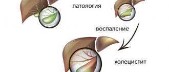
The cause of the disease is a violation of the natural outflow of bile. The secretion stagnates, corrodes the walls of the gallbladder, blood supply to tissues deteriorates, pathogenic microflora is activated. When the bile ducts become clogged with a stone, the patient becomes inflamed.
Factors that increase the likelihood of disease:
- obesity;
- sudden weight loss;
- starvation;
- long-term parenteral nutrition;
- taking medications belonging to the category of synthetic hormones, antibiotics (contraceptives, estrogen- and progesterone-containing drugs, ceftriaxone).
The likelihood of occurrence and exacerbation of the disease is observed during pregnancy and childbirth. Risk factors also include advanced and middle age, hereditary predisposition, a history of diabetes mellitus, liver cirrhosis, Crohn's disease, etc.
Treatment
Non-surgical treatment. It is based on diet and medication. Patients with calculous cholecystitis refuse foods that lead to excessive secretion of bile and cause an exacerbation of the disease. Alcohol and carbonated water are completely excluded. The doctor prescribes hepatoprotectors, antispasmodics, enzymes, herbal medicines, antibiotics (if there is an infection). Treatment of chronic calculous cholecystitis is possible with small stones (up to 15 mm), consisting of cholesterol, and normal motor activity of the gallbladder.
Surgical treatment. Treatment is often carried out surgically. As a rule, the entire gallbladder is removed along with the stones. Depending on the indications, access can be carried out either laparoscopically or laparotomically. The operations are performed under general anesthesia.
To make an appointment at the ABC-Medicine clinic to clarify symptoms and treat chronic cholecystitis, call +7.
Types of diseases and their symptoms
There are acute and chronic calculous cholecystitis. In the chronic form, symptoms manifest themselves moderately, discomfort occurs in the acute phase.
The chronic form is characterized by the following symptoms:
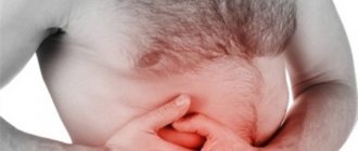
- Drawing, aching and dull pain in the right hypochondrium. The pain can radiate to the lower back, under the shoulder blade, right shoulder, or side of the neck.
- Hepatic colic is a symptom of the onset of an exacerbation of the disease. The patient experiences acute cramping pain in the right hypochondrium.
- Due to intoxication with bile acids, the patient exhibits signs of jaundice: the sclera, skin and mucous membranes become yellow, the urine darkens, and the feces become discolored.
- Gastrointestinal disorders: unstable stool, heartburn, belching, vomiting, bitter taste.
- Low-grade fever.
Acute calculous cholecystitis begins when stones block the bile ducts. As a result of obstruction, an inflammatory process begins, which is accompanied by severe symptoms:
- sharp pain in the right side;
- nausea, bouts of vomiting;
- increased temperature;
- weakness.
In an acute process, the injured mucous membrane secretes a larger amount of fluid, stretching the walls of the bile duct. In this case, inflammation intensifies and the likelihood of a secondary infection increases. An untreated acute process either worsens or subsides, as a result of which the gallbladder tissue degenerates and it ceases to fully cope with its functions. This is how the patient develops chronic cholecystitis.
The disease is also classified depending on the nature of the inflammation. There are the following types:
- purulent;
- catarrhal - the patient complains of severe pain in the right side of the body;
- phlegmonous calculous cholecystitis manifests itself in the form of increased pain when coughing and changing body position;
- gangrenous - a dangerous form that develops from phlegmonous;
- mixed type.
Acute calculous cholecystitis
Acute calculous cholecystitis is an acute inflammation of the gallbladder containing stones. Prevalence. Acute calculous cholecystitis ranks second in frequency among acute surgical diseases of the abdominal organs. In 12-24% of cases it is complicated by choledocholithiasis, in 26-49% by obstructive jaundice, in 23-47% by cholangitis.
In the development of acute calculous cholecystitis, the leading role is played by the penetration of infection into the gallbladder and disruption of the outflow of bile. Microbial flora (intestinal, Pseudomonas aeruginosa, staphylococci, enterococci) enters the gallbladder ascending (from the duodenum) or descending (from the liver) by hematogenous or lymphogenous route. The outflow of bile is disrupted when stones obstruct the neck of the gallbladder or cystic duct, common bile duct, or pathological processes in the periampullary zone. The development of calculous cholecystitis is facilitated by changes in the vessels of the gallbladder wall during atherosclerosis, damage to its mucous membrane by pancreatic enzymes during pancreatobiliary reflux.
Morphologically, three types of acute calculous cholecystitis are distinguished: catarrhal, phlegmonous and gangrenous. With catarrhal cholecystitis, the gallbladder is slightly enlarged, its wall is thickened due to edema and swelling of the mucosa. The mucous membrane is cloudy due to desquamation of the epithelium and its infiltration with leukocytes. Inflammation also spreads to the submucosal layer. With phlegmonous cholecystitis, the wall of the gallbladder is significantly thickened as a result of abundant impregnation with inflammatory exudate. The mucosa is sharply hyperemic, with fibrin deposits. The bladder is much enlarged in volume, filled with purulent exudate, and covered with fibrin on the outside. In case of occlusion of the cystic duct by a stone or due to swelling of its wall, acute empyema of the gallbladder develops. With gangrenous cholecystitis, which occurs against the background of cystic artery thrombosis, partial or total necrosis of the gallbladder wall occurs. Gangrene usually occurs on the 3-4th day of the disease. Often there is perforation of the bladder wall (gangrenous-perforated cholecystitis) with the flow of bile into the abdominal cavity and the development of biliary peritonitis. Perforation occurs more often in the area of the neck of the gallbladder or Hartmann's pouch, i.e. in the places where stones are most often localized. All described pathomorphological forms of acute calculous cholecystitis are accompanied by pericholecystitis, which is characterized by a local or widespread adhesive process that limits the spread of infection to the area of the right hypochondrium.
Signs of acute calculous cholecystitis are pain in the right hypochondrium, vomiting, nausea. The pain occurs suddenly, most often after a diet violation, and radiates to the right shoulder, collarbone, lower back, and iliac region. The intensity of pain increases with the slightest physical stress. Vomiting in patients with acute calculous cholecystitis is of a reflex nature, often repeated, and does not bring relief. During an objective examination of patients, dryness of the tongue, some bloating of the abdomen and limitation of its participation in the act of breathing, severe pain and muscle tension in the right hypochondrium, especially at the point of projection of the gallbladder: the intersection of the outer edge of the right rectus abdominis muscle with the costal arch (Ker's point) are revealed. . In the absence of pronounced muscle tension in the abdominal wall in patients, an enlarged, tense, sharply painful gallbladder is often palpated (Parturier's symptom). At the same time, characteristic symptoms are determined in patients: the appearance or intensification of pain when lightly tapping the inner edge of the hand along the right costal arch (Grekov-Ortner symptom); pain on palpation in the right hypochondrium, increasing with inspiration (Keur's symptom); pain when pressing on the xiphoid process (Pekarsky's symptom); pain on palpation between the legs of the sternocleidomastoid muscle on the right above the collarbone (Mussy symptom, phrenicus symptom); the appearance or intensification of pain in the right hypochondrium when pressing with the index finger between the legs of the sternocleidomastoid muscle on the right above the collarbone (Georgievsky’s symptom); pain on palpation to the right of the navel and slightly higher in the projection of the common bile duct (Janover's symptom); pain when pressing on the right near the spinous processes of the VIII-X thoracic vertebrae (Boas symptom); irradiation of pain to the heart area (Botkin’s cholecystocardial symptom). The body temperature in patients with acute calculous cholecystitis is elevated. Neutrophilic leukocytosis and increased ESR are observed in the blood.
The severity of clinical manifestations of the disease is directly proportional to the degree of morphological changes in the gallbladder. Thus, the most rapid course is observed with gangrenous cholecystitis. However, in elderly patients, acute calculous cholecystitis occurs atypically, which is due to the lack of a clear relationship between structural changes in the gallbladder and the existing clinical manifestations. This is due to a decrease in the overall reactivity of the body and the presence of concomitant diseases. Acute calculous cholecystitis in this category of people occurs with erased local symptoms and is accompanied by rapid generalization of the main process, symptoms of intoxication, and a high incidence of complications. The body temperature of patients is usually low-grade. Tachycardia is pronounced. A discrepancy is determined between heart rate and temperature. Moderate leukocytosis with a shift to the left is detected in the blood.
The duration of acute calculous cholecystitis ranges from 3-5 days to several weeks. In most cases, the disease becomes chronic or is accompanied by complications: obstructive calculous cholecystitis - empyema or hydrocele of the gallbladder; perforation of the gallbladder (perforative, calculous cholecystitis) with the development of bile peritonitis, subhepatic and subphrenic abscesses; cholangitis; liver abscesses, etc. Complications, as a rule, accompany phlegmonous and gangrenous forms of acute calculous cholecystitis.
Empyema of the gallbladder in acute calculous cholecystitis occurs due to blockage of the cystic duct with a stone in the presence of a virulent infection in the gallbladder. Due to the progression of inflammation, pus accumulates in the lumen of the bladder. The bladder increases in size, becomes tense, and sharply painful on palpation. In the right hypochondrium, patients feel intense, constant pain that lasts for several days. The temperature rises to 38-40 °C, often taking on a hectic character with chills and frequent heavy sweating. During intensive therapy, most symptoms disappear, but patients continue to be bothered by a feeling of heaviness and pain in the projection of the gallbladder (chronic empyema). Violation of the diet and physical stress lead to an increase in body temperature to 38-39 ° C and increased pain. Hydrocele of the gallbladder most often forms after an attack of catarrhal acute calculous cholecystitis caused by microbial microflora with little virulence and preserved occlusion of the neck of the gallbladder or cystic duct. In such cases, due to the absorption of bile pigments and the death of microorganisms, colorless mucous contents are formed in the gallbladder. In the projection of the gallbladder, an elastic formation with a smooth surface is determined, which moves with inhalation along with the liver. Dropsy can exist for a long time, accompanied by a feeling of heaviness in the right hypochondrium, nausea, and vomiting. When the stone blocking the duct comes out, it disappears. In some cases, gallbladder rupture is possible.
The diagnosis of acute calculous cholecystitis is based on data from a physical examination, general and biochemical blood tests, the results of ultrasound examination of the gallbladder, liver, biliary tract, less often - laparoscopy, etc. When acute calculous cholecystitis is complicated by obstructive jaundice, ERCG is informative. Differential diagnosis of acute calculous cholecystitis. Most often, acute calculous cholecystitis is differentiated from a perforated ulcer, acute pancreatitis, right-sided renal colic, Botkin's disease in the preicteric period, and biliary dyskinesia. An attack of right-sided renal colic develops suddenly and is characterized by intense pain in the lumbar region, radiating to the groin. Urine tests determine hematuria. An ultrasound examination of the kidneys and a plain radiograph of the abdominal cavity reveal stones in the projection of the kidneys and urinary tract. In the pre-icteric period of Botkin's disease, general weakness, loss of appetite, nausea, a feeling of heaviness in the epigastrium and right hypochondrium, and an increase in body temperature up to 38 ° C prevail. Then parenchymal jaundice gradually increases: first, the sclera, soft palate, and later the skin become jaundiced. The liver and spleen enlarge. In the blood, the content of direct bilirubin is increased and cholesterol is decreased, and the activity of transaminases is increased. Urobilinuria is detected. Manifestations of biliary dyskinesias are varied, which is associated with the existence of several of their forms: 1) atony and hypotension of the gallbladder; 2) hypertensive gallbladder; 3) spasm of the sphincter of Oddi; 4) atony of the sphincter of Oddi. The atonic gallbladder is accompanied by a constant feeling of heaviness, pain in the right hypochondrium, and vomiting mixed with bile.
Ultrasound and X-ray contrast studies revealed a sharply enlarged gallbladder. With a hypertensive gallbladder, attacks of hepatic colic are frequent. By increasing the muscle tone of the wall, the gallbladder acquires a spherical shape. Hypertension of the sphincter of Oddi occurs in four forms - icteric, painful with colic, febrile and asymptomatic. Atony and insufficiency of the sphincter of Oddi are characterized by weak contrast of the bile ducts during a conventional X-ray contrast study of the biliary system with the simultaneous accumulation of a large amount of contrast in the duodenum. However, after subcutaneous administration of morphine, the shadow of the gallbladder projection becomes clear. When diagnosing acute calculous cholecystitis, one should remember some similarity of the clinical manifestations of the disease with those of acute intestinal obstruction, thrombosis of mesenteric vessels, myocardial infarction, pneumonia and pleurisy.
The question of tactics for managing patients with acute calculous cholecystitis should be decided individually based on the existing clinical picture and the general condition of the patients. Most surgeons adhere to active expectant tactics, the essence of which is as follows. In case of acute calculous cholecystitis complicated by peritonitis, the operation is performed immediately after the patient’s admission or short-term preoperative preparation (emergency surgery). Also, urgent cholecystectomy (in the vast majority of cases laparoscopic) is performed if the duration of the disease, from the moment of the attack, does not exceed 72 hours. In all other cases, intensive complex drug treatment is prescribed with the aim of performing the operation in the cold period, i.e. after acute inflammation in the gallbladder and bile ducts has subsided, eliminating intoxication and metabolic disorders, functional changes in vital organs and systems (planned surgery) . However, if the treatment is ineffective, jaundice increases, or signs of destructive cholecystitis occur, the operation is performed 48-72 hours after admission (urgent surgery). However, cholecystectomy, especially when the disease lasts more than 3-5 days, is associated with certain technical difficulties due to inflammatory tissue edema in the area of the gallbladder and hepatoduodenal ligament, and the formation of a dense infiltrate in the subhepatic space. They disrupt topographic-anatomical relationships and increase tissue bleeding in the surgical area, increasing the risk of developing intraoperative and postoperative complications of cholecystectomy.
For the conservative treatment of acute calculous cholecystitis, analgesics, antispasmodics, anticholinergics, antihistamines, broad-spectrum antibiotics, antioxidants, etc. are used. Novocaine blockades (subxiphoidal, round ligament of the liver, perinephric, etc.), gastric lavage and local hypothermia are performed. Treatment of concomitant diseases is prescribed according to indications. Abnormalities of acid-base balance and electrolyte metabolism are corrected (polarizing mixtures, panangin, 4% sodium bicarbonate solution); dysproteinemia (albumin, protein, amino acid mixtures, plasma); detoxification therapy (forced diuresis) is carried out. For infusion-transfusion therapy, it is advisable to catheterize the central vein, which allows for correction of hemostasis in a short time.
Patients with a significant degree of surgical risk. It is advisable to carry out two-stage treatment. The risk group consists of patients over 60 years of age, sub- and decompensated forms of acute and chronic vascular, pulmonary, renal or liver failure; acute or previous myocardial infarction; acute or residual effects of a previous cerebrovascular accident; bronchial asthma; pronounced changes in the lungs (emphysema, acute and chronic pneumonia, acute and chronic bronchitis, etc.); phlebothrombosis of the deep veins of the lower extremities; moderate to severe diabetes mellitus; heart disease with circulatory failure stage 2-3; atrial fibrillation; acute cholecystitis, complicated by obstructive jaundice, cholangitis; acute cholecystitis lasting more than 5 days. At the first stage, decompression of the gallbladder is performed using one of the well-known methods: percutaneously transhepatic (through the edge of the liver) under ultrasound or laparoscopy control; using laparoscopic microcholecystostomy or cholecystostomy from minilaparotomy access. Decompression of the gallbladder in combination with general treatment makes it possible to prevent the progression of the destructive process in its wall, as well as in the bile ducts, and remove patients from a state of severe intoxication within 3-5 days. In addition, transdrainage cholecystocholangiography allows one to establish the surgical anatomy of the gallbladder and bile ducts, knowledge of which is necessary to determine the scope of subsequent surgery.
During the second stage, after relief of acute inflammation in the gallbladder wall, a complete examination and comprehensive preoperative preparation, patients undergo a planned cholecystectomy. If patients refuse surgery, the stones remaining in the lumen of the gallbladder create a real danger of developing another attack of acute cholecystitis. However, patients with an extremely high degree of surgical and anesthetic risk are prescribed symptomatic conservative treatment. The operation of choice for acute calculous cholecystitis is cholecystectomy: laparoscopic or open in combination with corrective interventions on the extrahepatic bile ducts (if necessary). The surgical approach for open cholecystectomy is an upper midline laparotomy or an oblique incision in the right hypochondrium. The choice of method for removing the gallbladder depends on the experience of the surgeon, the technical equipment of the operating room, and the nature of inflammatory changes in the gallbladder and surrounding tissues.
According to generalized literature data, laparoscopic cholecystectomy in patients with acute cholecystitis can be performed in 73.8-97.2%. The following are considered contraindications to its implementation: 1) inflammatory infiltrate or abscess in the gallbladder area; 2) expansion of the common bile duct (more than 8 mm); 3) the thickness of the gallbladder wall is more than 1 cm; 4) “shrinked” gallbladder; 5) increased levels of bilirubin and amylase in the patient’s blood; 6) pulmonary heart diseases in the stage of sub- and decompensation; 7) uncorrectable disorders of the hemocoagulation system; widespread peritonitis; 9) third trimester of pregnancy; 10) biliodigestive and biliobiliary fistulas; 11) portal hypertension syndrome; 12) unclear anatomical situation in the area of the gallbladder neck and hepatoduodenal ligament. However, due to the constant progress of endoscopic surgery, contraindications to laparoscopic cholecystectomy for acute calculous cholecystitis are constantly narrowing. But keep in mind that the consistent rule of modern gallbladder surgery is immediate conversion to open cholecystectomy in case of difficulties in manipulation in the subhepatic space, rough adhesions and extensive dense infiltration in the area of the gallbladder that is not amenable to blunt preparation, and unclear anatomical situation.
The results of cholecystectomy for acute calculous cholecystitis largely depend on the condition of the bile ducts. Therefore, all patients with this disease must undergo a comprehensive examination of the biliary tract before surgery: ultrasound, ERCG, intravenous cholecystocholangiography. In the event that it is not possible to carry out a comprehensive examination of patients in the preoperative period (emergency surgery), the condition of the biliary tract should be assessed during the operation. The most informative method for intraoperative assessment of the patency of the bile ducts during laparoscopic cholecystectomy is intraoperative cholangiography. Indications for its implementation in patients with acute calculous cholecystitis are: 1) a history of jaundice with a common bile duct diameter of up to 7-8 mm according to ultrasound; 2) dilation of the extrahepatic bile ducts more than 7-8 mm, discovered during surgery; 3) the diameter of the cystic duct is more than 2 mm; 4) small (up to 1-2 mm) stones in the gallbladder; 5) changes in the anatomy of the extrahepatic bile ducts and difficulties in preparation in the area of Calot’s triangle; 6) lack of visualization of the bile ducts due to swelling of the hepatoduodenal ligament; 7) the impossibility of using ERCG in the preoperative period if there are appropriate indications. If a pathology of the biliary tract or major duodenal papilla is established, an appropriate corrective operation is performed. Thus, in patients with stenosis of the papilla of Vater and choledocholithiasis, endoscopic pagkillosphincterotomy is performed in the postoperative period. In the case of cicatricial stricture of the common bile duct, a transition to open surgery is performed with the formation of one of the options for biliodigestive anastomosis.
Medicines
Conservative treatment involves the use of broad-spectrum antibiotics (Norfloxacin, Cephalexin, Spiramycin) and intravenous administration of antispasmodics (No-Shpa, Papaverine, Spazmalgon).
To support digestive function, enzyme-containing supplements are used: Pancreatin, Mezim, Festal.
To support liver function, hepatoprotectors are indicated: Gepabene, Chofitol.
Doctors also recommend choleretic agents of natural origin Allochol, Cholenzym and sorbents Activated carbon, Atoxil, etc.
Popular questions about calculous cholecystitis
What is calculous?
The term means that the cause of an acute or chronic inflammatory process lies in stones formed inside the gallbladder.
How much water should you drink if you have cholecystitis?
During an attack, it is recommended to drink as much mineral water as possible without carbon. Water is especially useful after a bout of vomiting.
What can you eat with acute cholecystitis?
Doctors recommend limiting food intake and eating foods that do not cause increased bile secretion.
Diagnostics
Diagnosis of cholelithiasis is based on the clinical picture, as well as data obtained during an instrumental examination. To make a diagnosis with sufficient accuracy, it is enough to perform an ultrasound of the upper abdominal cavity. With its help, you can identify stones in the ducts, gall bladder, determine the size of the bladder, its walls, the condition of the pancreas and liver. Gastroduodenoscopy is also performed to determine the condition of the mucous membrane of the stomach, esophagus and duodenum. If there are complications, retrograde cholangiography or transgastric ultrasound of the bile ducts is prescribed to detect choledocholithiasis.