The heart begins to beat long before we are born. Surprising and even magical, right? However, this means that this organ works longer and harder than others. And not only the quality of our life, but also life itself depends on how carefully we pay attention to his condition!
More than 17 million people die every year from heart and vascular diseases. It has been proven that 80% of premature heart attacks and strokes could be prevented!
Fortunately, most cardiovascular diseases are successfully diagnosed thanks to modern innovative equipment and the professionalism of doctors. And as you know, establishing an accurate diagnosis means taking an important step towards healing.
One of the most informative and safe studies is cardiac echocardiography (EchoCG) or, in other words, ultrasound of the heart.
1 ECHO-KG in "MedicCity"
2 Echocardiography at MedicCity
3 Ultrasound of the heart at MedicCity
The essence of echocardiography
Ultrasound of the heart is the process of studying all the main parameters and structures of this organ using ultrasound.
When exposed to electrical energy, the echocardiograph transducer emits high-frequency sound that travels through the structures of the heart, is reflected from them, captured by the same transducer and transmitted to the computer. He, in turn, analyzes the received data and displays it on the monitor in the form of a two- or three-dimensional image.
In recent years, echocardiography has been increasingly used for preventive purposes, which makes it possible to identify cardiac abnormalities at an early stage.
What does an ultrasound of the heart show:
- heart size;
- integrity, structure and thickness of its walls;
- sizes of the cavities of the atria and ventricles;
- contractility of the heart muscle;
- operation and structure of valves;
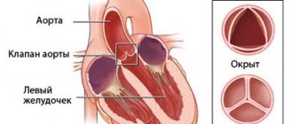
- condition of the pulmonary artery and aorta;
- pulmonary artery pressure level (to diagnose pulmonary hypertension, which can occur with pulmonary embolism, for example, when blood clots from the veins of the legs enter the pulmonary artery);
- direction and speed of cardiac blood flow;
- condition of the outer shell, pericardium.
1 ECHO-KG in "MedicCity"
2 Echocardiography at MedicCity
3 Ultrasound of the heart at MedicCity
What does the specialist see?
During an echocardiogram, the doctor can evaluate the functioning of the heart using several criteria. Each of them has certain norms, and a deviation in one direction or another indicates the presence of various pathologies.
Ultrasound allows you to evaluate the following indicators:
- main characteristics of the heart chambers;
- characteristics of the ventricles and atria;
- functioning of valves and their condition;
- condition of the walls of blood vessels;
- direction and intensity of blood flow;
- characteristics of the heart muscles during the period of relaxation and contraction;
- is there exudate in the pericardial sac?
To make a diagnosis, doctors use certain standards of echocardiography, but sometimes minor deviations in one direction or another are allowed. It depends on the age, weight of the patient and other individual characteristics.
Important! The interpretation of the results obtained should be carried out exclusively by a cardiologist. Once you have the conclusion in your hands, you should not try to establish a diagnosis yourself.
Who is prescribed echocardiography?
The following are routinely examined:
- infants - if congenital defects are suspected;
- teenagers are a time of intensive growth;
- pregnant women with existing chronic diseases - to decide on the method of childbirth;
- professional athletes - to monitor the state of the cardiovascular system.
An echocardiogram is required if:
- anomalies of the endocardium and valve apparatus:
- heart tumors;
- arrhythmias;
- threat of a heart attack or a heart attack;
- IHD;
- failures of cardiac activity due to various intoxications;
- attacks of angina pectoris;
- pericarditis of various origins;
- hypertension;
- heart failure.
And also in the process of treating heart diseases, before and after cardiac surgery.
What abbreviations are used in the protocol?
Having received the EchoCG protocol completed by a specialist, the patient is faced with abbreviations that are incomprehensible to him. For example, MPAP is the average pressure in the pulmonary artery, CO and DO are the short and long axis. The most commonly used abbreviations can be seen in the figure.
Basic abbreviations that are indicated in the ultrasound protocol
In most cases, it is impossible to make a diagnosis based only on the results of the protocol. The specialist takes into account such features as ultrasound indicators, the patient’s medical history, the chronology and intensity of the development of symptoms, and other nuances. Taken together, these data help to accurately determine a particular pathology.
When is a cardiac ultrasound performed?
Indications for cardiac ultrasound are:
- alarming changes in health (increased or interrupted heartbeat, shortness of breath, swelling, weakness, prolonged fever, chest pain, cases of loss of consciousness);
- changes detected in the last ECG;
- increased blood pressure;
- heart murmurs;
- cardiomyopathy;
- manifestations of coronary heart disease;
- heart defects (congenital, acquired);
- pericardial diseases;
- lung diseases.
Indications and contraindications for EchoCG
Echocardiography may be recommended to patients both if the doctor suspects they have any cardiovascular pathology, and during therapy to assess the effectiveness of the drugs used.
Indications for echocardiography are:
- Hypertension.
- Suspicion of the presence of congenital or acquired heart disease, including with a hereditary predisposition to this disease.
- Frequent dizziness, fainting, shortness of breath and swelling.
- Complaints about a “fading” heart, about “interruptions” in its work.
- Pain behind the sternum, especially if it radiates to the area of the left shoulder blade or the left half of the neck.
- Myocardial infarction, diagnosis of angina and cardiomyopathy, suspected heart tumor.
- Preventive examination of patients who often experience emotional and physical overload.
- Changes on the ECG and chest x-ray that require clarification of morphological changes in the heart.
It is worth mentioning separately in what cases echocardiography is recommended for expectant mothers. Pregnant women should undergo echocardiography if:
- The expectant mother has pain in the precordial area.
- The patient has congenital or acquired heart defects.
- Weight gain has stopped or sudden weight loss has occurred.
- Unreasonable swelling of the lower extremities and shortness of breath appeared with a slight antiepileptic load.
- Hemodynamic disturbances during pregnancy.
It should be noted that there are practically no absolute contraindications to echocardiography. However, certain types of this research are not recommended in certain situations, which will be discussed below.
Preparation for cardiac echocardiography
Ultrasound of the heart does not require special preparations. On the eve of the procedure, the patient is free to eat as usual and perform normal activities. The only thing that will be asked of him is to give up alcohol, caffeine-containing drinks, and strong tea.
If the patient is constantly taking medications, this must be warned in advance so that the results of the study are not distorted.
For each subsequent ultrasound of the heart, you should take a transcript of the previous one. This will help the doctor see the process over time and draw the right conclusions about your condition.
The examination itself takes from 15 to 30 minutes.
The patient, undressed to the waist, is lying on his back or side. A special gel is applied to his chest, ensuring easier sliding of the sensor over the area under study (the patient does not experience discomfort).
The specialist performing echocardiography has access to any areas of the heart muscle - this is achieved by changing the angle of the sensor.
Sometimes standard ultrasound of the heart does not provide complete information about the functioning of the heart, so other types of echocardiography are used. For example, fat on the chest of an obese person can interfere with the passage of ultrasound waves. In this case, transesophageal echocardiography is indicated. As the name suggests, the ultrasound probe is inserted directly into the esophagus, as close to the left atrium as possible.
And to screen cardiac function under stress, the patient may be prescribed stress echocardiography. This study differs from the usual one in that it is performed with a load on the heart, achieved by physical exercise, special medications or under the influence of electrical impulses. It is used primarily to identify myocardial ischemia and the risk of complications of coronary artery disease, as well as for certain heart defects to confirm the need for surgery.
Basic concepts and norms of ultrasound for adults
The heart consists of several sections, each of which plays an important role. Malfunction of any of the chambers can provoke heart failure and other serious complications. The organ consists of the left and right atria, ventricles and valves.
The echocardiographic diagnostic method allows you to visualize the condition of this organ, see the functioning of the valves, the thickness of the myocardium, the speed and direction of blood flow, the presence of vasoconstriction and blood clots in them.
There are no clear boundaries in this area, since each organism is individual. But certain standards still exist. For an adult, the indicators should be as follows:
- in the systole and diastole phase, the wall thickness of the left ventricle is 10–16 and 8–11 mm;
- the wall of the right ventricle should not be dilated and extend beyond the boundaries of 3 to 5 mm;
- interventricular septum in diastole and systole – 6–11 and 10–15 mm;
- aortic circumference – from 18 to 35 mm;
- in women and men, the total myocardial mass should be between 90–140 g and 130–180 g;
- heart rate – 75–90;
- the ejection fraction should not be less than 50%.
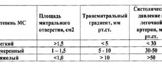
In addition, such parameters are assessed in adult patients as the volume of fluid in the heart sac (35 sq. ml), the diameter of the aortic valve should not exceed one and a half centimeters, and the opening of the mitral valve (4 sq. cm).
Decoding EchoCG
After the examination, the doctor draws up a conclusion. First, a visual picture with the presumed diagnosis is described. The second part of the study protocol indicates the patient’s individual indicators and their compliance with standards.
Decoding the data obtained is not a final diagnosis, since the study can be done not by a cardiologist, but by an ultrasound diagnostic specialist.
It is the cardiologist, based on the collected medical history, examination results, interpretation of tests and data from all prescribed studies, who can draw accurate conclusions about your condition and prescribe the necessary treatment!
Preparing and performing the procedure for children
Like adult patients, children do not need any preliminary preparation. It is advisable that the child does not eat anything for three hours before the test, because with a full stomach, the diaphragm is high, which can distort the result.
Parents should take with them the results of the electrocardiogram performed the day before, as well as the results of studies that were carried out previously. Without fail, the baby should be psychologically prepared for the procedure, explaining that no one is going to hurt him.
In order to carry out the procedure, the baby is undressed to the waist and placed on the left side on the couch. After moving the sensor across the chest, the doctor examines the resulting image.
EchoCG norm: what some parameters indicate
There is a range of normal values for one or another cardiac ultrasound indicator for adults (in children the norms are different and directly depend on age).
Thus, along with other important parameters, echocardiography helps to obtain information about the ejection fraction of the heart - this indicator determines the efficiency of the work performed by the heart with each beat.
Ejection fraction (EF) is the percentage of blood volume ejected into the vessels from the heart ventricle during each contraction. If there were 100 ml of blood in the ventricle, and after the heart contracted, 55 ml entered the aorta, it is considered that the ejection fraction was 55%.
When the term ejection fraction is heard, we are usually talking about the left ventricular (LV) ejection fraction, since it is the left ventricle that ejects blood into the systemic circulation.
A healthy heart, even at rest, pumps more than half of the blood from the left ventricle into the vessels with each beat. As the ejection fraction decreases, heart failure develops.
The normal left ventricular ejection fraction for an adult is 55-70%. A value of 40-55% indicates that EF is below normal. An ejection fraction of less than 40% and even lower ejection fractions indicate heart failure in the patient.
Deviations from the norm
There are a huge number of indicators indicating deviations from the norm. Eg:
- Stenosis is said to occur when the valve opening is reduced in diameter, making it difficult to pump blood.
- If the valve leaflets interfere with the reverse blood flow, perform their natural function inadequately, insufficiency is suspected.
- Pericarditis is detected when, with a normal fluid level of 20-30 ml, all 500 ml are traced.
The list of deviations from the norm and possible diagnoses associated with this can be endless. But you shouldn’t take deciphering an ultrasound of the heart so lightly. Here it is inappropriate to guess and assume without the help of a doctor. Mistakes lead to irreparable consequences. Therefore, experiments should be abandoned.
Is cardiac echocardiography safe?
During this study, there is no radiation or other load on the organ. Therefore, if necessary, it can be prescribed even several times a week.
This study is characterized by the absence of complications and side effects.
EchoCG does not harm either the expectant mother or the fetus during pregnancy.
Limitations for the procedure may include inflammatory diseases of the skin of the chest, chest deformities and some other reasons.
Research methodology
For transthoracic echocardiography, the patient is placed in the left lateral position. When a person lies in this position, the apex of the heart and the left side of the chest are brought closer together. This makes it possible to provide the most accurate visualization of the heart - as a result, all four of its chambers are visible on the monitor at once.
The doctor applies a gel to the sensor, which improves contact between the electrode and the body. After this, the sensor is alternately installed first in the jugular fossa, then in the area of the fifth intercostal space, where the apex beat of the heart can be monitored as clearly as possible, and then under the xiphoid process.
Of course, every doctor strives to ensure that the results of the study are as accurate as possible. It should be noted that how informative the procedure will be depends on three main factors.
First of all, the anatomical features of the patient should be taken into account. Serious obstacles to ultrasound are obesity, chest deformation and other similar factors. As a result, the resulting image may not be clear and cannot be interpreted properly. In order to clarify the diagnosis, doctors in such cases offer a transesophageal examination or MRI.
The quality of the equipment should also be taken into account. Of course, more modern equipment will provide the doctor with more opportunities to obtain sufficient information about the patient's heart.
Finally, the competence of the person conducting the examination should be taken into account. In this case, not only his technical skills are important (the ability to position the patient in the correct position and place the sensor at the right point), but also the ability to analyze the data obtained.
When performing stress echocardiography, the patient first undergoes a regular echocardiography, and then special sensors are applied that record parameters during physical activity. For this purpose, bicycle ergometers, treadmill test, transesophageal electrical stimulation or medications are used. In this case, the initial load is minimal, and then it is gradually increased, monitoring blood pressure and pulse indicators. If the patient's health worsens, the examination is stopped.
All this time, an electrocardiogram is continuously performed, which makes it possible to quickly respond if any extreme situations arise. During exercise, the patient may feel dizziness, increased heart rate, and discomfort in the heart area. After stopping the exercise, the heart rate slows down. Sometimes, in order for the heart to function completely back to normal, it is necessary to administer other medications. In this case, the patient's condition is carefully monitored until complete recovery.
Typically, the entire procedure lasts about an hour.
Transesophageal echocardiography begins with irrigating the patient's mouth and pharynx with lidocaine solution. This is intended to reduce the gag reflex during insertion of the endoscope. After this, the patient is asked to lie on his left side, a mouthpiece is inserted into his mouth and an endoscope is inserted through which ultrasound will be received and delivered.
How to do an ultrasound of the heart at the MedicCity clinic?
It is advisable for every person to have an echocardiogram at least once in their life. The fairly low cost of cardiac ultrasound is another reason to prefer this particular diagnostic method.
At MedicCity you can undergo all types of cardiac diagnostics - ECG, EchoCG, bicycle ergometry, HOLTER, ABPM, etc.
Just type in a search engine: “Ultrasound of the heart, Savelovskaya metro station, Dynamo metro station.” Or call us at: +7 (495) 604-12-12.
The doctors of our multidisciplinary clinic will do everything possible to relieve your heart pain!