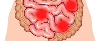
Hidradenitis is a purulent inflammation of the apocrine sweat glands. The disease develops gradually. For the first few days, the inflamed gland appears as a dense, painful nodule the size of a pea. After 5-7 days it increases in size to 2-3 centimeters and takes on a pear-shaped shape. The abscess looks like a nipple, which is why the disease is popularly called “bitch udder.” The armpits are predominantly affected. The groin area is less commonly affected: the labia majora in women and the scrotum in men, as well as the area around the anus. There are cases of inflammation of the sweat glands around the nipples and in the scalp. Hidradenitis is never observed in children and the elderly. This is due to the fact that the functioning of apocrine sweat glands begins only at puberty and fades away in old age. The majority of cases of hidradenitis (about 85%) occur in women aged 16 to 55 years. Hidradenitis most often occurs during puberty, when hormonal imbalance occurs and during menopause.
Causes of hidradenitis (axillary, inguinal and other localizations)
The main triggering factor for this pathology is personal untidiness. If a person ignores the rules of personal hygiene and does not take a shower or bath in a timely manner, the skin becomes a favorable environment for the rapid growth of microorganisms, including Staphylococcus aureus. In recent years, the main cause of suppuration of the sweat glands in the axillary and groin areas is shaving. Microcuts that occur during hair shaving become entry points for infection.
In addition to uncleanliness, obesity plays an important role, due to which the folds of the skin do not dry out in a timely manner (in a humid environment, microbes multiply tens of times more actively). Diaper rash and scratching also reduce the protective functions of the skin. Finally, diabetes mellitus, which impairs the nutritional function of blood vessels, is also a favorable background for the development of purulent skin infections.
The use of antiperspirants is based on blocking the function of sweat glands. Because of this, sweat does not flush their ducts, Staphylococcus aureus is not washed out and inflammation develops in the sweat gland itself.
Chronic hidradenitis suppurativa (HSS) is a multifactorial disease with a chronic relapsing course [1]. Apocrine glands, involved in the pathological process in CHG, begin to function in puberty. Previously, some glands, for example in the areola, were considered modified apocrine glands; now it is recognized that all of them are true apocrines [2].
The exact pathogenetic mechanism of the disease remains unclear. Possible causes of CHG are associated with genetic, environmental, endocrine and microbiological factors. Several studies have indicated higher incidence rates in smokers (70–90%), and have also noted a correlation between body mass index and the severity of CHG. Also, some bacteria and their associations isolated from lesions are considered one of the causes of the inflammatory process. However, CHG is not a classic infectious disease, since it is still not clear what role bacteria play in pathogenesis: primary or secondary [3]. Currently, the number of scientific publications on this aspect of the disease is growing.
The histological picture of CHG varies. Thus, according to G. Jemec et al. [4], in a study of 60 samples of biopsy material, only 17 of them showed follicular occlusion, while in the axillary region apocrine glands were involved in the inflammatory process more often than in the inguinal region. Other biopsy studies have shown the presence of occlusion of the hair follicle due to hyperkeratinization, which led to its expansion, the formation of nodules or cysts and, ultimately, tissue rupture with the formation of sinuses, fistulas and fibrosis [5]. Perhaps the heterogeneity of the histological picture causes difficulties in the treatment of CHG.
Dermatologists and surgeons usually make a diagnosis of CHG based on the characteristic localization and clinical symptoms, but, unfortunately, often with a long diagnostic delay, when the disease already reaches III-IV severity. On average, the time from the onset of the disease to diagnosis takes about 7 years [6]. It has been established that patients turn to doctors of various specialties (surgeons, dermatologists, urologists, proctologists, immunologists, homeopaths), self-medicate for a long time, which also contributes to late diagnosis and delay of timely adequate treatment, and, ultimately, aggravation of the clinical picture and deterioration quality of life.
There are three main diagnostic criteria for CHG [7]:
1) typical anatomical localization of foci of inflammation: axillary and inguinal areas (symmetrical lesions);
2) typical clinical symptoms: moderate pain, deep nodes, conglomerates of nodes, “heavy” tissue infiltration, abscess formation, fistula tracts with purulent discharge, fibrosis, scars;
3) recurrence and chronic course.
Additional criteria include hereditary predisposition, the presence of CHG in relatives; pilonoid sinus, a history of suppurating sacrococcygeal cyst; metabolic syndrome, hyperhidrosis, smoking. A. Jørgensen et al. [8] studied the clinical presentation of patients with different body mass index (BMI) and found phenotypic heterogeneity in individuals with high and low BMI. The authors emphasized that these two phenotypes may reflect different etiologies and prognosis and, therefore, require different approaches to management tactics.
To facilitate the assessment of the severity of the clinical picture and the severity of the process, various rating scales are used around the world to help the attending physician choose the optimal treatment algorithm and tactics for managing a patient with hidradenitis.
In existing European clinical guidelines [7], the choice of a particular treatment method is based on a staged approach, taking into account the severity of the disease according to H. Hurley. Later, other rating scales were proposed that allow the doctor to determine the indications for a therapeutic or surgical method of treating a given disease, depending on the severity of the clinical picture.
In accordance with the classification according to H. Hurley, there are three degrees of severity of CHG [9]:
— I degree of severity is characterized by the formation of one or several painless or slightly painful isolated nodes and/or abscesses that are not prone to acute progression and suppuration; fistula formation and scarring are absent;
- II degree of severity - intensification of the inflammatory process limited to one localization, recurrent nodes and abscesses are delimited from each other, do not merge, open up, form fistulous tracts with purulent-suppurative discharge, some elements are scarred;
- III degree of severity - a diffuse conglomerate of inflammatory infiltrates is formed, consisting of merging nodes and abscesses, fluctuation intensifies, many interconnected fistulas are formed, scarring continues; Limitations of joint mobility due to pain sometimes lead to contractures.
There is also a more complex system for determining the severity index of CHG - the modified Sartorius score, which involves quantitative scoring depending on the presence of the main clinical parameters of the disease: the number of certain anatomical zones involved in the process, the number of painful nodes, abscesses, fistula tracts with purulent discharge, scars, superficial pustules and folliculitis, the distance between damaged areas, the presence of healthy skin in the lesion [10].
Some authors [11] use the “Physician General Assessment of the Severity of CHG” (HS-PGA) criteria. The minimum degree is one or more non-inflamed, painless nodes; mild degree - 1-4 inflamed formations, one abscess or fistula; moderate degree - less than 10 inflammatory elements, 1-5 abscesses and fistulas; severe degree - more than 10 nodes, 2-5 abscesses and fistulas; very severe severity - more than 5 abscesses and fistulas.
There is also a scale for assessing the HSSI severity index in points: mild - from 0 to 7 points, moderate - 8-12, severe - 13 or more. When calculating, the number of affected areas, pain syndrome, number of inflammatory elements, socialization, etc. are taken into account [12, 13].
The Hidradenitis Suppurativa Clinical Response Scale (HiSCR) was one of the latest to be developed to assess CHG. This scale was proposed in the treatment of CHG with adalimumab; it shows a decrease of 50% or more in the number of inflammatory elements and no signs of an increase in abscesses or fistulas with CHG compared with the baseline [14, 15].
A multidisciplinary approach to solving the problem of CHG requires not only the study of severity and the development of rating scales, but also the analysis of laboratory parameters and the development of the most optimal tactics in the treatment of such patients by both dermatologists and surgeons. N. Yaşar et al. [16] focused on changes in hematological parameters in CHG depending on the severity of the disease. It was found that hemoglobin level was higher in stage I than in stage III, while hematocrit, mean platelet volume, corpuscular hemoglobin concentration level and platelet count did not differ between stages. The number of leukocytes and neutrophils was higher in stage III than in stage II, and the number of lymphocytes was lower in stage III than in stage II. These changes can be further used to assess the severity of CHG.
Treatment of CHG can be divided into conservative and surgical. Conservative methods include local, systemic and other types of therapy. Considering the impact of the disease on the quality of life, increased attention should be paid to the psychosocial adaptation of patients. The doctor's recommendations should also be aimed at reducing risk factors for the disease. Since smoking and obesity show the strongest correlation with the severity of the pathological process, patients with CHG should be advised to quit smoking and take measures to reduce body weight [2].
In our country, the approach to the treatment of CHG is similar to the treatment of deep pyoderma and is determined by the use of anti-inflammatory and antibacterial drugs [17, 18]. Recent publications by Russian authors show a positive effect of therapy with TNF-α inhibitors, but, unfortunately, these studies are rare [19].
In European recommendations [20], antibiotics are mentioned as first-line drugs in the treatment of CHG, taking into account the severity according to H. Hurley. However, antibacterial therapy, especially long-term, is often accompanied by complications from parenchymal organs. In addition, if it is impossible to identify the species of bacteria in culture and determine the sensitivity of the microflora from the lesion, antibacterial therapy may be ineffective and cause only a short-term effect. Antibiotics, as a rule, are used for all degrees of severity of CHG and without fail in the pre- and postoperative periods. Most often these are lincosamides, tetracyclines, rifampicin, cephalosporins, etc.
The use of steroid drugs and dapsone was abandoned due to their low clinical effectiveness [21–24].
Therapy with biological drugs and tumor necrosis factor-α blockers against CHG is effective, but has a number of limitations: it is not used in severe forms of the disease, profuse suppuration and abscess formation, the risk of spreading infection, initiation of immunosuppressive therapy, and some diseases of internal organs [25, 26]. Adalimumab, which is used at a dose of 40 mg/week, has been more thoroughly studied [27]. Less popular is etanercept, which has shown little effectiveness in treating CHG [28].
The results of a number of studies have been published [29, 30] based on small clinical material on the use of new immune methods of exposure to the drug apremilast, a molecular selective inhibitor of phosphodiesterase 4 (PDE4). Some effectiveness has been shown in the treatment of CHG.
Recent publications show that timely, early and wide surgical excision of purulent foci in the treatment of any stages of CHG is the most effective way to achieve recovery, prevent complications and relapses, and quickly improve the quality of life of patients [31-33]. The volume and type of surgical intervention are determined individually in accordance with the age, severity, localization of the inflammatory process and general status of the patient. Preoperative preparation is important because it affects the outcome of surgery and subsequent recovery. Z. Alharbi et al. [30] conducted a retrospective analysis of 50 operations with wide surgical access in 32 patients; 81.25% had no relapses after treatment.
Other methods of intervention for CHG have been described: cryoinsufflation, botulinum therapy, photodynamic therapy (PDT). However, the results are heterogeneous and isolated [33].
Along with traditional surgical methods, laser surgery is used. Carbon dioxide (CO2) laser ablation has shown fairly high efficiency. P. Hazen et al. [34], using a sample of 22 patients, showed positive results in the treatment of hidradenitis using a long-pulse neodymium diode yttrium aluminum garnet laser (Nd:YAG laser) - 72.7 compared to 22.9%.
In the context of growing interest in the problem of CHG in our country, a search is being carried out for new medications and optimal surgical methods for treating this disease, algorithms for patient management are being created depending on the duration of the disease, localization, severity and prevalence of the purulent-inflammatory process, rules for preoperative preparation and postoperative preparation are being developed. management of patients with CHG.
A necessary condition for radical excision of foci of inflammation and the success of the operation is careful preoperative preparation of the patient. In order to maximize the relief of perifocal inflammation, reduce purulent discharge from fistulas, delimit infiltrates from healthy tissue, increase the overall resistance of the body, normalize the temperature reaction, peripheral blood parameters and immunological parameters, antibiotic and actinolysate therapy are carried out, as well as evacuation of purulent masses with microorganisms from the fistula tracts by washing them with antiseptic solutions [35, 36].
In our country, there is no single algorithm that would allow us to determine the indications for conservative and surgical treatment of CHG. The search for new treatment methods, as well as the development of a unified clinical classification and rating scales are urgent tasks at the present stage.
Thus, future research should focus on optimizing treatment.
Information about authors
Burova S.A. — https://orcid.org/0000-0003-0017-621X
Borodulina K.S. – https://orcid.org/0000-0001-7670-1450
Corresponding author:
Borodulina K.S. — email
Where does hidradenitis occur?
Typical localization is the axillary region (the same in both women and men), hidradenitis in the groin (in women more often due to shaving of the intimate area), in the areola of the nipple and under the mammary glands. Hidradenitis genitalis occurs less frequently (in men - the scrotum, in women - the labia majora), as well as on the face (mainly in men).
Such localizations are due to the fact that apocrine sweat glands located in these places are most often susceptible to inflammation. Their peculiarity is the tortuosity of the ducts, which impairs the outflow of sweat and contributes to its inflammation when Staphylococcus aureus penetrates into the gland.
2. Reasons
Hidradenitis in most cases is caused by Staphylococcus aureus (sometimes streptococci, Escherichia coli, pseudomonas, etc. are identified as the causative agent). The predominant route of penetration into the apocrine gland is lymphogenous; Often the source of infection is an infected hair follicle. The most significant risk factors are:
- female gender (women have significantly more apocrine glands);
- microtrauma of the skin due to shaving, hair removal, etc.;
- scratching, diaper rash, abrasions, irritation and other skin lesions that facilitate the penetration of bacterial infections;
- endocrine and metabolic disorders, especially accompanied by obesity, sweating, increased humidity and oily skin;
- weakness of the immune system due to one reason or another (general exhaustion, previous abdominal surgery, hypovitaminosis, etc.).
It should be noted, however, that most often the immediate cause is a simple failure to comply with the rules and procedures of aseptic hygiene.
Visit our Microsurgery page
Stages of the disease hidradenitis
There is no clear stage in the development of the disease. It all starts with the appearance of a painful small nodule under the skin in a typical location. It is not visible at first, but can be felt. After 24-48 hours, it increases significantly in size, the skin over it changes color to red, and then to purple-bluish. The pain becomes very severe and often prevents active movements of the limb.
At this moment, in the thickness of the node there is a process of tissue melting, which is manifested by the appearance of fluctuations (when you feel the node, you feel that its center seems to fall under your finger). The skin over the abscess gradually thins until a small hole appears in it, through which thick pus begins to flow. This usually occurs 7-10 days from the onset of the disease. Often, in the absence of treatment for hidradenitis suppurativa, the process ends in the formation of a fistula, through which serous-purulent contents constantly leak.
Subcutaneous tissue is penetrated by a large number of lymphatic vessels. Through them, the infection can spread to neighboring sweat glands. In this case, the general condition of the patient may also suffer. Symptoms of chronic hidradenitis: body temperature rises, fatigue, weakness appear, sleep and performance are disturbed. Due to massive infection, the subcutaneous tissue swells greatly, foci of infection protrude above the skin, which together forms a picture similar to a “bitch udder” (this has become the popular name for the disease).
Causes of hidradenitis development
Apocrine glands do not take a major part in the thermoregulation of the body. They are activated during stressful situations, affecting the smell and viscosity of sweat. The main function of the organ is to remove heavy metals, water, salts and various metabolic products from the human body. Improper functioning of the glands and the penetration of bacteria into them lead to hidradenitis.
Hidradenitis is a type of pyoderma and the reasons for its appearance are directly related to the presence of pyogenic cocci. Inflammation of the apocrine glands in hidradenitis is the result of the destructive effects of staphylococcus.
Risk factors that provoke the disease include:
- Excessive sweating or hyperhidrosis.
- Diseases, skin injuries.
- Lack of personal hygiene.
- Incorrect use of deodorizing agents.
- Lipid metabolism disorders.
- Conditions of high humidity or hot climate, etc.
Treatment of hidradenitis
At the stage of infiltration formation, the doctor may try to treat hidradenitis in the groin area, armpits and other locations conservatively. On the first or second day, the use of local remedies is recommended. Antibiotics for hidradenitis are used in the form of ointments, most often tetracycline, erythromycin ointments, and clindamycin lotions are used. Since it is most often impossible to cure acute hidradenitis with conservative methods, it is necessary to resort to surgical intervention.
Adequate outflow is a key point in the treatment of any purulent process. Hidradenitis surgery is the best way to achieve this. According to surgeons, the most appropriate method of pain relief is anesthesia. This avoids systemic effects from anesthetics that would have to be administered in too large quantities.
Progress of the operation:
- The skin above and around the abscess is treated with antiseptics.
- A small incision is made throughout the entire depth of the skin until the abscess cavity is opened.
- The cavity is treated with antiseptics, then drained with gauze turunda soaked in water-soluble antibiotic ointment.
- With multiple lesions of the sweat glands, each abscess is opened immediately. If the lesions are located very close to each other, it is better to remove the entire inflammatory conglomerate within healthy tissue. In this case, the wound is sutured. In all others, it heals on its own.
After the operation, daily dressings are performed, during which the skin around the wound is treated with antiseptics. They also wash the abscess cavity, change the turundas with ointment. At the end of the dressing, bandages with antimicrobial ointment are applied to the wound.
Turunda is removed after pus stops flowing from the wound. After this, it quickly heals on its own. Of course, until complete healing, dressings continue.
General conservative treatment is usually carried out after surgery in cases where hidradenitis recurs (some call this form of the disease chronic, which does not reflect its actual course) or many sweat glands are affected. In this case the following applies:
- courses of antibiotic therapy with cephalosporins and semisynthetic penicillins;
- transfusion of antistaphylococcal plasma;
- UV irradiation of blood;
- laser irradiation of blood;
- vitamin therapy.
For severe pain, non-narcotic analgesics are prescribed; for sleep disturbances, appropriate sleeping pills are prescribed.
4.Treatment
As a rule, the course and treatment of hidradenitis takes 10-14 days. Antibiotics are prescribed (only and exclusively by a doctor), nitrofuran derivatives, and in some cases autohemotherapy and immunostimulation. In some cases, the abscess is opened surgically. UHF, medicinal (in particular, ichthyol) applications, ultraviolet irradiation, etc. are used as local treatment. There are also numerous folk remedies that are considered effective for hidradenitis. It is important to remember, however, that hidradenitis is dangerous due to complications, incl. as severe as phlegmon or sepsis; in addition, it easily acquires a relapsing course. Therefore, self-medication in this case is too risky, and consultation with a doctor is necessary. Finally, it will not be difficult to eliminate most of the risk factors listed in the corresponding paragraph, which will serve as preventive protection against this unpleasant, painful and unsafe disease.
Methods of treating the disease
At the initial stages of the disease, the site of inflammation should be wiped with antiseptics - salicylic or boric alcohol, chlorhexidine, a solution of calendula alcohol tincture, octenisept. Next, you need to apply any ointment to the site of inflammation - Vishnevsky, levomekol or ichthyol, which must be applied under the bandage and changed several times a day.
In addition, a visit to a doctor is required, who will assess the size and severity of the inflammation and prescribe appropriate treatment, which may include surgery. The operation is performed under local anesthesia and consists of dissecting the abscess, draining and treating adjacent tissues with an antiseptic. After which a healing bandage is applied.
Preparation, diagnostics
Upon examination, the surgeon differentiates hidradenitis from other purulent lesions of the skin and soft tissues and determines the stage of the disease. For adequate antibiotic therapy, bacterial culture of purulent discharge is carried out to determine microflora and sensitivity to antibiotics. For prolonged and recurrent pathologies, an immunogram is prescribed. Surgical intervention does not require specific preparation. All you need to do is pass a standard package of tests. Thanks to our own laboratory, the answers will be ready within an hour, after which the surgeon will carry out the treatment.
Indications for surgery
Indications for surgical treatment are:
- the formation of a painful inflammatory-purulent infiltrate in the armpit, near the navel, on the labia, in the inguinal fold;
- destruction of the apocrine gland with the formation of an abscess;
- recurrent hidradenitis.
The decision on the possibility of surgical intervention on an outpatient basis is made by the surgeon based on the characteristics of the clinical situation. In some cases, hospitalization may be required.
Stages of the disease
There are 3 stages of development of hidradenitis:
- The initial stage is characterized by the appearance of nodular neoplasms, their enlargement, and the development of inflammatory processes.
- The empyema stage consists of an increase in the symptoms of inflammation, the growth of the inflammation focus, the formation of pus in the neoplasm and its release using surgical excision or independently.
- The final stage of healing is characterized by the formation of scars at the site of the ulcers.
The entire process from the initial stage to the healing stage lasts on average 2 weeks, and proceeds with successful resolution only when seeking medical help. If treatment is delayed, sepsis may occur.
Symptoms of hidradenitis
This disease is also called “bitch udder”, since in the area of penetration of staphylococcus an abscess is formed that looks similar to the mammary gland of a dog. Hidradenitis is most often found in the armpits and groin area. It may also occur in the genital area and under the breasts.
There may be one abscess; several of them form at once, then later they will merge into one large infiltrate. The patient may sometimes have a fever, accompanied by weakness and malaise. Boils are painful.
The lesion is often multiple, when several abscesses develop at once. The patient's condition worsens, fever occurs, redness and severe pain appear in the affected area. Sometimes infiltrates suppurate and burst with the release of large amounts of liquid pus. Then they tighten, but soon the neighboring glands are involved in the process and everything starts again. The disease has a protracted course and can lead to sepsis - blood poisoning.