How does the liver work?
The liver is the largest gland in humans. It is located in the upper floor of the abdominal cavity on the right. It consists of two main lobes - right and left, which in turn are divided into 8 segments.
In an adult, it weighs about 1/40 of the total body weight (approximately 1.6 kg in men and 1.2 kg in women). The liver has a dual blood supply: approximately 80% of the blood entering the liver comes from the portal vein (venous blood collected from all unpaired abdominal organs), the rest (oxygenated arterial blood) comes from the hepatic artery. Having entered the portal of the liver, both vessels give rise to multiple branching smaller vessels. At the exit from the liver, 3-4 hepatic veins are formed, which flow into the inferior vena cava.
The main structural unit of the liver is the hepatic lobule, which consists of liver cells - hepatocytes. This cell is one of the most important biochemical laboratories of the body. The hepatocyte takes part in the metabolism of proteins, carbohydrates, fats, bile acids, and vitamins. Neutralizes toxic substances coming from the intestines, followed by the release of associated products into the intestinal lumen. An important function of the hepatocyte is the synthesis of bile, which is involved in digestion.
What is bile and why is it needed?
Bile is a secretion produced by hepatocytes. The color of bile ranges from light yellow to dark green. Bile contains bile pigments - bilirubin and biliverdin, cholesterol, fatty acids, lecithin, mucin.
Bile is the main participant in digestion. It neutralizes hydrochloric acid that enters the duodenum from the stomach, thereby creating favorable conditions for the action of pancreatic juice and parietal digestion in the jejunum.
Bile acids emulsify fats, allowing them to be absorbed in the intestines.
Bile has a bactericidal effect that prevents the proliferation of pathogenic microflora.
What and how to eat to avoid stones?
Promotes the formation of stones. Fast food, meat (especially fatty meat), sugar and other easily digestible carbohydrates (sweets, flour, soda), low fiber intake, very low-calorie diets (up to 1500 kcal per day), high fructose intake (diabetic foods, honey, fructose soda), beans and other legumes, vitamin C deficiency, overeating (excess calorie intake), prolonged fasting.
Protects against the formation of stones. Monounsaturated and polyunsaturated fats (vegetable oils), nuts, fiber (vegetables, fruits and whole grains), calcium (dairy products), moderate alcohol consumption containing no more than 20-40 g of pure alcohol per day, coffee and caffeine, vitamin C and foods rich in it, a regular diet, a vegetarian diet, fish and fish oil, with low-calorie diets it is important to consume enough fat (7-10 g per day).
How are the bile ducts arranged and how do they work?
The bile ducts are a system of ducts that originate from hepatocytes and gradually gather into interlobular, segmental and right and left lobar ducts, which unite into the common hepatic duct.
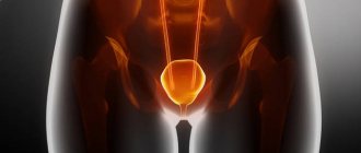
One of the stages of bile removal is its accumulation in the reservoir - the gallbladder. The main function of the gallbladder is to concentrate bile and release it into the lumen of the duodenum during digestion. The volume of this organ is about 80 ml.
The gallbladder is located on the lower surface of the liver. It is a hollow muscular organ, externally covered with a serous membrane and internally lined with mucous membrane. The structure of the gallbladder is divided into the bottom, body and neck of the bladder, which passes into the cystic duct.
Anatomy and location of the organ
The gallbladder is shaped like a pear and is located on the visceral surface of the liver in a special depression that separates the two lobes of the liver. In the anatomy of the gallbladder, there are three sections: the bottom, the body, and the neck. The bottom of the organ is located near the lower edge of the liver, and the neck faces the gate of the gland and is located together with the ducts in the duplication of the hepatoduodenal ligament.
In the area where the body meets the neck, a bend is formed, so the neck lies at an angle to the body. Between the transition to the cystic duct there is a depression called Hartmann's pouch. The normal size of the gallbladder in adults is: length 8–14 cm, width 3–5 cm. The organ holds 60–100 ml of liver secretion. In children, normal organ sizes are in an even larger range.
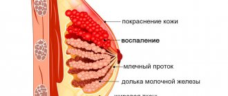
So, in a child 2–5 years old, the gall bladder is 3–5.2 cm long and 1.4–2.3 cm wide, and in a teenager it is 3.8–8 cm long and 1.3–2.8 cm wide. If the organ is larger, then this indicates obstruction of the bile ducts or acute cholecystitis. A decrease in size occurs with viral hepatitis (liver inflammation) or chronic cholecystitis. The organ wall includes the mucous membrane, muscular, subserous and serous layers. The mucous tissue is sensitive to adverse events occurring in the body, which is why it looks swollen and flaky.
Bundles of muscle fibers are located in the longitudinal and circular directions. There are gaps between them, and then in these places the mucous tissue connects with the serous tissue. This structure of the gallbladder increases the risk of bile leaking into the abdominal cavity (peritonitis) without compromising the integrity of the organ. There is less muscle tissue in the bottom area, and more in the neck area.
The photo shows the location of the organ relative to the liver
The blood supply to the organ occurs through the cystic artery, which comes from the right branch of the hepatic artery and at the neck of the bladder divides into two branches, one of them goes to the upper surface of the bladder, and the other to the lower. Lymph nodes are located to the left of the bladder neck and near the duodenum. When the bladder becomes inflamed, the nodes enlarge and block the common bile duct.
The innervation of the biliary system comes from the celiac, inferior phrenic plexuses and the anterior trunk of the vagus nerve. This means that diseases of the stomach, small intestine or irritation of the vagus nerve (which happens with a diaphragmatic hernia) can provoke malfunction of the sphincter of Oddi and inflammatory disorders in the bladder itself, and vice versa.
Patients often ask which side the gallbladder is on. The gallbladder is located on the right side of the body, under the ribs. In front of the gallbladder is the liver, on the left side is the pylorus, and on the right are the loops of the small intestine.
The bottom of the bladder, as a rule, extends from under the lower edge of the liver closest to the peritoneum by 2–3 cm and touches the anterior abdominal wall. This arrangement of the gallbladder and its ducts gives a projection of pain to the right hypochondrium and epigastric region.
Why do gallstones form?
The exact cause of cholelithiasis has not yet been fully elucidated. Only a few factors are known that increase the likelihood of its occurrence.
- Genetic: family predisposition increases the risk of cholelithiasis by 4-5 times;
- Congenital anomalies of the biliary tract;
- Gender - the risk of developing gallstones in women is approximately 2-3 times higher, which is associated with the influence of estrogens on the ability to form gallstones;
- Age - the maximum frequency of clinical manifestations of cholelithiasis is recorded at the age of 40-69 years;
- Dietary (high-calorie diet, poor in plant fiber, as well as with an excess of simple carbohydrates, animal proteins, foods containing cholesterol);
- Use of medications (oral contraceptives, fibrates, ceftriaxone, somatostatin, nicotinic acid);
- Concomitant diseases and conditions (obesity - a tendency to form gallstones in persons with a body mass index of more than 30 is 3 times higher, type 2 diabetes mellitus, hypothyroidism, cirrhosis of the liver, etc.);
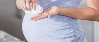
- Pregnancy - the risk of developing cholelithiasis increases during pregnancy, especially with repeated pregnancies (the likelihood of stone formation increases by 10-11 times). During pregnancy, bile heterogeneity, or the so-called biliary sludge, develops in 20-30% of patients, stones - in 5-12% of cases.
There is a so-called 5F rule that describes patients at greatest risk of developing stones:
1. Female (female);
2. Fat (overweight/obesity);
3. Forty (age 40 years or more);
4. Fertile (fertile women - that is, pregnant and giving birth);
5. Fair (light-skinned blondes).
The list of gallbladder diseases also includes less common pathologies:
- Congenital anomalies of the biliary tract - changes in the size, shape of the biliary tract, size of the gallbladder. Pain in these cases can occur when the bladder is torsion or volvulus2.
- Congenital anomalies of the bile ducts - atresia (congenital absence), underdevelopment (hypoplasia). These anomalies usually appear in early childhood2.
- Bile duct cysts. They develop very slowly, so in about half of cases symptoms appear decades later2.
- Choledocholithiasis is the formation of stones in the common bile duct, usually occurring in old age. It may not manifest itself in any way for several years2.
How are gallstones formed?
The process of stone formation is quite well studied. The key links are:
- An increase in the lithogenicity of bile (the ability to form stones) means an increase in the content of cholesterol and bilirubin in it.
- Impaired contractile function of the gallbladder.
- Increased pressure in the bile duct system.
Mucin is a natural gel, constantly secreted by the wall of the gallbladder; when lithogenicity increases, it begins to “absorb” cholesterol crystals, thereby forming a “fossilization nucleus.” The formed suspension, with impaired contractile function, continues to attach salt crystals encrusted with calcium salts. Thus, gallstones can grow up to 5-7 mm per year.
How does gallstone disease occur?
The main factors influencing the course of the disease are the nature of nutrition and the size of the stones. Gallstone disease can occur both in the form of chronic stone carriage and in acute, and sometimes in complicated, form.
Where does pain come from with gallstone disease?
By themselves, gallstones do not cause any pain and can be an accidental finding during medical examination. When food enters the duodenum, the mechanoreceptors are irritated and cause contraction of the gallbladder. The stones wedge into the cervix, causing a prolonged spasm, which manifests itself as pain. When taking antispasmodic drugs, the calculus can “move” back from the cervix and the attack of hepatic colic is stopped. If the stone remains in the cervix, then inflammation begins to develop against the background of obstruction.
Chronic calculous cholecystitis is an asymptomatic stone carrier. The size of the stones can vary from 2 – 3 mm to 5 cm. This stage is characterized by an almost complete absence of clinical manifestations of the disease. Stones can remain in the lumen of the gallbladder for years without causing any symptoms. However, if there is an error, discomfort may occur in the right hypochondrium, which can be relieved independently or while taking antispasmodic drugs.
Hydrocele of the gallbladder - occurs against the background of obstruction of the neck of the gallbladder by a calculus, cutting off the latter from the entry of bile. The bladder is filled with mucus secreted by the mucous membrane and is not involved in digestion.
Acute calculous cholecystitis - occurs with prolonged obstruction of the neck of the gallbladder by a calculus. The pain syndrome persists against the background of ineffective conservative treatment. The patient is bothered by vomiting, which does not bring relief; pain can be felt behind the sternum - cholecystocardial syndrome, in the right shoulder blade. Body temperature rises and chills appear. Inflammatory changes begin to develop in the wall of the gallbladder, which progress from catarrhal, phlegmonous and gangrenous forms. Against the background of the latter, perforation of the gallbladder or abscess may develop.
Choledocholithiasis and acute biliary pancreatitis. Choledocholithiasis develops as a result of migration of biliary sludge or small stones from the gallbladder into the common bile duct. Once in the common bile duct, small stones freely descend to the sphincter of Oddi, which is a release mechanism - a circular muscle cuff. Sludge and stones “wedge” into the sphincter, causing its long-term spasm, causing a violation of the outflow of bile - obstructive jaundice. The patient's skin and sclera become a rich yellow color. This condition requires emergency decompression of the bile ducts - restoration of bile outflow.
An increase in pressure in the common bile duct promotes the reflux of bile into the main pancreatic duct and intraductal activation of pancreatic juice enzymes - a trigger for the development of acute biliary pancreatitis.
Mirizzi syndrome . A fairly rare complication of gallstone disease. Develops as a result of prolonged stone bearing. First, a stricture forms, then a bedsore between the gallbladder and the common bile duct.
Mirizzi syndrome presents significant difficulties for surgical treatment. This is due to the fact that in the human body there is no plastic material to close the defect of the common bile duct. Surgical treatment of Mirizzi syndrome is often accompanied by the formation of an anastomosis between the small intestine and the common bile duct. When a stone migrates into the small intestine, acute intestinal obstruction may develop, requiring emergency surgical treatment.
In the photographs, in the lumen of the resected section of the small intestine there is a calculus that caused intestinal obstruction.
When the gallbladder is inflamed. We talk about cholecystitis
Modern medicine has to deal with pathology of the biliary tract quite often. And the number of such diseases continues to grow. Today we will talk about cholecystitis: what kind of disease it is, what are its symptoms, the causes of its appearance, how it is detected and treated.
Nikolai Borisovich Patrushev, a gastroenterologist at Clinic Expert, Perm, answers our questions.
— Nikolai Borisovich, there is evidence that today 10-20% of the adult population suffers from cholecystitis, and there is a tendency for this pathology to further increase. Please tell us what it is?
— In one sentence, cholecystitis is an inflammation of the gallbladder. Pathology of the biliary tract remains a pressing problem for today's medicine. Yes, there is a tendency towards an increase in incidence. Moreover: according to scientists, the number of diseases of the digestive system (which includes pathology of the biliary tract) will increase by 30-50% in the world in the next 15-20 years.
— What types of cholecystitis are known to modern medicine?
— There are two of them: acute and chronic cholecystitis. The first develops quickly, against the background of complete well-being. Abdominal pain appears, the pain is localized in the right hypochondrium. Nausea and vomiting may occur, and the temperature may rise. As a rule, acute cholecystitis is most often a manifestation of cholelithiasis. Such patients are hospitalized by ambulance in a surgical hospital.
Read more about gallstone disease here: Remove or leave? What to do if gallstones are found?
Chronic cholecystitis initially develops as an independent process, gradually and imperceptibly. Often, a patient is diagnosed with cholecystitis during an examination for completely different diseases. It should also be taken into account that diseases of the gallbladder and biliary tract are characterized by a variety of clinical manifestations, duration of course, and prolonged exacerbations - this causes frequent seeking of medical help, making these ailments a social problem.
- What are the causes of cholecystitis? What causes it?
— Various factors are involved in the development of this pathology. In first place I would put a violation of the contractile function of the gallbladder. This leads to stagnation of bile, slowing down its evacuation from the gallbladder. Most often, the contractile function of the gallbladder is affected by psycho-emotional overload, neurotic reactions and prolonged stressful situations, physical inactivity.
The infectious factor also matters. Infectious agents penetrate the gallbladder from chronic foci of inflammation in the body - for example, in diseases of the ENT organs, stomach, duodenum, and from other parts of the intestinal tube.
Helminthic infestations, such as opisthorchiasis, also contribute to the development of cholecystitis. Previous hepatitis A (Botkin's disease) can also lead to inflammation of the gallbladder.
This disease can also be provoked by the reflux of pancreatic juice into the cavity of the gallbladder - a so-called chemical burn of the gallbladder mucosa occurs, which can lead to its inflammation.
Read materials on the topic:
How to protect yourself from Botkin's disease? Is there a stress vaccine? Childhood is for movement! What does physical inactivity lead to?
— Please tell us about the signs of cholecystitis. How does it manifest itself?
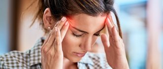
— Cholecystitis can occur with varying degrees of severity. The symptoms will depend on this, as well as on the stage of the disease. First of all, the pathology manifests itself as pain. These pains occur due to spasm of the gallbladder or due to its stretching.
Pain appears in the right hypochondrium, usually 40 minutes - 1.5 hours after an error in diet (for example, when eating spicy, fatty foods, fried foods, overeating). The pain is felt for about half an hour and, in mild cases, goes away on its own. In severe forms, the pain is more intense and prolonged.
Pain can also be provoked by a person spending a long time in a sitting position - driving a car, at a computer. From the right hypochondrium, such pain can radiate upward - to the right shoulder, neck, right shoulder blade.
In addition to pain, the patient may be bothered by so-called dyspeptic disorders: belching, nausea, metallic taste in the mouth, sometimes vomiting (if it occurs repeatedly, bile may appear in it). Abdominal bloating and alternating constipation and diarrhea may occur.
Also, cholecystitis may be accompanied by irritability, insomnia, and decreased performance.
The more severe the form of cholecystitis, the brighter and longer the listed symptoms will be expressed.
— Is there asymptomatic cholecystitis? That is, nothing bothers the person, and the disease is detected only when conducting any diagnostic studies - for example, as part of a preventive examination
- Yes, this happens. In 50% of cases, cholecystitis occurs latently and asymptomatically. Signs of gallbladder pathology before clinical manifestations of the disease can only be indicated by ultrasound data. Therefore, this study must be included in the screening program for diseases of the digestive system.
In addition, there are atypical clinical forms that can mislead the doctor, simulating various diseases of other organs and systems, for example, cardiovascular, endocrine and others (so-called “masks”). And when we start to understand, it turns out that we are talking about the pathology of the gallbladder. The most well-known and studied is the so-called cardiac mask of cholecystitis (or cholecystocardiac syndrome): every third or second patient with cholecystitis may complain of heart problems. These are rapid heartbeat, shortness of breath, pain in the heart area. The electrocardiogram in such patients is without any abnormalities.
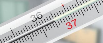
Cholecystitis can also occur under the guise of thyroid pathology - here the patient’s complaints will be similar to thyrotoxicosis (irritability, excessive sweating, the same rapid heartbeat, trembling of the fingers, increased body temperature to 37-37.5 degrees). In this case, the thyroid gland may turn out to be somewhat enlarged, and here it is necessary to figure out whether the symptoms are caused by its pathology, or whether the problem is still in the gallbladder.
Cholecystitis may also have an allergic mask, and a number of others. To understand this, the doctor must have considerable practical experience and correctly build a diagnostic search.
- Let's talk about diagnosing cholecystitis. What does it include? How can a doctor detect this pathology?
— Today, the most accessible and common method for diagnosing cholecystitis is ultrasound of the abdominal cavity. Ultrasound diagnostics allows you to assess the condition of the walls of the gallbladder: if they are thickened by more than 4 mm, this is already a clear sign of cholecystitis, if less, perhaps the pathology is only at the beginning of development. The doctor also evaluates the contractility of the gallbladder.
An ultrasound will also help identify gallstones. If they are detected and cholecystitis is also present, then they speak of stone (calculous) cholecystitis.
You can sign up for an abdominal ultrasound here. ATTENTION: the service is not available in all cities
There are other research methods. For example, probing of the duodenum (duodenal intubation). It allows you to evaluate the biochemical composition of bile, test it for infection and identify microbes that cause inflammation in the gallbladder.
— How can doctors help a patient with this disease? How is cholecystitis treated?
“We can only help after, during a full examination, we find out as fully as possible all the reasons that led to the appearance of cholecystitis in the patient. The treatment program is structured as follows. In the first place is therapeutic nutrition. First of all, this is a fractional meal, five to six times a day. The gallbladder “loves” that we take food at the same hours: by doing this we train it and prevent stagnation of bile in it. It is important that the food is not hot or cold, but warm.
The selection of medications is carried out taking into account the function of the gallbladder, in particular its contractility. If the function is increased, if there are sharp spastic pains, antispasmodic drugs are recommended. When, during normal contractile function of the organ, we find signs of viscous, stagnant bile, drugs with a choleretic effect are used.
If studies show poor contractility of the bladder, then the treatment program includes drugs that stimulate its function and help it work correctly.
In certain cases, antiparasitic, antibacterial, and anti-inflammatory drugs are used. The doctor decides all this, depending on the results of the study.
Let's not forget about other methods of treating cholecystitis - the same sanatorium-resort treatment. Chronic patients need to go to resorts for at least three or four years in a row - as they used to say, “to the waters.” It is very useful. But you need to go there with all medical documents, then the patient will be given the right diet and water intake (type, quantity and temperature).
— What can you eat with cholecystitis and what can’t you eat?
— Proteins, fats and carbohydrates should be exactly as much as necessary for the normal functioning of the body. The diet should include lean meats (beef, rabbit), fish (preferably boiled or steamed), low-fat cottage cheese, and wheat bran. Among cereals, preference is given to oatmeal and buckwheat.
Let's not forget about fresh vegetables and fruits (it's better to have melon, pumpkin, watermelon, carrots, apples).
Fatty, fried foods, smoked foods, marinades, pickles, and alcoholic drinks, including beer, are excluded. This list also includes carbonated drinks, hot seasonings and egg yolks (the latter for periods of exacerbation).
Fasting days (1-3 per week) will not hurt: these can be rice-compote, cottage cheese, watermelon days. In addition, drinking plenty of fluids is recommended.
— How to prevent the development of cholecystitis? Please tell us about prevention
— Probably, much is clear already from the above. Basic prevention differs little from the prevention of many other diseases. This is a healthy lifestyle, rational nutrition - we eat often, not on the run, we chew food thoroughly, slowly.
Agree, seemingly simple, truisms. But, believe me, first of all, they are important in terms of preventing cholecystitis.
Interviewed by Igor Chichinov
If you need a consultation with a gastroenterologist, you can make an appointment here. ATTENTION: the service is not available in all cities
The editors recommend:
Biliary dyskinesia: what is hidden behind this diagnosis? Why do people turn yellow? MRI of the liver: pros and cons
For reference:
Patrushev Nikolay Borisovich
Gastroenterologist, Candidate of Medical Sciences Graduate of the Perm State Institute in 1988. Primary specialization in gastroenterology - 1995. From 2005 to 2014. worked at the Central Research Institute of Gastroenterology (Moscow). First - a doctor in the department of chronic liver diseases, then the head of the consultative and diagnostic department of the institute. Since 2021 – gastroenterologist at “Clinic Expert” Perm. Receives at the address: Perm, st. Monastyrskaya, 42a.
How to diagnose gallstone disease?
The gold standard for diagnosing gallstone disease is ultrasound examination of the abdominal cavity. This method allows you to identify stones in the gallbladder. Assess their size, shape and location. An important indicator is the thickness of the gallbladder wall. Normally, it does not exceed 2-3 mm. Thickening of the wall is an absolute sign of inflammatory changes - acute cholecystitis. An important criterion is the diameter of the common bile duct. If there are stones, its diameter increases from 3 – 6 to 20 mm.
Magnetic resonance imaging allows you to see the intra- and extrahepatic bile ducts, as well as the main pancreatic duct. This diagnostic method is used when there is a suspicion of the presence of stones in the common bile duct. This is necessary for treatment planning.
Multislice computed tomography is indicated for the development of acute biliary pancreatitis. This diagnostic method allows you to identify inflammatory changes in the pancreas and surrounding tissues.
Gastroduodenoscopy - this diagnostic method is especially valuable for complicated cholelithiasis, as it allows not only to evaluate the stomach, duodenum, but the flow of bile through the major duodenal papilla. If necessary, gastroduodenoscopy can easily be transformed from a diagnostic procedure into a therapeutic one. Using special instruments, you can “paint” the bile ducts and remove stones.
In the figure, the arrow indicates a stone in the common bile duct. Using a duodenoscope, a dissection of the sphincter of Oddi was performed in the duodenum. A radiopaque substance was introduced to allow visualization of the calculus. Using a special basket, the stone is captured and removed.
Diagnostics
A careful collection of medical history and eating habits plays a major role in determining the cause of pain. During a physical examination, a gastroenterologist checks for cystic symptoms that indicate an inflammatory process. The diagnostic search involves a comprehensive laboratory and instrumental examination of the biliary system. The following methods are used:
- Ultrasound of the gallbladder.
According to sonography, the size and contours of the gallbladder and the condition of the bile ducts are assessed. During the examination, stones and adhesions may be noticed. To study the contractile activity of the organ, a test with a choleretic breakfast is indicated. - ERCP.
The technique of reverse contrasting of the bile ducts is necessary for detailed visualization of their condition, finding small stones that were not visible during sonography. ERCP is performed using an endoscope, so it is both a diagnostic and therapeutic method. - Duodenal sounding.
Obtaining several portions of bile is necessary for its microscopic and bacteriological analysis. An increased number of leukocytes and mucus are found in the bile. During bacterial culture, a mixed bacterial flora is usually found. - X-ray techniques.
A plain radiography of the abdominal cavity is performed to identify complications - calcification of the gallbladder wall, free gas under the diaphragm. To clarify the diagnosis, cholecystography, MSCT of the abdominal organs, and dynamic scintigraphy of the hepatobiliary system are recommended. - Laboratory research.
In the hemogram for inflammatory diseases of the gallbladder, leukocytosis and increased ESR are detected. A biochemical blood test is performed to look for signs of cholestasis (increased alkaline phosphatase and cholesterol levels) and to assess the content of direct bilirubin.
Treatment of gallstone disease
Treatment of gallstone disease is only surgical. Conservative treatment, taking choleretic drugs promotes the migration of stones from the gallbladder into the common bile duct and can cause acute biliary pancreatitis and obstructive jaundice.
Depending on the access and equipment used, cholecystectomy can be performed openly, laparoscopically through punctures in the anterior abdominal wall, and robotically using the DaVinci apparatus. Each method has its own indications and limitations.
Open cholecystectomy, this type of surgical intervention is indicated if there are contraindications for laparoscopic cholecystectomy. Such as severe cardiovascular or respiratory failure, abdominal adhesive disease, the presence of internal biliodigestive fistulas, Mirizzi syndrome.
Laparoscopic cholecystectomy is the gold standard in the treatment of gallstone disease. This method is low-traumatic and provides excellent visualization and functionality. Modern high-tech equipment reduces the risk of intraoperative complications.
One of the options for laparoscopic surgery is robotic cholecystectomy using the DaVinci device. Allows you to isolate extrahepatic bile ducts with pinpoint accuracy, thereby protecting the patient from a dangerous complication - intersection of the common bile duct.
Special categories of patients
Elderly and senile patients require special attention. Many concomitant diseases can cause refusal of planned surgical treatment in favor of surgical treatment “for health reasons” in other clinics. Our clinic has accumulated extensive experience in providing care to patients of the older age group. After preoperative evaluation and preparation, older patients can expect to undergo surgical intervention.
Patients suffering from diabetes with a high body mass index require increased attention. Against the background of diabetes mellitus, the body's reparative capabilities are significantly reduced, which, along with severe pain and prolonged bed rest, can lead to a number of undesirable complications.
In both groups of patients, laparoscopic cholecystectomy is the operation of choice, because minimally invasive access can significantly reduce surgical trauma and significantly speed up the process of patient activation. As a rule, you can get out of bed 3-4 hours after surgery. Thanks to early activation, the risk of developing cardiovascular complications and abdominal adhesive disease is significantly reduced. The use of special extended instruments allows laparoscopic surgery to be performed on patients with a body mass index of more than 40 kg/m2.
How is laparoscopic cholecystectomy performed?
The photographs show in detail the stages of laparoscopic cholecystectomy.
The picture shows the stage of mobilization of the neck of the gallbladder, its isolation from the adhesions with the duodenum.
In this image, using a laparoscopic instrument, the peritoneum in the area of the neck of the gallbladder is opened and the cystic duct is mobilized.
In this image, the cystic duct is highlighted and a clip is applied to it.
After applying two clips to the remaining part of the cystic duct and one to the outgoing part, it can be crossed.
After crossing the cystic duct, the cystic artery is isolated and clipped.
Now that the cystic artery has been crossed, all that remains is to separate the gallbladder from the bed and remove it from the abdominal cavity.
This image shows the separation of the gallbladder from its bed in the cervical region. Seemingly simple to perform, it requires very careful work to avoid liver damage.
The final stage of the operation. The gallbladder is separated from the bed and removed from the abdominal cavity.
What do gallbladder stones look like?
The photographs show removed gallbladders with extracted stones. The appearance of stones is varied. They differ in shape, size, appearance and structure.
Complications after laparoscopic cholecystectomy
The most common intraoperative complications are the intersection of the cystic artery and the common bile duct.
Bleeding from the transected cystic artery is stopped by clipping and does not cause technical difficulties for an experienced surgeon.
Damage to the common bile duct requires reconstructive intervention, which is associated with significant difficulties. In the long term, such a surgical injury can lead to a significant decrease in quality of life and disability.
Performing cholecystectomy using minimally invasive and high-tech equipment can significantly reduce the risks of intraoperative complications.
Postoperative period
In case of an uncomplicated course of the postoperative period, early activation of the patient is practiced, 3-4 hours after the operation. Eating is allowed on the second day. Discharge from the hospital occurs within 3-4 days, and depends on the individual threshold of pain sensitivity. Removal of sutures is not required, as skin wounds are sutured with cosmetic sutures and absorbable sutures.
A return to normal lifestyle and physical activity is possible within 10–14 days, after complete healing of skin wounds.
Appearance after laparoscopic cholecystectomy:
What does heart rate depend on and how to measure it correctly
The heart of a healthy person works like a clock, but, unlike the movement of the second hand, the frequency of its contractions either slows down or speeds up. This is influenced by many factors:
- physical exercise;
- Body temperature;
- stress, intense joy, excitement;
- dehydration;
- drinking drinks that contain alcohol and caffeine;
- smoking;
- taking certain medications.
So how do you know what your “real” heart rate is?
To do this, you need to measure it at rest, and before doing so, do not take alcohol, caffeine, take any medications, or smoke. The best time is immediately after waking up, while you are still in bed. It is most convenient to feel the pulse on the radial artery. Place your thumb on the inside of your wrist and your index finger on the outside of your wrist, under your thumb. To accurately determine your pulse, you need to use a stopwatch. You may come across recommendations that require taking measurements for 15 or 30 seconds, and then multiplying by four or two, respectively. But it’s better to spend a minute - it will be more accurate.
It is also important to remember that each person’s heart rate is individual, and it is also influenced by body size and age. Also, heart rates tend to be low in well-trained athletes.
What questions can you ask the doctor at your appointment?
- Do I need surgery?
- Is it possible to dissolve stones?
- Why is it better not to dissolve stones?
- Is it possible to simply remove the stones and leave the bubble?
- How will my life change after cholecystectomy?
- Will there be dietary restrictions?
- Will it hurt after the surgery?
- What kind of anesthesia will it be?
- When can I eat fatty heavy foods and alcohol?
- Are dressings necessary after discharge from the hospital?
You can contact the clinic of coloproctology and minimally invasive surgery for diagnosis and treatment of gallstone disease. Thanks to modern operating rooms and highly qualified staff, the clinic provides all types of treatment for gallstone disease, including robotic surgeries.
Treatment under the compulsory medical insurance policy is possible.
Sign up for a consultation and receive quality treatment as soon as possible.