Otitis
Symptoms of otitis media
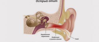
There are a number of signs of otitis media that may indicate the onset of the disease: hearing loss, pain and purulent or putrefactive discharge from the auricle, fever, headache, nausea, vomiting.
It is worth considering several symptoms together. Separately, mild pain may not indicate otitis media, but, for example, dental disease.
Symptoms of otitis media in infants are more difficult to recognize. If a hungry baby suddenly stops sucking on the breast or bottle, starts turning his head and crying, the process may be painful. In addition, the baby’s temperature rises, vomiting may occur, and when you press on the tragus, the baby will begin to break free and cry. All this may indicate otitis media.
External limited otitis
Otitis externa is characterized by:
- Severe itching in the ear and ear canal.
- Painful sensations that increase when chewing and can radiate to the occipital region, jaws, and also involve the half of the head on the side where the otitis media occurred.
- Hearing loss.
External diffuse (diffuse) otitis media
Diffuse external otitis, in which the inflammatory process occurs in the area of the external auditory canal, has a number of characteristic symptoms:
- Severe itching in the area of the external auditory canal.
- Purulent discharge.
- Painful sensations when pressing on the tragus.
- Redness and swelling in the area of the membranous-cartilaginous region of the auditory canal.
Upon examination, the membrane is covered with epidermis.
Catarrhal otitis media
With catarrhal otitis media, the inflammatory process usually spreads from the mucous membrane of the nasopharynx to the auditory tube, as a result of which its lumen is significantly reduced or completely blocked. This, in turn, leads to retraction of the eardrum and the formation of effusion.
The disease has the following symptoms:
- Noise and congestion in the ears.
- Hearing impairment.
- Heaviness in the head.
- Minor pain, tingling sensations.
It may feel like there is water in the ear. If the temperature rises, it is only slightly.
Serous (exudative) otitis media
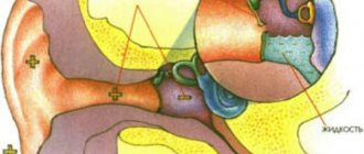
Serous exudative otitis media can occur without signs of inflammation and is characterized only by fluid accumulating in the middle ear cavity.
Symptoms of the disease are:
- Hearing loss.
- The patient feels stuffy in the ears.
- Difficulty breathing and nasal congestion.
- The pain is minor and short-lived.
Incorrect and untimely treatment of this disease can ultimately lead to adhesive otitis media, which is much more difficult to get rid of.
Otitis media
Purulent otitis media is an inflammatory process of the mucous membrane of the tympanic cavity, which is characterized by the following symptoms:
- Pain in the ear and on palpation of the mastoid process.
- Decreased or complete loss of hearing.
- Noise and congestion in the ears.
- The patient's body temperature rises to 38-39 degrees.
- Suppuration is possible, sometimes mixed with blood discharge.
Internal otitis (labyrinthitis)
Internal otitis is a fairly serious, but rare disease. The inflammatory process in tissues is bacterial in nature and has the following symptoms:
- Deterioration of hearing, in rare cases - complete loss.
- Noise.
- Nausea, vomiting.
- Sweating.
- Dizziness.
- Signs of partial facial nerve paralysis.
- Balance imbalance.
- Interruptions in the functioning of the heart.
- Pale skin.
Forms
The disease has several forms, whose course and symptoms may vary. There is external otitis, middle and internal, which is also called “labyrinthitis”.
Otitis externa
With external otitis, an inflammatory process occurs in the area of the external auditory canal. It may be diffuse or limited. The most common type of disease is diffuse.
With this disease, most often not only the ear canal becomes inflamed, but also the outer ear, eardrum, and pinna. Treatment of otitis of the external ear requires constant monitoring by a competent specialist, otherwise it may lead to more serious health consequences.
Otitis media
The most common otitis media has several variants of the course and is most often accompanied by complications. With this disease, inflammation occurs in the cavity of the eardrum.
Average could be:
- purulent;
- catarrhal;
- in the acute phase;
- chronic;
- perforative;
- imperforate.
In advanced stages, chronic otitis media can lead to: meningitis, mastoiditis, brain abscess.
Internal otitis (labyrinthitis)
Labyrinitis can be limited or diffuse. The disease occurs in acute and chronic form, which, in turn, can be latent and overt.
Depending on how the infection got into the middle ear, the disease can be:
- hematogenous;
- tympanogenic;
- traumatic;
- meningogenic.
In addition, there are three forms of the disease:
- purulent;
- seasonal;
- necrotic (one of the most dangerous).
Most often, doctors diagnose tympanogenic limited serous internal otitis.
Diagnostics
It is not possible to diagnose otitis media on your own. An accurate diagnosis can only be made by a specialist after conducting the necessary research.
The ear is examined using an otoscope. This device allows you to notice any contractions of the eardrum.
In addition, tympanometry is sometimes used for diagnosis. The method allows you to determine whether there is fluid in the middle ear, as well as detect the presence of an obstruction in the Eustachian tube.
Sometimes a specialist will resort to analyzing middle ear fluid.
Reflectometry is also used for diagnostics. This method is capable of measuring reflected sound.
In children
Most often, otitis media in a child is accompanied by other diseases, so at the first symptoms, the child should be shown to a specialist who will conduct a full otolaryngological examination.
Children are examined using a special device - an otoscope. Exudate is often taken for analysis to conduct a detailed bacteriological study.
Sometimes the child is referred for an X-ray examination of the temporal region.
If the disease recurs or becomes chronic, a hearing test using audiometry is required, and the auditory tube is also checked for patency.
In adults
Diagnosing otitis in an adult is much easier than in a child. Doctors collect and analyze the patient’s complaints, and examine the auricle using an otoscope. If the disease becomes chronic, a bacteriological culture of the mucus is prescribed to determine the flora and its sensitivity to antibiotics.
If the disease does not go away, and the symptoms of otitis in adults increase, a computed tomography scan is prescribed.
In pregnant women
Otitis media during pregnancy is diagnosed using standard methods. The only exception can be tomography. Additionally, the patient is prescribed a hearing test, as well as an ear smear.
Treatment of otitis media
Since the disease can have unpleasant and even dangerous consequences, it can only be treated under the supervision of specialists who will prescribe all the necessary studies and also prescribe adequate treatment.
Medical has all the equipment necessary for high-quality diagnostics. Our specialists have many years of experience in treating such diseases, which allows us to reduce the risk of complications to a minimum. This applies to both unilateral and bilateral otitis, in all forms and types.
Prevention of otitis
However, it is better to prevent any disease than to treat it later. First of all, to prevent otitis media, proper hygiene of the auricle is necessary.
Children under one year old are prohibited from being in a draft without a hat. It is forbidden to clean a child’s ears using objects that penetrate deeply into the ear canal.
ENT diseases must be treated promptly and under the supervision of a physician.
CLASSIFICATION
Currently, exudative otitis media is divided into three forms based on the duration of the disease: • acute (up to 3 weeks); • subacute (3-8 weeks); • chronic (more than 8 weeks). In accordance with the pathogenesis of the disease, various classifications of its stages have been adopted. M. Tos (1976) distinguishes three periods of development of exudative otitis media: • primary, or the stage of initial metaplastic changes in the mucous membrane (against the background of functional occlusion of the auditory tube); • secretory (increased activity of goblet cells and epithelial metaplasia); • degenerative (decrease in secretion and development of the adhesive process in the tympanic cavity). O.V. Stratieva et al. (1998) distinguish four stages of exudative otitis media. • Initial exudative (initial catarrhal inflammation). • Pronounced secretory; According to the nature of the secretion, it is divided into: - serous; — mucous (mucoid); — serous-mucosal (serous-mucoid); • Productive secretory (with a predominance of the secretory process). • Degenerative-secretory (with a predominance of fibrosclerotic process); According to the form, they are distinguished: - fibrous-mucoid; - fibrocystic; — fibrous-adhesive (sclerotic). N.S. Dmitriev et al. (1996) proposed an option based on similar principles (the nature of the contents of the tympanic cavity according to physical parameters: viscosity, transparency, color, density), and the difference lies in the choice of treatment tactics depending on the stage of the disease. Pathogenetically, four stages of the course are distinguished: • catarrhal (up to 1 month); • secretory (1-12 months); • mucosal (12-24 months); • fibrous (more than 24 months).
Etiology
The most common theories of the development of exudative otitis media: • “hydrops ex vacuo”, proposed by A. Politzer (1878), according to which the disease is based on reasons that contribute to the development of negative pressure in the cavities of the middle ear; • exudative, explaining the formation of secretion in the tympanic cavity by inflammatory changes in the mucous membrane of the middle ear; • secretory, based on the results of a study of factors that contribute to hypersecretion of the mucous membrane of the middle ear.
Pathogenesis
Exudative otitis media begins with the formation of a vacuum in the tympanic cavity (hydrops ex vacuo). As a result of dysfunction of the auditory tube, oxygen is absorbed, the pressure in the tympanic cavity drops and, as a result, transudate appears. Subsequently, the number of goblet cells increases, mucous glands are formed in the mucous membrane of the tympanic cavity, resulting in an increase in the volume of secretion, which is easily removed from all parts through a tympanostomy. The greater density of goblet cells and mucous glands contributes to greater viscosity and density of the secretion, turning it into exudate, which is more difficult or even impossible to evacuate through the tympanostomy. In the fibrous stage, degenerative processes predominate in the mucous membrane of the tympanic cavity: goblet cells and secretory glands undergo degeneration; mucus production decreases, then stops completely, fibrous transformation of the mucous membrane occurs, involving the auditory ossicles in the process. The predominance of shaped elements in the exudate determines the development of the adhesive process, and the increase in shapeless elements determines the development of tympanosclerosis. The trigger, as mentioned above, is dysfunction of the auditory tube, often caused by mechanical obstruction of its pharyngeal opening. More often this occurs with hypertrophy of the pharyngeal tonsil, juvenile angiofibroma. Obstruction also occurs with inflammation of the mucous membrane of the auditory tube, which is provoked by a bacterial and viral infection of the upper respiratory tract and is accompanied by secondary edema.
