The Innovative Vascular Center has all the necessary technologies for the treatment of acute and chronic occlusion of the inferior vena cava. Our specialists have successful experience in solving this complex problem.
Acute thrombosis of the inferior vena cava requires treatment in a specialized vascular surgery hospital. The goal of treatment is to restore the patency of the IVC. This problem is successfully solved using endovascular surgery methods. There are modern thrombolytic drugs and endovascular probes for removing thrombotic masses.
Thrombolysis
Dissolution of venous blood clots with special drugs - thrombolytics. These include streptokinase, urokinase and actilise. Only direct administration of a thrombolytic through a catheter into a thrombus with regular monitoring of the patency of the affected segment is effective. Thrombolysis options may include the use of a special Angiojet device. The thrombolytic solution is supplied through a special probe under high pressure, and then the blood clots are sucked out by a special suction. Another similar device used in our clinic is the Aspirex probe. This is a special spiral suction that gently removes thrombotic masses. The use of Aspirex in the inferior vena cava is limited due to its large diameter, so catheter thrombolysis is the most acceptable method. Thrombolysis is possible only in the first 10 days from the onset of the disease, while the blood clots have not yet healed.
Anatomy of the vena cava
The upper one is located in the chest cavity, namely in its upper part. It is formed by the fusion of two veins - the brachiocephalic veins (right and left). It originates at the level of the first rib to the right of the sternum, goes down, flows at the level of the third right rib into the right atrium. It is adjacent to the right lung, and the aorta passes to the left of it. Behind the superior hollow is the root of the right lung, at the level of the second right rib it is covered by the pericardium. Before its entrance into the pericardial cavity, two veins flow into it: the azygos and the accessory hemi-unpaired.
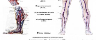
The inferior vena cava begins in the abdominal cavity. It is formed at the confluence of the iliac veins, goes up, deviates to the right from the aorta towards the diaphragm. It is located in the retroperitoneal space behind the internal organs. Through an opening in the diaphragm it is directed into the chest cavity, from there it goes to the pericardium and flows, like the superior hollow, into the right atrium. The following veins drain into the IVC:
- hepatic;
- diaphragmatic lower;
- adrenal gland right;
- renal;
- right ovarian or testicular;
- lumbar
The inferior vena cava is usually divided into three sections: infrarenal, renal and hepatic.
Angioplasty and stenting
The chronic form of NVC syndrome is more difficult to treat. When the venous outflow is decompensated, it becomes necessary to restore the patency of the vessel. Open operations involving the isolation of the IVC and its replacement with a vascular prosthesis are feasible, but very traumatic and ineffective. The artificial vena cava prosthesis often thromboses again and the complex operation becomes completely useless. With the advent of new composite materials for large-diameter stents, our clinic began to perform endovascular methods for restoring the patency of the vena cava. Angioplasty and IVC stenting are performed by experienced endovascular surgeons at the Innovative Vascular Center. The point of the intervention is to restore the patency of the closed segment of the inferior vena cava with a special conductor, a high-pressure balloon and the installation of a metal frame - a stent.
Links[edit]
- ^ abcdefgh Moses, GEZA; Gloviczki, PETER (1 January 2007), Bergan, John J (ed.), "Chapter 2 - Venous Embryology and Anatomy", Vienna Book
, Burlington:. Academic Press, pp. 15-25, DOI: 10.1016/b978-012369515-4/50005-3, ISBN 978-0-12-369515-4, retrieved November 22, 2020 - ^ ab Dardis, Ronan M.; Saxena, Amar; Shad, Amjad; Chitnavis, Bhupal; Gullan, Richard (1 January 2012), Quiñones-Hinojosa, Alfredo (ed.), "Chapter 154 - Disc Replacement Technologies in the Cervical and Lumbar Spine", Schmidek and Sweet Operative Neurosurgical Techniques (Sixth Edition)
, Philadelphia: WB Saunders ., from 1777-1788, DOI: 10.1016/b978-1-4160-6839-6.10154-6, ISBN 978-1-4160-6839-6, retrieved November 22, 2021 - ^ab Blumgart, Leslie H.; Schwartz, Lawrence H.; DeMatteo, Ronald P. (January 1, 2021), Jarnagin, William R. (ed.), "Chapter 2 - Surgical and Radiological Anatomy of the Liver, Biliary Tract and Pancreas", Blumgart Surgery of the Liver, Biliary Duct and Pancreas, 2-
volume
set (sixth edition)
, Philadelphia: Elsevier, pp. 32–59.e1, doi: 10.1016/b978-0-323-34062-5.00002-9, ISBN 978-0-323-34062-5, received 22 November 2021 - Turhan Yavuz; Nazli, K; Kinay, O; Kutsal, A (2002). "Giant Eustachian valve: with echocardiographic appearance of a divided right atrium". Journal of the Texas Heart Institute
.
29
(4): 336–8. PMC 140300. PMID 12484622. - Stavropoulos, S. William; Solomon, Geoffrey A. (1 January 2011), Pretorius, E. Scott; Solomon, Jeffrey A. (ed.), "Chapter 30 - N Filters", Radiology Secrets Plus (Third Edition)
, Philadelphia:. Mosby, pp. 223-227, DOI: 10.1016/b978-0-323-06794-2.00030-4, ISBN 978-0-323-06794-2, retrieved November 22, 2021 - ^ a b Susan Standring; Neil R. Borley; et al., eds. (2008). Gray's Anatomy: Anatomical Foundations of Clinical Practice
(40th ed.). London: Churchill Livingstone. ISBN 978-0-8089-2371-8. - Brophy, SM; Evans, L; Sumpio, B. E. (1993). "Fecal syncope secondary to functional obstruction of the inferior vena cava during the Valsalva maneuver." Annals of Vascular Surgery
.
7
(4): 374–7. DOI: 10.1007/BF02002893. PMID 8268080. S2CID 42135883. - Geehan DM, Inferior Caval Thrombosis, emedicine.com, URL: https://www.emedicine.com/med/topic2718.htm, accessed August 3, 2005.
Conservative therapy
The most common treatment option is conservative therapy with anticoagulants. Drugs used for inferior vena cava syndrome include the anticoagulant warfrin or Xarelto. Detralex or phlebodia are used to improve blood flow from the legs. The main means of conservative treatment is the constant wearing of compression tights of compression class 2-3. They need to be changed every 3 months, as they lose their properties with prolonged wear.
Treating the symptoms of inferior vena cava syndrome with medications and compression can reduce chronic venous insufficiency. Considering the technical complexity of surgical treatment, conservative methods are predominant in modern medical practice.
Varicose veins of the pelvis - symptoms and treatment
Pelvic varicose veins (PVD) is a disease characterized by dilation of the ovarian veins and intrapelvic venous plexuses [3].
The ovarian vein is a paired vessel that carries venous blood from the ovaries. The right ovarian vein drains into the inferior vena cava, which directly opens into the right atrium. Blood from the left ovarian vein is sent first to the left renal vein and then to the inferior vena cava.
The intrapelvic venous plexuses include a set of veins of various calibers that envelop certain organs and are repeatedly connected to each other. These are the pampiniform plexus of the ovary, uterovaginal, vesical, presacral and rectal plexuses, which also interconnect and drain venous blood from the corresponding organs. Blood from the pampiniform plexus flows mainly into the ovarian veins, from other plexuses - into the internal iliac veins, from where through the common iliac veins it enters the inferior vena cava.
The diversity of views on the development of varicose veins of the pelvis also determines the presence of significant “scatter” in the terminology of this disease. In the domestic literature, the following names are used to denote the disease: pelvic varicose veins, pelvic varicose veins, pelvic varicose veins, pelvic venous congestion syndrome, varicose ovarian veins, left ovarian vein syndrome, pelvic organs overflow syndrome. In the English-language literature, the following terms are used: pelvic congestion syndrome (pelvic venous congestion syndrome), pelvic varicies (pelvic varicose veins), pelvic venous incompetence (pelvic venous insufficiency), pelvic venous disorders (pelvic venous pathology), Iliac vein insufficiency syndrome (iliac vein insufficiency syndrome). veins), pelvic varicocele (pelvic varicocele), pelvic venous stasis (pelvic venous stasis) [1][2].
The disease occurs in all biological periods of women’s lives and does not tend to decrease [4]. VBT is observed in 6-15% of women of reproductive age [5].
This pathology is traditionally associated with the female gender, and therefore it is often called pelvic varicose veins in women. However, recently this concept has begun to extend to males. In this case, the gonadal vein is also damaged, which in men is represented by the testicular vein [6].
Pelvic varicose veins, better known in English literature as pelvic congestion syndrome, is relatively “young”, since it has only recently been identified. Nevertheless, this is a very relevant and independent form of the disease, and its manifestations are witnessed not only by vascular surgeons, phlebologists and gynecologists, but also by a fairly wide range of doctors of other specialties. That is, patients with VBT are treated by various specialists who find something familiar from the arsenal of complaints of this category of patients.
Due to the lack of knowledge of the disease, the diversity and non-specificity of clinical manifestations hidden behind the masks of various diseases, patients with VBT are often subject to long-term and not always adequate observation by urologists, proctologists, vertebroneurologists, nephrologists, gastroenterologists, surgeons, orthopedists, endocrinologists and infectious disease specialists [7] . At the same time, long-term and unsuccessful treatment of symptoms, when efforts are aimed not at the cause of the disease, but at combating its destructive consequences, leads patients into a vicious circle, leaving them alone with their disease. The inability to lead a normal lifestyle, constant pain leading to a decrease in physical and social capabilities, conflicts in the family associated with an inferior sex life, suspicions of faking and exaggerating their symptoms worsen the psychosomatic state of sick women, increasing anxiety and irritability. Ultimately, all this becomes the reason for their referral to a psychiatrist or sex therapist, which, unfortunately, only aggravates the disease [8].
Causes of the disease
Pelvic varicose veins are associated with chronic stagnation of venous blood in the pelvis. The conditions for the occurrence of this process arise against the background of various provoking factors that occur over the course of a woman’s life. The main ones are multiple pregnancies and childbirths . The incidence of the disease is directly proportional to the number of pregnancies. Other risk factors:
- age (most often found in active reproductive age - from 21 to 31 years) [48];
- unfavorable working conditions (forced long periods of sitting or standing, heavy physical labor);
- hereditary predisposition: collagenosis (diseases characterized by damage to connective tissue, especially fibers containing collagen) and angiodysplasia (various structural anomalies of arterial, venous, lymphatic vessels);
- conditions and diseases that increase intra-abdominal pressure (chronic respiratory diseases, constipation, strength sports, etc.);
- sexual dysfunction;
- excess body weight;
- hormonal contraception.
In addition, various gynecological pathologies (inflammatory diseases, endometriosis, ovarian tumors, genital prolapse, bending of the broad ligament of the uterus due to uterine retroflexion), menstrual disorders and hormonal imbalances pose a risk for the development of VBT [9][10].
One of the main causes of the disease is a genetic predisposition caused by impaired development of connective tissue, which is the basis of the structure of the venous wall. In this case, the strength of the connective tissue decreases due to a decrease in various types of collagen in its composition or a violation of the ratio between them [11][12].
No less important predisposing factors for the development of VVT are various anomalies in the development of the venous system (venous dysplasia), previous thrombotic lesions of the inferior vena cava system , as well as the so-called venous compression syndromes . These compression syndromes include May-Turner syndrome (compression of the left common iliac vein by the right common iliac artery) and aorto-mesenteric compression syndrome of the left renal vein - Nutcracker syndrome (compression of the left renal vein in the space between the aorta and superior mesenteric artery), in which external compression of venous vessels, preventing the normal outflow of venous blood from the pelvis [13][14][15].