Polycythemia vera
(IP) is a clonal myeloproliferative disease of a chronic nature with a relatively benign course, affecting hematopoietic stem cells and accompanied by the proliferation of three hematopoietic lineages. It is characterized by an increase in the number of red blood cells, an increase in the concentration of hemoglobin, blood cells (leukocytes, erythrocytes, platelets) and total blood volume.
Polycythemia vera is also known as Vaquez's disease, true, or red, erythremia. Belongs to orphan (rare) diseases. It mainly affects people over 60 years of age, more often men. In young people, the disease is less common but has a more unfavorable course.
According to ICD-11, IP is assigned code 2A20.4.
What is polycythemia vera and why is it dangerous?
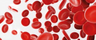
Polycythemia vera (PV) is a tumor blood disease that belongs to the group of chronic myeloproliferative diseases. Essentially, a breakdown occurs in a person’s bone marrow, the very place where peripheral blood cells are produced. As a result, the activity of the bone marrow increases, and more such cells (red blood cells, platelets and white blood cells - leukocytes) are produced than in a healthy person. The main diagnostic sign is an increase in the number of red blood cells in the blood and an increase in hemoglobin concentration.
With PV, blood viscosity increases. There is such an indicator - hematocrit (Ht), which shows the ratio between blood cells and its liquid part, plasma. An increase in this ratio leads to the risk of complications (arterial and venous thrombosis, in particular stroke and myocardial infarction). This condition is especially dangerous for people who already have some other vascular diseases (arterial hypertension, dyslipidemia), diabetes mellitus, as well as risk factors such as smoking, excess body weight, and physical inactivity. When the number of platelets in the blood is very high, the risk of bleeding rather than thrombosis becomes higher. There are no exact data on incidence in Russia, but estimates are about one or two cases per 100 thousand population.
Causes
The exact causes of the disease are still unknown. Scientists suggest that hereditary predisposition, exposure to ionizing radiation and toxic substances play a significant role in the development of pathological changes.
In 20-30% of patients with erythremia, chromosomal abnormalities are detected ( https://cyberleninka.ru/article/n/klinicheskie-rekomendatsii-po-diagnostike-i-terapii-ph-negativnyh-mieloproliferativnyh-zabolevaniy-istinnaya-politsitemiya-1/viewer ).
The likelihood of developing erythremia increases in funeral home employees who embalm bodies, in people who are forced to frequently come into contact with gasoline and other petroleum products, and in those exposed to low doses of radiation. In people who survived the atomic bombing of Nagasaki and Hiroshima, a high risk of developing IP was not observed.
Can this disease be controlled?
With regular therapy, which protects against thrombotic complications, bleeding and infections, the life expectancy of people with PV is almost the same as that of their healthy peers. The essence of therapy is to reduce the production of blood cells and prescribe drugs that reduce the risk of thrombosis. The doctor selects the regimen, having previously calculated which risk group for thrombosis the patient belongs to.
The simplest method of treatment is the use of regular aspirin to prevent arterial thrombosis, which provokes the development of stroke and myocardial infarction. Another traditional method is conventional blood exfusion, that is, bloodletting. Periodically, 500 milliliters of blood are taken from a person two to three times a week until the hematocrit level decreases to normal.
Read also
Invisible War
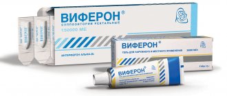
Finally, there are treatment methods based on modern medicines. Patients under 70 years of age are prescribed interferon drugs, which are administered subcutaneously three times a week. Special pen syringes look like those used by people with diabetes to inject insulin. These drugs belong to the so-called cytokines - regulatory protein molecules that are normally produced by cells of the human immune system. Older people are prescribed a cytostatic drug, hydroxyurea.
Polycythemia vera (polycythaemia rubra vera)
Polycythemia vera
is a rare myeloproliferative neoplasm in which the bone marrow
produces
too many red blood cells. It can also lead to overproduction of white blood cells and platelets.
Most health problems associated with polycythemia are caused by blood becoming thicker as a result of an increase in the number of red blood cells. It is more common in older people and may be symptomatic or asymptomatic. Common signs and symptoms include itching (itching) and severe burning pain in the arms or legs, which is usually accompanied by a reddish or bluish discoloration of the skin. Patients with polycythemia are more likely to have gouty arthritis. Treatment consists primarily of phlebotomy.
Patients with polycythemia may have no symptoms. The classic symptom of polycythemia vera is itching, especially after exposure to warm water (such as taking a bath), which may be due to abnormal histamine release or prostaglandin production. This itching is present in approximately 40% of patients with polycythemia. Gouty arthritis may occur in up to 20% of patients. Peptic ulcer disease is also common in patients with polycythemia vera; most likely due to increased histamine from mast cells, but may be associated with increased susceptibility to infection with the ulcer bacterium H. pylori. Another possible mechanism for the development of peptic ulcers is the increased release of histamine and increased gastric acidity associated with polycythemia vera.
Polycythemia vera (PCV), which is a primary polycythemia, is caused by neoplastic proliferation and maturation of erythroid, megakaryocyte and granulocytic elements to form what is called panmyelosis. Unlike secondary polycythemia, PCV is associated with low serum levels of the hormone erythropoietin (EPO). Instead, PCV cells often carry an activating mutation in the tyrosine kinase gene (JAK2), which acts in the EPO receptor signaling pathways, causing these cells to proliferate independently of EPO
The classic symptom of polycythemia vera (and the related myeloproliferative disease, essential thrombocythemia) is erythromelalgia. This is a burning pain in the arms or legs, usually accompanied by a reddish or bluish discoloration of the skin. Erythromelalgia is caused by an increased platelet count or increased platelet stickiness (aggregation), which leads to the formation of tiny blood clots in the vessels of the limb; Moreover, she responds quickly to treatment with aspirin.
Patients with polycythemia vera are prone to developing blood clots (thrombosis). A serious thrombotic complication (such as heart attack, stroke, deep vein thrombosis, or Budd-Chiari syndrome) can sometimes be the first symptom or sign that a person has polycythemia vera.
Headaches, poor concentration and fatigue are common symptoms that also occur in patients with polycythemia.
Physical examination findings are not specific, but may include an enlarged liver or spleen, congestion, or gouty nodules. The diagnosis is often suspected based on laboratory tests. Common findings include increased hemoglobin levels and hematocrit, reflecting an increase in the number of red blood cells; The number of platelets or white blood cells in the blood may also be increased. The erythrocyte sedimentation rate (ESR) decreases due to an increase in zeta potential. Because polycythemia vera results from a significant increase in red blood cell production, patients have normal blood oxygenation and low erythropoietin levels. (EPO) level.
In primary polycythemia there may be 8 to 9 million and sometimes 11 million red blood cells per cubic millimeter of blood (the normal range for adults is 4-6), and the hematocrit can reach 70-80%. In addition, the total blood volume sometimes increases to twice normal. The entire vascular system may become noticeably engorged with blood, and the time it takes for blood to circulate throughout the body may double its normal value. An increase in the number of red blood cells can lead to an increase in blood viscosity up to five times normal. The capillaries can become clogged with very viscous blood, and the flow of blood through the vessels tends to be extremely sluggish.
As a consequence of the above, patients with the untreated version of polycythemia are at risk of various thrombotic events (deep vein thrombosis, pulmonary embolism), heart attack and stroke, and have a significant risk of Budd-Chiari syndrome (hepatic vein thrombosis) or myelofibrosis. The disorder is considered chronic; There is no cure. Symptomatic treatment can normalize blood counts, and most patients can live a normal life for years.
The disease is more common in Jews of European descent than in most non-Jewish populations. Some familial forms of polycythemia have been reported, but the mode of inheritance is unclear.
A mutation in JAK2 kinase (V617F) is closely associated with a version of polycythemia. JAK2 is a member of the Janus kinase family and renders erythroid progenitors hypersensitive to erythropoietin (EPO). Identification of this mutation may be useful in making a diagnosis or as a target for future therapy. After collecting anamnesis, it is recommended to perform the following tests: complete blood count
- complete blood count (increased hematocrit; neutrophils, basophils, platelets, elevated in half of patients)
- JAK2 mutation
- serum ferritin
- kidney and liver tests
If the JAK2 mutation is negative and there are no obvious secondary causes, the following tests are suggested:
- red blood cell mass
- arterial blood oxygen saturation
- Abdominal ultrasound
- serum erythropoietin level
- bone marrow aspirate and trephine
- cytogenetic analysis
- burst-forming unit erythroid (BFU-E) culture
Other signs that may occur with polycythemia include low ESR and elevated leukocyte alkaline phosphatase.
The diagnostic criteria for polycythemia vera have recently been updated. This replaces the previous criteria for polycythemia vera.
JAK2-positive polycythemia vera—diagnosis requires both criteria to be present
Without treatment, polycythemia can be fatal. Research has shown that “1.5 to 3 years of median survival without therapy has been extended to at least 10 to 20 years by new therapeutic tools. The first line of therapy is venesection, reducing the hematocrit to <0.45. Aspirin reduces the risk of thrombosis, the most common complication of primary polycythemia vera. Cytoreductive drugs such as hydroxyurea may also be used. Radioactive phosphate is limited to elderly patients as changes in leukemic transformation are enhanced.
Phlebotomy is a form of treatment that can often be combined with other treatments. Removing blood from the body causes iron deficiency, thereby lowering hemoglobin/hematocrit levels and reducing the risk of blood clots. Phlebotomy is usually performed to lower the hematocrit (percentage of red blood cells) below 45 for men or 42 for women. [19] Phlebotomy has also been noted to improve cognitive impairment.
How to Diagnose Polycythemia Vera
With IP, the hematocrit, which normally does not exceed 45%, sharply increases to 52-56%. The hemoglobin concentration in the blood is also higher than normal (for men the upper limit of normal is 185 g/l, for women - 165 g/l). However, if the patient has recently had any bleeding symptoms, the hemoglobin concentration may appear normal due to concomitant iron deficiency. In addition, an increase in hemoglobin is also characteristic of other pathological conditions in which the body increases the production of red blood cells to improve tissue saturation with blood (from the hypoxia of smokers to some tumors that produce the hormone erythropoietin).
Therefore, in addition to a general blood test, a test is prescribed to identify the specific genetic mutation JAK2 V617F. This mutation appears in PV in more than 90% of cases and is quite strong evidence of the presence of the disease. In order to definitively confirm the diagnosis, a bone marrow biopsy is required.
Symptoms of the disease
Clinical manifestations of IP are divided into 2 groups:
- Plethoric (“plethora” - plethora) syndrome - the mass of red blood cells increases, due to which:
- headaches and dizziness occur;
- pressure increases;
- the skin and mucous membranes acquire a bluish or red-cherry tint;
- disturbing skin itching;
- the conjunctiva turns red;
- vision deteriorates, spots appear before the eyes;
- erythromelalgia develops - small vessels periodically dilate, which provokes redness of the fingers and toes, unbearable burning pain and burning;
- thrombosis and cerebrovascular insufficiency occur;
- angina attacks are observed.
2. Myeloproliferative syndrome is caused by hyperplasia of three hematopoietic lineages: erythroid, granulocytic, megakaryocyte
With polycythemia vera, myeloid metaplasia of the spleen and splenomegaly often develop (the size of the spleen increases). The most common complications are thrombosis (occurring in 39% of patients), thromboembolism and bleeding (gingival, nasal, gastrointestinal, menorrhagia).
The risk of transformation of polycythemia vera into acute myeloid leukemia is 5%, and into myelofibrosis - less than 10% ( https://cyberleninka.ru/article/n/istinnaya-politsitemiya-sovremennye-predstavleniya-o-pathogeneze-diagnostike-lechenii/viewer ) .
With polycythemia vera symptoms
depend on the stage:
- Stage I (asymptomatic). The main signs (plethora, erythrocytosis, skin hyperemia) are moderate, the number of leukocytes and platelets, the size of the spleen is normal, there are no vascular complications or manifestations of fibrosis. Blood pressure increases and blood circulation is disrupted. Duration – 5 years or more.
- Stage IIA (erythraemic). There is no myeloid metaplasia of the spleen. Skin hyperemia, hepatosplenomegaly, and scleral injection are evident. A general blood test shows thrombocytosis, erythrocytosis, neutrophilia, and bone marrow examination shows focal myelofibrosis, total three-line hyperplasia, and often reticulin fibrosis. Thromboembolic complications are common.
- Stage IIB (erythraemic). Myeloid metaplasia is present. Hepatosplenomegaly develops - the liver and spleen simultaneously increase in size. A general blood test reveals leukocytosis, shifted to myelocytes, and a possible decrease in the number of platelets and red blood cells. When diagnosing bone marrow, focal myelofibrosis and panmyelosis are noticeable.
- Stage III (posterythremic, anemic) – terminal. The concentration of blood cells and hemoglobin sharply decreases. Symptoms of tumor intoxication become noticeable, and hemorrhagic complications occur. Myelofibrosis develops and blast crisis occurs.
Diagnostics
Diagnosis of erythremia includes the collection of the following tests:
- analysis of the medical history and complaints;
- life history analysis;
- physical examination to determine skin color;
- blood test, which makes it possible to determine the number of red blood cells and hemoglobin level;
- urine analysis, which is carried out to identify concomitant pathologies;
- biochemical blood test, which determines the level of cholesterol, glucose, creatinine, uric acid, electrolytes to identify concomitant organ damage;
- bone marrow examination is performed;
- trephine biopsy is performed by taking a column of bone marrow with bone and periosteum for analysis;
- Ultrasound of internal organs;
- spiral computed tomography and magnetic resonance imaging are necessary to obtain an accurate image of organs, which will allow assessing the extent of the tumor process;
- Magnetic resonance imaging.