Author:
- Elena Vitalievna Naumova, otolaryngologist, leading doctor of the clinic
(Voted by: )
Acoustic neuritis and central auditory disorders. This group of diseases includes inflammatory and atrophic-degenerative lesions of the peripheral and central parts of the auditory analyzer.
Peripheral diseases include both lesions of the cochlea and lesions of the auditory nerve. There are toxic and infectious neuritis, damage to the auditory nerve in cardiovascular diseases, kidney diseases, metabolic disorders, etc.
As a rule, hearing damage comes to the fore; less often, a disease of the vestibular apparatus is simultaneously observed. The predominant damage to the auditory function is explained by the fact that the auditory apparatus is a much later, and therefore less resistant, formation than the vestibular apparatus.
Initially, the inflammatory process affects the connective tissue surrounding the nerve fibers, then they disintegrate, and last of all, the neuroepithelium of the spiral organ is involved in the degenerative-atrophic process.
Prices
| Disease | Approximate price, $ |
| Prices for diagnosis and treatment of sinus pathologies | 10 370 — 17 560 |
| Prices for the treatment of thyrotoxic goiter | 22 590 — 22 670 |
| Prices for treatment of laryngeal cancer | 6 170 — 77 000 |
| Disease | Approximate price, $ |
| Prices for diagnosing migraine | 7 060 — 8 260 |
| Prices for diagnosing childhood epilepsy | 3 100 — 4 900 |
| Prices for brain shunting for hydrocephalus | 33 180 |
| Prices for treatment of Parkinson's disease | 58 600 |
| Prices for migraine treatment | 9 680 |
| Prices for the diagnosis of amyotrophic lateral sclerosis | 6 550 |
| Prices for diagnosing epilepsy | 3 520 |
| Prices for rehabilitation after a stroke | 78 300 — 82 170 |
| Prices for treatment of childhood epilepsy | 3 750 — 5 450 |
| Prices for treatment of multiple sclerosis | 4 990 — 17 300 |
Otolaryngology in Israel receives extremely positive reviews, and the reason for this is not only the high effectiveness of the treatment, but also its affordable cost for most medical tourists. Prices for diagnostic and treatment procedures in Israeli hospitals are at least a third lower than in similar medical institutions in Western Europe, the USA, Canada and other countries with developed medicine.
The cost of treatment for an ENT disease in Israel depends on its nature, location, stage of development, medications used, the number and type of diagnostic procedures required, therapeutic methods, as well as the individual characteristics of the patient. You can find out the approximate cost of solving a particular ENT problem from a medical representative of the clinic during a free consultation. The final cost will be clear on the spot, after all examinations have been completed and the treatment plan has been approved by the doctor.
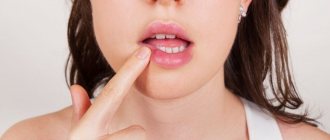
Symptoms of acoustic neuritis
Clinical picture of acoustic neuritis. First of all, patients begin to worry about tinnitus and hearing loss such as impaired sound perception. Hearing loss is progressive. Very rarely, complete deafness can occur within a few hours or days. In cases where, along with damage to the auditory fibers, there is damage to the vestibular analyzer, symptoms of irritation of the vestibular apparatus (dizziness, imbalance, the appearance of spontaneous nystagmus) may be expressed in the acute stage.
2. Reasons
Cochlear neuritis can develop under the influence of many factors:
- infectious diseases (flu, measles, ARVI, herpes, scarlet fever, etc.);
- intoxication with salts of heavy metals, industrial hydrocarbons, drugs (antibiotics, chemotherapy, antiarrhythmics, diuretics, etc.);
- industrial hazards (vibration, noise);
- acute and chronic conditions associated with cerebrovascular disorders (blood supply to the brain);
- traumatic brain, acoustic, radiation, birth injuries;
- cancer;
- neurodegenerative processes and age-related changes.
As can be seen from the above, in most cases, cochlear neuritis is acquired. However, there is also congenital sensorineural hearing loss, which is usually caused by hereditary factors and is one of the elements of one or another chromosomal syndrome.
Visit our Otolaryngology (ENT) page
Causes of auditory neuropathy
During the process of sound conduction, sound passes through the outer, middle and inner ear.
The inner ear contains hair cells, these are sensitive cells that perceive sound vibrations, then convert it into an electrical impulse, so that this impulse is subsequently transmitted, through the auditory nerve, to the human brain. The hair cells of the inner ear are divided into inner and outer. The outer hair cells help amplify the sound vibration entering the inner ear, and the inner hair cells convert it into an electrical impulse. In auditory neuropathy, the outer hair cells are not damaged.
The exact cause of auditory neuropathy is still being determined, but scientists have already identified the following number of causes:
- Damage to inner hair cells
- Poor communication between hair cells in the inner ear and the nerve that carries impulses to the brain
- Nerve damage
- A combination of these problems
Causes of neuritis
In order to correctly prescribe treatment for neuritis, it is necessary to correctly identify the causes that caused it. These are bacteria and viruses, as well as:
- intoxication due to poisoning with alcoholic beverages, medications, food;
- nerve compression;
- injury.
The reasons may lie in a number of diseases:
- diabetes;
- rheumatism;
- metabolic disease;
- excess body weight.
Dizziness and tinnitus
S.Ya.Kosyakov, G.Z.Piskunov Dizziness
Introduction
Dizziness is a general term that characterizes a large number of symptoms.
In general, it means a pathological sensation of movement, it can also mean imbalance, lightheadedness, darkening of the eyes, disorientation, weakness and other sensations. Symptoms can vary in intensity from mild and short in duration to severe attacks of rotation accompanied by nausea and vomiting. To more accurately define symptoms, the following definitions are used: Dizziness:
a general concept describing symptoms of imbalance and stability.
Balance problems:
Difficulty maintaining balance, especially while standing and walking.
Presyncope:
A feeling of being “switched off”, similar to the sensation that occurs when holding your breath for a long time.
Systemic dizziness:
sensation of rotation, spinning, twisting of surrounding objects.
The ability to maintain balance is the result of a complex interaction of various organs and systems. The brain is the main center for processing all information about balance coming from the senses to the muscles that maintain balance. Information in the form of nerve impulses comes from the main systems: visual, vestibular, proprioceptive and tactile (joints and feet). Visual information is the most important for the brain and signals movement in relation to surrounding objects. Anatomy
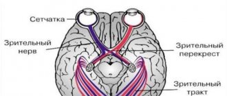
There are two components of hearing: mechanical and electrical (neural).
The mechanical component ensures the delivery of sound waves through the external auditory canal, the movement of the eardrum and the three auditory ossicles in the middle ear. The inner ear is represented by a cochlea, consisting of two halves connected to each other and filled with liquid. The cochlea is responsible for the electrical component of hearing and converts a mechanical signal into an electrical one, which in turn goes to the brain. The other part of the inner ear is responsible for balance and the vestibular system. The three semicircular canals are located in mutually perpendicular planes. Depending on the direction of movement of the head, the fluid moves in the channels, the resulting electrical impulse is transmitted to the brain through the vestibular nerve, transmitting information about the direction of movement. The fluid in the inner ear is renewed daily. Its source of origin is cerebrospinal fluid, absorption occurs in the endolymphatic sac. In Meniere's disease, the absorption capacity of the endolymphatic sac deteriorates. Increased pressure in the inner ear leads to dizziness and decreased hearing. The facial nerve exists in close relationship with the ear. The facial nerve carries out movements of the facial muscles and allows the tip of the tongue to distinguish taste. When it is affected, the eye closes poorly, fluid pours out of the corner of the mouth, and facial movements on the affected side are impossible. Balance function
The balance function is ensured by the interaction in the brain of nerve impulses coming from the inner ear, neck muscles, muscles and joints of the lower extremities.
Disturbances in any of these systems can lead to a subjective feeling of dizziness and instability. General problems with body functions (such as low or high blood pressure, nearsightedness, and many others) can lead to dizziness by affecting the coordination of impulses in the brain. The brain's response to distorted or inconsistent impulses can lead to false sensations of movement (dizziness), which in turn leads to unsteady gait and falls. Dizziness is often accompanied by cold sweats, nausea and vomiting. Visual and muscle and joint signals (tactile and proprioceptive) to the brain warn us that we are moving on the right path or that our head is tilted. The brain interprets this information along with information from the vestibular system and gives the appropriate command to the muscles to maintain balance. Dizziness occurs when sensory information is distorted. Some people experience dizziness when they are in a high place, for example. This is partly due to the inability to focus on nearby objects. While standing on the ground, a person may sway slightly. A person maintains balance, identifies his body position relative to something. When standing in a high place, it is difficult for a person to correlate the position of his body relative to objects in the distance and, accordingly, it is more difficult to maintain balance. As a result, anxiety, fear, and dizziness may occur, which sometimes forces the person to sit down. There is an opinion that motion sickness, a disorder that occurs during sea motion, in a car, or in flight, occurs when the brain receives conflicting sensory information about the movement and position of the body. For example, when reading while driving in a car, the inner ear perceives the movement of the vehicle, but the gaze is fixed on a stationary book that does not move. As a result, sensory conflict can lead to typical symptoms of motion sickness, dizziness, nausea, and vomiting. Another form of dizziness occurs when you spin repeatedly and suddenly stop. Rotation causes movement of the endolymph. The movement of the endolymph causes impulses, which in turn tells the brain that we are moving, but other sensory systems report that we have stopped, so the patient feels dizzy. Causes of Dizziness
Dizziness can be classified into types depending on the part of the vestibular system that is not working properly.
Disturbances can occur at the level of the inner ear, brain, eyes and limbs (the muscles of the back, neck, legs and joints that react to maintain our position). Dizziness due to the inner ear
Part of the inner ear (cochlea) is used for hearing, the other part is used for balance (labyrinth).
If there are disturbances in the labyrinth or in the nerve connecting it to the brain, this leads to dizziness. Various types of disorders in the inner ear can lead to vertigo, including Meniere's disease, labyrinthitis, positional vertigo, vestibular neuronitis, and nerve tumors. These disorders typically cause problems with balance, a sensation of spinning objects, and nausea. Also, these phenomena may be accompanied by tinnitus and hearing loss on the corresponding side. Dizziness of a central nature
The cause of dizziness of a central nature is usually a disturbance in the area of the brain responsible for balance.
Symptoms may include lightheadedness, confusion, unsteadiness, and sometimes loss of consciousness. Causes of central vertigo include low blood sugar, low blood pressure, stroke, multiple sclerosis, migraines, head injuries, tumors and age-related changes. Treatment of this type of dizziness is usually associated with the elimination of problems leading to disruption of brain function. Muscular-articular dizziness
This type of dizziness is rare.
If there are diseases of the muscles, joints, or the sensitivity of the lower extremities is impaired, then difficulties arise in the body’s reaction to movement and in maintaining an upright position. Musculo-articular dizziness can be caused by: atrophic changes in muscles (muscular dystrophy), severe forms of diabetes, arthritis, joint implantation and trauma. Symptoms: Typically unsteadiness and poor balance. Visual dizziness
Unsteadiness of the eye muscles and poor vision can impair balance function.
The brain relies on visual information to maintain balance. Motion sickness in a car or at sea are examples of visual vertigo because the eyes are constantly fixated on a moving object and “confuse” the vestibular part of the brain. This leads to dizziness, nausea and vomiting. Dizziness is not a fatal condition and may improve with treatment, but balance problems may remain. Diagnosis of dizziness
Dizziness can be caused by various disorders in the body.
Based on the medical history and examination data, the doctor selects the required scope of examination to obtain a more complete picture of the disease. The usual set of examinations includes testing of hearing and vestibular function, computed tomography and nuclear magnetic resonance, blood tests, and ultrasound examination. The most commonly used test for dizziness is electronystagmography (ENG). This test measures inner ear endurance and eye coordination. The method involves observing eye movements while cold and warm air is blown into the external auditory canal. This usually causes a brief feeling of dizziness. It is important not to take medications before the test that could affect the test results (for example, Valium, alcohol, etc.). When ordering such an examination, it is necessary to find out from the doctor the effect of the medications taken on the test results. Transcranial Doppler sonography is another test specific for the examination of dizziness of vascular origin. It is a safe, quick way to see disturbances in blood flow in the parts of the brain responsible for balance. In addition, a computed tomography (CT) scan of the temporal bones and, in some cases, magnetic resonance imaging (MRI) may be performed. The purpose of these examinations is to achieve confidence in the absence of a life-threatening pathology and to determine the exact location of the disorder. This is the basis for effective treatment. The scope of the examination is determined by the doctor in each specific case. Several tests are necessary to diagnose the cause. Perseverance and understanding are necessary for both the doctor and the patient, which is also the basis for effective treatment. The most common types of vertigo Benign paroxysmal positional vertigo (BPPV)
BPPV is the most common type of vertigo.
With this disease, dizziness occurs only when the head position changes (usually when turning in bed, tilting the head backwards or forwards). This type of dizziness is caused by microcrystals that float in the fluid of the inner ear and cause a spinning sensation. The most common cause of BPPV is head trauma or viral infections, but sometimes it begins without any obvious cause. Treatment for BPPV consists of certain exercises to return the crystals to a place where they will not cause dizziness. When left at rest in a certain position for 48 hours, they often lock into place. Exercise may reduce symptoms. If these actions are ineffective, then surgical treatment (for example, occlusion of the posterior semicircular canal) may be necessary. Vestibular neuronitis
Neuronitis (nerve inflammation) usually occurs due to a viral infection and can affect the balance centers or the vestibular nerve.
When this happens, the balance centers in the brain are overstimulated, resulting in significant imbalance and systemic dizziness. Fortunately, vestibular neuronitis usually subsides over time and does not recur. Drugs such as betaserc help in the initial stage and reduce the severity of the main symptoms; later, vestibular rehabilitation exercises can speed up the recovery process. In some cases of persistent disease, surgical treatment is recommended. Meniere's disease (Endolymphatic hydrops)
Meniere's disease is a consequence of disorders in the inner ear due to increased pressure in the endolymphatic space.
This is usually due to increased sodium concentration in the fluids of the inner ear. In addition to imbalance that lasts for hours, patients may experience hearing fluctuations, tinnitus, and a feeling of fullness in the affected ear. Sometimes the lesion affects both ears. The full cause of this disorder is not fully known. Sometimes attacks can be caused by excessive salt intake, anxiety, changes in weather, and other reasons. Treatment usually includes limiting salt intake and using diuretics, fluid restriction, sedatives and some other vestibular suppressants. Betaserc is the only drug created for long-term treatment of dizziness. Treatment helps reduce the severity of attacks, but complete cure of the disease cannot be achieved. Vestibular rehabilitation exercises can speed up the recovery process and increase the patient's resilience to vestibular disorders. All prescriptions of drugs should be carried out only by a doctor. For severe cases of Meniere's disease, surgical treatments are available. The list of these methods is long and more often they are destructive in nature for the structures responsible for balance. Vascular vertigo
The correct functioning of the balance system requires not only the flow of information into the inner ear, but also the appropriate transmission of impulses along the nerves to the brain.
If there is not enough blood flow to the areas of the brain responsible for balance, even for a short time, dizziness can occur. The causes of vascular dizziness are different. The phenomena of osteochondrosis in the cervical spine can lead to compression of the arteries leading to the brain; atherosclerotic plaques can narrow the arteries, also causing a decrease in blood flow. Often, blood pressure in the vessels leading to the brain may temporarily decrease when standing up suddenly, especially in older patients receiving blood pressure-lowering medications. Special examinations such as MRI or Doppler sonography help in diagnosing such diseases. Another rather rare cause of dizziness is Perilymphatic Fistula
. The inner ear is a space filled with fluid located in the temporal bone.
If fluid leaks from the structures of the inner ear, hearing loss may occur, which may be greater or less, and dizziness. Most often, fluid leaks through the membranes of the windows of the inner ear, which can occur after physical activity or injury. In some cases, there are congenital disorders that characterize an enlarged connection between the inner ear and the brain ("enlarged vestibular aqueduct"). Sometimes this can be seen with a special x-ray examination - computed tomography. Sometimes the site of the membrane rupture heals on its own, sometimes minor surgery is required. A perilymphatic fistula, or as it is also called, a labyrinthine fistula, can occur as a result of chronic inflammation of the middle ear, especially with cholesteatoma. Cholesteatoma is compacted skin scales. If there is a hole in the eardrum, the skin grows into the cavity of the middle ear, and its metabolic products, like the formation of a pearl, form a lump of cholesteatoma, which presses on the walls of the middle ear cavities and destroys the bone, in particular, the semicircular canal. Therefore, the treatment of chronic otitis media is very important, and when the hole is localized in the upper part of the eardrum (epitympanitis), it must be surgical, because Most often, cholesteatoma is found in these cases. Tumors
Rarely, tumors may be the cause of dizziness.
Most tumors are benign. Acoustic neuroma is a benign tumor of the vestibular nerve. The presence of a neuroma can lead to instability, hearing loss and noise. The most effective treatment method is surgery. Treatment of dizziness All questions regarding the treatment of dizziness and, in particular, taking medications should be discussed with your doctor.
Treatment in each specific case is selected individually and depends on age, severity of dizziness, concomitant diseases and many other factors.
Ear noise
Ear noise is a very common symptom.
The noise can be constant or periodic, of varying severity and frequency. The noise can be subjective (audible only to the patient) or objective (audible to others), and may or may not be associated with hearing loss. Noise is a symptom and not a disease and can occur in various diseases, such as pain in the arm or leg are symptoms of various diseases. Noise appears when the auditory nerve is irritated for various reasons. The noise may or may not be accompanied by hearing loss. Hearing is measured in decibels (dB). A hearing level of 0 to 25 dB is considered normal for the perception of spoken language. Mechanisms of Hearing
To understand the possible causes of tinnitus, it is necessary to have some understanding of the mechanisms of hearing.
The mechanism of auditory perception is provided by five main components: the outer ear, the middle ear, the inner ear, the pathways and the brain. Outer ear
The outer ear consists of the pinna and the external auditory canal.
These structures collect sound waves and transmit them to the eardrum. Middle Ear
The middle ear is located between the eardrum and the inner ear.
This space contains three auditory ossicles: the malleus, the incus and the stapes. Vibrations from the eardrum are transmitted through the ossicles to the fluid in the inner ear. The middle ear is lined with mucous membrane identical to the nose and contains mucous glands and blood vessels. The tympanic cavity is connected to the posterior parts of the nose using the Eustachian tube. The Eustachian tube serves to maintain equal pressure between the middle ear and the outside atmosphere. The sensation of clicking or congestion when changing height is a demonstration of the ventilating function of the eustachian tube. Inner Ear
The inner ear is encased in a dense bony capsule and contains fluids and auditory cells.
The cells are covered with a delicate membrane with microscopic blood vessels. In the inner ear, fluid vibrations resulting from movements of the stapes are converted into electrical impulses in the nerve. Electrical impulses generated in the inner ear are transmitted to the brain via the auditory nerve. The auditory nerve to the brain is located in a small bony canal along with the vestibular and facial nerves. Brain
The auditory nerve reaching the brain is divided into many internal connections.
In the brain, nerve impulses are recognized as recognizable sounds. Ear noise
Most ear noises are audible only to patients - this is a subjective noise.
The noise that the patient himself and anyone else hears is called objective. Objective noise may be the result of muscle spasms in the middle ear or auditory tube, or due to abnormalities in the blood vessels surrounding the ear. Ear noise of a muscular nature
The noise can be the result of a spasm of the muscles attached to one of the auditory ossicles or the result of a spasm of the muscles attached to the auditory tube.
There are two muscles in the middle ear: the stapedius, which is attached to the stapes, and the tensor tympani muscle, which is attached to the malleus. Typically, these muscles contract quickly in response to loud noise or fear. Sometimes one or two of these muscles begin to contract rhythmically for no apparent reason. These contractions can cause recurring noises in the ear. The annoying clicking noise usually goes away on its own. Tinnitus of a muscular nature as a result of spasm of various muscles of the pharynx is quite rare, but sometimes it can occur. If the muscle spasm is long-lasting, then drug treatment (muscle relaxants) or surgical treatment (intersection of the spasmed muscles) is used. Tinnitus of a vascular nature
There are two large blood vessels closely connected to the middle and outer ear: the jugular vein and the carotid artery.
These are large blood vessels that supply the brain with blood and carry out its outflow. It is not normal to hear your own heartbeat or the sound of blood passing through these large vessels. Sometimes this phenomenon can occur with high fever, middle ear infection, or after intense physical activity. The circulation noise in these situations is temporary and is not audible to others. Sometimes the noise of blood circulation becomes audible to others. This may occur due to a thickening of the blood vessel wall, a bend, or a narrowing in the vessel. Further examination is necessary to identify the cause and choose treatment for this pathology. Ear noise due to the external ear.
Closure of the external auditory canal with wax, foreign body, and edema leads to hearing loss and pressure on the eardrum.
This often results in a pulsating type noise. Ear noise due to the middle ear
Impaired function of the middle ear can be the result of an allergic reaction, infection, trauma, scarring and limited mobility of the auditory ossicles.
These disorders often lead to hearing loss and tinnitus. However, there is no direct relationship between the degree of hearing loss and noise intensity. Tinnitus due to the inner ear
Any condition that disrupts the balance of fluid pressure in the inner ear can lead to tinnitus.
This may be the result of an allergic reaction, infection, or circulatory disorders, which lead not only to changes in the fluids of the labyrinth, but also in the membrane structures of the inner ear. Ear noise due to damage to the conduction pathways
The pathways are the most delicate structures of the mechanism responsible for hearing.
Hair cells convert fluid vibrations into nerve impulses. The most insignificant edema and violation of interference in the hair cells, regardless of the cause, lead to function and irritation. This can happen for various reasons: an allergic reaction, infection, edema, systemic diseases, both acute and chronic, toxic effects, sudden loud sounds and sensitive subjects, injuries, the effects of medications, minute changes in blood supply and diet changes. Pressure changes can cause swelling both outside and inside the nerve when passing in the bone tunnel to the brain. In these cases, ear noise occurs on one side. Because The bone tunnel cannot stretch, then due to compression, not only the auditory and vestibular functions suffer, but also the facial nerve. The gap or spasm of a small vessel that occurred somewhere in the auditory path causes compression and violation of circulation. Accordingly, under such conditions, a sudden noise may occur with complete or partial loss of auditory function. If the blood clot, then it can resolve with minimal consequences. Auro noise of brain nature
as a result of edema, pressure or circulation disorders for hypertension, atherosclerosis, as a result of the consequences of injuries, one or more complexes of the passing paths at the entrance and end of their brain can be involved.
In such situations, symptoms are usually localized on the one hand, in addition, the development of symptoms and signs can tell the doctor the place and prevalence of the lesion. The ear noise accompanying the decrease in
ear noise can be associated or not with hearing impairment.
With the coexistence of ear noise and hearing loss, the intensity of ear noise is not an indicator of the further development of hearing loss. Many patients with the advent of ear noise are afraid of the progression of hearing loss. However, these are often not interconnected things. All issues of ear noise treatment should be discussed with your doctor.
Thus, the treatment of dizziness and ear noise is a difficult task, which is possible only by the joint efforts of the doctor and the patient. High -quality hearing diagnosis plays a very important role in such a treatment. The sequence in identifying the causes of these conditions, in treatment and rehabilitation is an integral condition for achieving success.
4.Treatment
Effective etiotropic therapy is possible only in cases where the causes of nerve damage are identified and when these causes are reversible. In different situations, neurological, antibiotic, antiviral, and physiotherapeutic treatment is prescribed (the role and possibilities of the latter are very large and have not been sufficiently studied to date). In some cases, hearing is restored after surgery, which eliminates, for example, the factor of mechanical pressure on the nerve.
According to data published today, significant improvement or complete restoration of hearing function can be achieved in approximately 80% of cases. If all measures taken are ineffective, a hearing aid is selected; The characteristics and design of modern high-tech digital models can radically reduce the psychological, social and professional limitations caused by hearing loss.
3. Symptoms and diagnosis
The most typical manifestations of cochlear neuritis, in addition to hearing loss itself, are sensations of constant extraneous noise in the head and asymmetrical hearing loss. Some patients also experience a complex of vestibular disorders (dizziness, coordination and balance disorders, unsteadiness of gait, etc.).
Obviously, any hearing loss, incl. caused by cochlear neuritis, is a socially significant problem, since it leads to disruptions in communication and social functioning, requires the use of hearing aids, in many cases limits or excludes professional suitability, and provokes the development of psychological disorders of the depressive register.
The degree of functional failure of the auditory analyzer is established in a series of audiometric, tuning fork and other similar tests. Otoscopy and microotoscopy are performed; Electrocochleagraphy, vestibulometric studies, consultations (and corresponding examinations) of specialized specialists, and a battery of laboratory tests are also prescribed.
About our clinic Chistye Prudy metro station Medintercom page!
Classification
According to the course of the disease in otorhinolaryngology, acute (up to 1 month), subacute (from 1 to 3 months), chronic (lasting more than 3 months), and recurrent (3 episodes or more acute external otitis within a year) forms of external otitis are distinguished.
The type of disease is determined by the location and nature of the inflammatory process. Among the nosological forms there are diffuse (eczema, dermatitis, erysipelas, herpes, perichondritis, chondroperichondritis, malignant external otitis and a number of other types and limited external otitis (furuncle, abscess).
According to the degree of severity, there is a mild degree (minor discomfort and itching in the ear, minimal swelling of the skin of the external auditory canal), moderate (pain and itching in the ear, narrowing of the external auditory canal due to severe swelling of the skin), severe (severe pain in the ear, external auditory meatus completely closed, periauricular erythema, regional lymphadenopathy and fever)
Privatdozent Dr. Parvis Mir-Salim
Otorhinolaryngology
Head of the Department of Otorhinolaryngology
Specialization
- Ear surgery
- Functional endoscopic sinus surgery, head and neck surgery
- Extensive experience in cochlear implantation in children
- Master of Medicine degree at University Hospital Kigali, Rwanda
- Cochlear Implant Training in Nairobi, Kenya and Tartu, Estonia
- Video consultation
Show doctor's personal profile
Treatment
There are no treatments for hereditary peripheral neuropathies. However, there are treatments for many other forms. First, the cause of the disease is treated and symptomatic treatment is carried out. Peripheral nerves have the ability to regenerate if the nerve cell itself is preserved. Symptoms can be managed, and addressing the causes of certain forms of neuropathy can often prevent recurrence of damage.
In general, healthy lifestyle choices—such as maintaining an optimal weight, eliminating toxins from entering the body, eating well with adequate vitamins, and limiting or eliminating alcohol intake—can reduce the physical and emotional effects of peripheral neuropathy. Active and passive exercise can reduce cramps, improve muscle elasticity and strength, and prevent muscle atrophy in paralyzed limbs. Various diets may improve gastrointestinal symptoms. Prompt treatment for injuries can help prevent permanent damage. Quitting smoking is especially important because smoking spasms the blood vessels that carry nutrients to peripheral nerves and can worsen neuropathy symptoms. Good nursing skills, such as careful care, of diabetic feet and wounds are essential because these patients have decreased pain sensitivity. Good care can relieve symptoms and improve quality of life and stimulate nerve regeneration.
Systemic diseases often require more complex treatment. Strict control of blood glucose levels has been shown in studies to reduce neuropathic symptoms and help patients with diabetic neuropathy avoid further nerve damage. Inflammatory and autoimmune diseases that lead to neuropathy can be treated in several ways. Immunosuppressants such as prednisone, cyclosporine, or imuran can be very effective. Plasmapheresis, a procedure that clears the blood of immune cells and antibodies, can reduce inflammation or suppress the activity of the immune system. Large doses of immunoglobulins, which function as antibodies, can also suppress abnormal immune system activity. But neuropathic pain is difficult to treat. Moderate pain can sometimes be relieved with analgesics. Some medications (used to treat other conditions) have proven helpful for many patients suffering from severe forms of chronic neuropathic pain. They include Mexilitine, a drug designed to treat irregular heart rhythms (but sometimes causes significant side effects); some antiepileptic drugs, including gabapentin, phenytoin, and carbamazepine; and some types of antidepressants, including tricyclics such as amitriptyline. Injecting a local anesthetic such as lidocaine or using patches containing lidocaine may relieve severe pain. In the most severe cases of pain, the nerves can be surgically destroyed; however, the results are sometimes temporary and the procedure can lead to complications.
Orthotics can help reduce pain and reduce the impact of physical disability. Various arm or leg orthoses can compensate for muscle weakness or relieve nerve compression. Orthopedic shoes can improve gait disturbances and help prevent foot injuries in people with decreased pain perception.
Surgery can often provide immediate relief for mononeuropathies caused by nerve entrapment or compression. Removal of a herniated disc causes decompression of the root. Removing tumors also reduces the impact of tumor tissue on the nerves. Additionally, nerve decompression can be achieved through ligament and tendon release.
Diagnostics
There are many pathologies that affect the quality of hearing of patients. That is why during the diagnosis of cochlear neuritis it is very important to exclude the presence of other pathological processes, namely:
- Meniere's disease;
- inflammation of the middle ear;
- otosclerosis;
- presence of wax plug or other foreign object in the ear.
To do this, the ENT doctor determines the degree and rate of hearing loss by conducting the following examinations:
- audiometry;
- Weber test;
- acoustic impedance measurement;
- electrocochleography, microotoscopy.
For accurate diagnosis, it is very important to inform the doctor about all the symptoms of the disease, talk about how and why the first signs arose.