Pneumothorax - what is it?
Pneumothorax is an accumulation of air/gas in the pleural cavity caused by a leak in the lung or damage to the chest.
The disease leads to collapse of lung tissue, displacement of the space located between the left and right pleural cavities (mediastinum) to the healthy side. As a result, compression of the blood vessels of the mediastinum occurs, and the dome of the diaphragm lowers. Disorders of circulatory and respiratory functions occur. Air during pneumothorax can pass between the layers of the parietal and visceral pleura through a defect in the chest or on the surface of the lung. This leads to an increase in intrapleural pressure (should normally be below atmospheric pressure), complete or partial collapse of the lung.
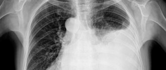
In the photo of the radiograph, pneumothorax looks like a clearing with a lightening of the pulmonary pattern. The long course of the pathology leads to atelectasis - the lung tissue collapses partially or completely, and ventilation is impaired.
The disease occurs not only in adults, but also in newborns. In infants, it is caused by overstretching of the alveoli or their rupture.
Types of pneumothorax
There are several classifications of pneumothorax, based on the provoking factor.
1. By origin, pneumothorax of the lungs is divided into:
- Traumatic. It is a consequence of closed/open chest injuries, due to which a lung ruptured.
- Spontaneous. It develops suddenly due to spontaneous disruption of the integrity of the lung tissue. May be:
- primary (idiopathic) - is a consequence of bullous disease of the lung or congenital weakness of the pleura, due to which the lung ruptures when coughing, diving, doing physical work, laughing, deep breathing;
- secondary (symptomatic) - occurs due to the destruction of lung tissue due to severe pathological processes (rupture of cavities due to tuberculosis, abscess, gangrene, etc.);
- recurrent (repetition of the spontaneous form).
- Artificial. Air is specially introduced into the pleural cavity during diagnosis or during treatment of lung disease.
2. Based on the volume of air trapped in the pleural cavity and the severity of lung collapse, pneumothorax occurs:
- total (full) - the lung is completely compressed;
- subtotal - lung collapse does not exceed 2/3 of the total volume;
- partial (limited, partial) - the lung collapses to 1/3 of its volume or less.
3. Based on distribution, pneumothorax is conventionally divided into:
- bilateral (compression of two lungs at once is observed);
- unilateral (only one lung collapses completely/partially - right or left).
The bilateral form of pneumothorax is characterized by critical impairment of respiratory function, which can lead to rapid death.
4. According to the complications that arise, pneumothorax occurs:
- uncomplicated (occurs as an independent disease);
- complicated (bleeding, pleurisy, subcutaneous or mediastinal emphysema, etc.).
5. By age factor:
- in newborns;
- in children;
- in adults.
6. Based on the presence of communication with the external environment, pathology is:
- Open. There is a defect in the chest through which air from the environment is “sucked” into the pleural cavity. When you inhale, air comes in, when you exhale, it comes out. The pressure inside the pleural cavity becomes equal to atmospheric pressure. As a result, lung collapse develops and the organ ceases to participate in the breathing process.
- Closed. The pleural cavity does not communicate with the environment. The volume of air trapped inside it does not increase. This is the mildest course of the disease, since a small accumulation of gas can resolve on its own.
- Valvular (tense). A special valve structure is formed in which air can enter the lung cavity, but cannot leave it. The volume of gas gradually increases. Acute respiratory failure, pleuropulmonary shock occurs, mediastinal organs are displaced, large blood vessels are compressed and cease to function normally.
A special form of pneumothorax is hydropneumothorax. It is characterized by the accumulation in the pleural cavity of not only gas/air, but also liquid.
Pneumothorax the pleural cavity, which leads to compression of the lung. Normally, the lung and the inner surface of the chest are covered with a membrane - the pleura. Between the layers of the pleura there is a small space - the pleural cavity. The layers of the pleura are smooth and covered with a special liquid, which reduces friction during breathing movements. The pleural cavity is sealed, the pressure in it is negative - such conditions are necessary for the functioning of the lungs. In pneumothorax, air enters the pleural cavity, causing the lung to compress.
Pneumothorax occurs with injuries to the chest and lungs, as well as with certain lung diseases (for example, tuberculosis, pneumonia). A small accumulation of air can resolve on its own; a larger pneumothorax threatens to impair lung function, cardiac activity, and compression of large vessels. Pneumothorax is an emergency condition and in some cases, if left untreated, can lead to the death of the victim.
Treatment of pneumothorax is surgical and consists of removing air from the pleural cavity.
Synonyms Russian
Air in the pleural cavity.
English synonyms
Pneumothorax.
Symptoms
- Chest pain on the side of the affected lung.
- Shortness of breath, the severity of which depends on the degree of impairment of lung function and the presence of lung diseases.
- Feeling of lack of air.
General information about the disease
Pneumothorax is the accumulation of air in the pleural cavity, which causes compression of the lung. The outer surface of the lungs and the inner surface of the chest are covered with a special membrane - the pleura. Between the layers of the pleura there is a pleural cavity - a small space in which the pressure is normally negative (below atmospheric), this is necessary for the lungs to carry out respiratory movements. With pneumothorax, the tightness of the pleural cavity is broken, air enters it, and the pressure in the pleural cavity increases (tends to approach atmospheric pressure). As a result, the lung is compressed and its respiratory function is impaired. With a large pneumothorax, large vessels and hearts are compressed, which can lead to respiratory arrest and cardiac activity.
There are several types of pneumothorax, depending on the causes that caused it.
- Primary spontaneous pneumothorax. It occurs more often in tall and thin young people without any lung diseases. Spontaneous pneumothorax can occur as a result of rupture of vesicles that are located closer to the surface of the lung. The reason for the appearance of such bubbles in people who do not have chronic lung diseases is not fully known. More often they rupture due to changes in pressure during scuba diving, flying, or stress from physical activity.
- Spontaneous pneumothorax in people with lung disease (secondary). It occurs as a complication of various lung diseases. Air enters the pleural cavity when stretched, damaged alveoli (pulmonary vesicles in which gas exchange occurs) rupture. It is more severe because lung function is already impaired by the existing disease.
- Secondary pneumothorax can occur with these and other lung diseases: Emphysema is a disease in which the airiness of the lung tissue increases. The small bronchi and alveoli retain air that does not participate in gas exchange. This leads to swelling of the lungs, decreased tissue elasticity, and disruption of their function. One of the reasons for the development of emphysema is a long-term inflammatory process in the lungs.
- Tuberculosis is an infectious disease caused by Mycobacterium tuberculosis. Transmitted by airborne droplets. Tuberculosis can cause damage to the lungs, bones, genitourinary system, eyes, nervous system and other organs.
- Pneumonia is an inflammation of the lungs that can be caused by various infectious agents.
- Lung cancer is a malignant tumor of the lungs that causes destruction of lung tissue.
If left untreated, a large pneumothorax causes serious problems that can lead to the death of the victim. These include:
- decreased oxygen levels in the blood;
- heart failure;
- breathing disorder.
If necessary, therapeutic measures aimed at eliminating pneumothorax are carried out immediately.
Who is at risk?
- Men (pneumothorax occurs more often in them than in women).
- People aged 20-40 years old who are tall or overweight.
- Suffering from lung diseases.
- Smokers.
- Having had pneumothorax in the past, it can occur again in the same or opposite lung.
Diagnostics
Diagnosis of pneumothorax is based on the patient’s characteristic complaints, medical history, and chest x-ray. If necessary, computed tomography is performed.
- X-ray of the chest organs. Allows you to identify pneumothorax, its size, the degree of compression of the lung, other lung diseases, and damage to the ribs. This is an inexpensive and very informative research method.
- Computed tomography of the chest organs. A more accurate research method based on the action of x-ray radiation. After computer processing of the data, layer-by-layer images of internal organs are obtained, which greatly helps in diagnosis.
Pneumothorax often occurs with various injuries and diseases of the chest organs. These conditions require laboratory diagnostics to identify the main indicators of the body’s activity. The following analyzes are carried out:
- General blood analysis. The number of red blood cells, platelets, leukocytes, and hemoglobin content in red blood cells are determined. This analysis allows you to assess the severity of anemia (a decrease in the number of red blood cells and hemoglobin in the blood, resulting in a decrease in the delivery of oxygen and nutrients to the tissues), which can occur as a result of bleeding. The level of leukocytes increases in various inflammatory diseases.
- General sputum analysis. May be required for lung diseases. Sputum is discharge from the lungs and respiratory tract, which appears in various diseases (for example, pneumonia, tuberculosis). Determining its basic properties (color, nature of sputum, environmental reaction) helps in diagnosing these diseases.
Treatment
Treatment tactics depend on the size of the pneumothorax. A small amount of air can be absorbed without any manipulation, then the patient’s condition is monitored and the necessary X-ray examinations of the lungs are performed. In other cases, surgical treatment is performed.
Pneumothorax is an emergency, so treatment begins as soon as it is identified. The goal is to eliminate air in the pleural cavity. To do this, a puncture is made and the air is removed. Air is also removed using a special tube (drainage): one end is inserted into the pleural cavity, and the other is connected to a device that removes excess air from it. Instead of a device, a valve mechanism is often used, allowing air to escape, but not allowing it into the pleural cavity. As a result, the lung expands.
In some cases, surgical treatment is required, which can be performed through small incisions using an endoscope (a special flexible tube equipped with a video camera and a set of instruments) or by opening the chest cavity.
Prevention
There is no specific prevention of pneumothorax, but quitting smoking reduces the risk of pneumothorax.
People with lung disease should have regular checkups.
Recommended tests
- General blood analysis
- General sputum analysis
Causes of pneumothorax
Pneumothorax is provoked by two groups of reasons:
- Mechanical damage to the lungs/chest.
- Diseases of the chest and lungs.
The first includes:
- closed chest injuries, which are accompanied by damage to the lungs from rib fragments;
- complications resulting from diagnostic/therapeutic measures (iatrogenic);
- open chest injuries;
- artificial pneumothorax, carried out for diagnostic (thoracoscopy) or therapeutic (pulmonary tuberculosis) purposes.
Diseases that cause pneumothorax of the lung include:
- rupture of caverns, breakthrough of caseous foci in tuberculosis;
- rupture of air cavities during bullous disease, spontaneous rupture of the esophagus (intestinal pneumothorax), rupture of a lung abscess into the pleural cavity (pyopneumothorax).
Causes and types of pneumothorax
The disease appears against the background of many reasons that determine its specific type. Based on this, the following classification of pathology is distinguished:
- Traumatic. Appears after trauma to the thoracic region, both from internal damage and penetrating wounds.
- Spontaneous pneumothorax. It is based on a lung defect or hereditary predisposition. In addition, pneumonia or tuberculosis may be the cause.
- Iatrogenic pneumothorax. Develops as a result of diagnostic or therapeutic manipulations (puncture, catheterization, biopsy).
Symptoms of pneumothorax
The severity of the symptoms of pneumothorax depends on the degree of compression of the lung and the causes of the disease. Spontaneous pneumothorax usually develops acutely:
- a severe coughing attack suddenly occurs;
- the patient begins to choke;
- a stabbing pain appears on the side of the affected lung, which radiates to the neck, arm, and behind the sternum;
- when breathing and making any movements, the pain intensifies;
- fear of death arises;
- the face turns pale.
After some time, shortness of breath and pain intensity decrease. The development of mediastinal/subcutaneous emphysema is possible: when exhaling, air penetrates into the subcutaneous tissue of the neck, face, mediastinum, and chest. A characteristic swelling forms in these areas. When palpated, a slight crunch is heard. On auscultation, breathing on the side of the affected lung is not heard or is greatly weakened.
Approximately 6 hours after the onset of spontaneous pneumothorax, the pleura becomes inflamed. After a few days, the pleural lobes begin to thicken due to edema and fibrin deposits. This prevents the expansion of lung tissue and promotes the formation of pronounced pleural contractions.
If we are talking about open pneumothorax, the patient takes a forced position - he lies all the time on the injured side, pressing the wound with his hand. Air is noisily sucked into the pleural cavity, blood with foam (air admixture) is released out.
If you experience similar symptoms, consult your doctor
. It is easier to prevent a disease than to deal with the consequences.
Folk remedies
Pneumothorax is a serious pathological condition that poses a danger to the life and health of the patient. Attempts at self-treatment using folk remedies and pharmaceutical drugs can lead to the death of the patient or the development of serious complications. If you suspect the development of this disease, you should immediately call an ambulance or take the victim to a specialized medical facility.
The information is for reference only and is not a guide to action. Do not self-medicate. At the first symptoms of the disease, consult a doctor.
Diagnosis of pneumothorax
Diagnosis of pneumothorax consists of an initial examination, during which characteristic symptoms of the disease are identified.
In the spontaneous form, percussion determines a box-shaped sound on the side of the pathology, a decrease in the size of absolute and relative cardiac dullness, and palpation determines the absence of vocal tremor.
In the valvular form, a shift of cardiac dullness towards the healthy lung, a sharp weakening of breathing (up to the complete absence of respiratory sounds) in the affected area are diagnosed.
The doctor makes the final diagnosis after an X-ray examination or tomography. X-ray in direct projection allows you to understand whether there is a pneumothorax and what its nature is. It is the basis for choosing additional diagnostic methods.
The main radiological sign of pneumothorax is an area of clearing in which there is no pulmonary pattern. It is located on the periphery of the pulmonary field and is separated from the collapsed lung by a clear boundary.
Radiography allows us to identify the presence of a connection between the formed pleural cavity and the external environment. With an open pneumothorax, the gas bubble increases during inspiration, the lung collapses, the dome of the diaphragm moves downward, and the mediastinal organs move to the healthy side.
In the closed form of the disease, the x-ray picture directly depends on the amount of gas accumulated in the pleural cavity and intrapleural pressure:
If the pressure is low, the amount of gas is minimal, the lung is slightly collapsed. During inhalation it increases in volume, and during exhalation it decreases.
If the pressure is high, the lung is sharply collapsed, the mediastinal organs are shifted towards the healthy organ, the diaphragm is shifted downwards, respiratory excursions are almost not noticeable.
In a situation where the pressure in the pleural cavity is equal to atmospheric pressure, the lung is only partially collapsed, the mediastinum is slightly displaced, and respiratory excursions are preserved.
With the development of valvular pneumothorax, the diseased lung does not change in size and configuration during breathing, the degree of its collapse is maximum, the mediastinum is shifted to the healthy side and moves slightly towards the lesion only on exhalation. With prolonged injection of air into the pleural cavity, flattening and drooping of the diaphragm and gas in the soft tissues of the chest are detected. In the total form, air occupies the entire pleural cavity, so in the picture the shadow of the mediastinum is moved to the healthy side, and the dome of the diaphragm is moved downwards.
Laboratory tests are not carried out when diagnosing pneumothorax, since they have no independent significance - the composition of urine and blood may not change at all against the background of this disease.
Diagnostics
Photo: twitter.com
Pathology is determined based on external examination data and the results of imaging techniques.
- Physical examination . The patient takes a forced position to facilitate breathing. The skin is moist, cool, pale or cyanotic. Excursions of the chest on the affected side are limited, the intercostal spaces are widened. Dyspnea, tachypnea, and tachycardia are observed.
- Radiography . The best conditions for confirming pneumothorax are created when the patient is in an upright position and using a direct projection. The images visualize an area of clearing without a pulmonary pattern, a displacement of the mediastinum, and a low dome of the diaphragm.
- CT scan . A more sensitive and informative test used when radiographic findings are ambiguous. Detects small limited pneumothorax, allows you to differentiate between gas in the pleural cavity and large air cysts in emphysema, and is carried out to clarify the cause of spontaneous pneumothorax.
Laboratory tests for diagnosis are not informative. Determination of the blood gas composition and acid-base balance is carried out to assess the severity of the patient’s condition, to determine the need and scope of resuscitation measures in the presence of extensive lesions and chronic background lung diseases.
Treatment of pneumothorax
Spontaneous pneumothorax requires emergency medical attention. It can be provided by a person who does not have special education. Necessary:
- calm the patient;
- ensure the flow of fresh air into the room;
- call a doctor.
If the pneumothorax is open, you need to quickly apply an occlusive dressing that will hermetically close the defect in the chest. To make a bandage, you can use pure polyethylene, cellophane, cotton wool and gauze for fixation.
In the valvular form of pneumothorax, when air accumulates in the cavity and does not come out, it is necessary to perform a pleural puncture and remove free gas.
Qualified medical care for pneumothorax
Patients with pneumothorax are hospitalized in a surgical hospital in the pulmonology department. Treatment consists of:
- performing a puncture of the pleural cavity;
- air suction;
- normalization of internal pressure.
As a result of treatment, negative pressure is created in the space between the visceral and parietal layers of the pleura surrounding the lung.
If the pneumothorax is closed, gas is aspirated through a puncture system in a small operating room. For this, a special long needle with a tube attached to it is used. The needle is inserted from the side of the damaged lung along the midclavicular line in the area of the second intercostal space, along the upper edge of the underlying rib.
Total pneumothorax requires the installation of drainage and passive air aspiration according to Bulau. It is also possible to carry out active aspiration using an electric vacuum apparatus. Manipulations are performed in order to prevent rapid expansion of the lung and prevent the patient's shock reaction.
Open pneumothorax is treated according to the following scheme: it is transferred to a closed form, suturing the existing defect on the chest, and then the same procedures are carried out as for the closed form of the pathology. A valve pneumothorax is first converted to an open pneumothorax to reduce the pressure inside the pleural cavity. To do this, use a thick needle. Then they resort to surgical treatment.
When treating the disease, it is important to carry out a competent selection of painkillers in order to alleviate the patient’s condition during the period of collapse and expansion of the lung. To prevent a relapse, the doctor may prescribe the patient pleurodesis with silver nitrate, talc, glucose and other sclerosing drugs that artificially cause adhesions in the pleural cavity.
If spontaneous pneumothorax due to bullous emphysema recurs, routine removal of air cysts is performed.
Danger of pneumothorax
Provided that competent medical care is provided in a timely manner and there is a sufficient amount of rehabilitation measures, pneumothorax in most cases has a favorable prognosis. Death with this pathology occurs only with the development of an extensive tense valvular form, which is accompanied by a disorder of central hemodynamics and severe hypoxia.
Among the most common life-threatening complications of pulmonary pathology are:
- exudative pleurisy (fluid accumulates in the pleural cavity);
- pleural empyema (infectious inflammation is associated);
- rigid lung (the lung cannot expand due to the formed connecting cords);
- acute respiratory failure;
- anemic syndrome (hemoglobin level drops sharply);
- hemopneumothorax (blood rushes into the pleural cavity).
According to statistics, these complications develop in 50% of patients diagnosed with pneumothorax.
Prevention of pneumothorax
There are no specific methods for preventing pneumothorax. Necessary:
- timely treatment for lung diseases;
- be examined for the presence of nonspecific lung diseases, tuberculosis;
- stop smoking;
- spend more time outdoors;
- do breathing exercises.
People who have had the disease are advised to avoid physical activity. Immediately after surgery to eliminate the pathology, you cannot fly on an airplane, dive, or parachute for two weeks. It is important to prevent chest injury.
This article is posted for educational purposes only and does not constitute scientific material or professional medical advice.
Medicines
Photo: zmina.info
Drug therapy plays a supporting role in the treatment of pneumothorax. In addition to the measures listed above, the following applies:
- Analgesics . In the first days after admission, they are administered intramuscularly. Depending on the intensity of pain, non-narcotic (analgin and analogues) or narcotic drugs are used. After reducing the severity of the pain syndrome, they switch to tablet forms of non-narcotic drugs.
- Local anesthetics . They are administered during cervical vagosympathetic blockade. Reduce pain, increase blood pressure, improve vascular tone, eliminate the cough reflex.
- Antibiotics . Necessary for open injuries and purulent processes. First, broad-spectrum medications are used. After receiving the results of the microbiological study, the antibiotic therapy regimen is adjusted taking into account the sensitivity of the pathogen.
- Bronchodilators . Indicated during the recovery stage to improve drainage of the bronchial system, prevent stagnation of sputum and the development of pneumonia.
An important role in the prevention of complications is played by breathing exercises, which are carried out from the first days of the patient’s stay in the hospital.


