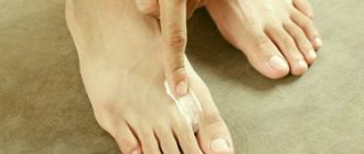
09.09.2021 Nail and skin fungus is a common problem. It is enough to swim in a pool, take a shared shower or visit a bathhouse to become infected with this disease. Diseased nails become yellow-gray and brittle, and noticeably thicken. The disease not only spoils the appearance, but also causes severe physical discomfort: itching and burning of the affected areas of the skin.
If the fungus is not treated, then soon its pathogen will spread throughout the body. The spores will enter the bloodstream, leading to allergic reactions. It is recommended to start treatment in the early stages and not stop therapy until all symptoms have passed.
Modern pharmacology offers many options for anti-fungal drugs in various dosage forms. The choice of remedy depends on the type of fungus, the degree of the disease and the individual reactions of the body.
Table of contents
- Etiology and pathogenesis
- Clinical manifestations
- Principles of treatment
Onychomycosis (fingernail fungus, toenail fungus) is a fungal infection of the toenails or fingernails that affects any component of the nail complex, including the base, bed or plate. The disease can be accompanied by pain, discomfort, and changes in the appearance of the nails, which in some cases leads to serious physical and professional limitations, anxiety for patients and a decrease in their quality of life.
In our company you can purchase the following equipment for the treatment of onychomycosis:
- AcuPulse (Lumenis)
In the USA, onychomycosis of the nails occurs in approximately 2–13% of the population, in Canada - in 6.5%, in England, Spain and Finland - in 3-8%. According to a systematic review of 1914 articles, the prevalence of onychomycosis is as follows:
- general population - 3.22%;
- children - 0.14%;
- elderly people - 10.28%;
- patients with diabetes mellitus - 8.75%;
- patients with psoriasis - 10.22%;
- HIV-positive patients - 10.40%;
- patients on dialysis - 11.93%;
- after kidney transplant - 5.17%.
Onychomycosis accounts for about half of all nail pathologies and is the most common nail disease in adults. As for the affected areas, statistically, the legs are affected much more often than the arms.
The main forms of onychomycosis are:
- Distal lateral subungual onychomycosis
- Proximal subungual onychomycosis
- White superficial onychomycosis
- Endonyx-onychomycosis
- Candidal onychomycosis
Often patients have a combination of these subtypes, or total dystrophic onychomycosis - damage to the entire nail complex.
How to choose drugs for fungus
To combat mild fungal infections, topical preparations—varnishes, ointments, sprays, shampoos, and special solutions—are sufficient. For advanced diseases, both local and systemic agents are used. Anti-fungal tablets serve as a supplement, ridding the subungual skin and other tissues of the pathogen. In the “Antifungal Drugs” section on our website you will find the medications listed in this article, as well as their analogues.
Any drugs for fungus should be selected with the help of a specialist who can determine the severity of the disease and take into account the individual characteristics of the body.
The offer is not an offer, the drugs presented are medicines, specialist consultation is required.
Etiology and pathogenesis of onychomycosis
Onychomycosis is caused by three main classes of fungi: dermatophytes, yeasts, and nondermatophytic molds.
Dermatophytes are the most common cause of the disease. Two main pathogens are responsible for 90% of all cases of onychomycosis: Trichophyton rubrum (70%) and Trichophyton mentagrophytes (20%). Onychomycosis, caused by non-dermatophytic mold fungi of the genera Fusarium and Aspergillus, as well as Scopulariopsis brevicaulis, is an anamorphic (asexual) representative of the genus Ascomycota, is gradually spreading throughout the world. Today they are responsible for 10% of cases of the disease. As for candidal onychomycosis, it is caused by Candida albicans and is quite rare.
In distal lateral subungual onychomycosis, Trichophyton rubrum is usually detected. Proximal subungual onychomycosis is typical in immunocompromised patients . The same type of onychomycosis, but with periungual inflammation, is caused by non-dermatophytic molds. White superficial onychomycosis of the nails is caused by Trichophyton mentagrophytes, and its deeper forms are caused by non-dermatophytic molds. Candida nail infection is often observed in premature infants, immunocompromised patients, and individuals with chronic mucosal candidiasis.
Risk factors for developing onychomycosis:
- family history;
- elderly age;
- warm and humid climate;
- weakened body;
- nail injury;
- regular fitness classes;
- immunosuppression (drug, HIV, etc.);
- visiting public swimming pools, baths and saunas;
- tight shoes.
Repeated microtrauma of the nails in tight and uncomfortable shoes provokes onycholysis (detachment of the nail from the soft tissues of the finger) and other degenerative conditions that contribute to the penetration of fungi into the nails.
Prevention
Provides, firstly, for the disinfection of floors, wooden flooring, benches, basins, gangs in bathhouses, showers, swimming pools, as well as the disinfection of impersonal shoes; secondly, regular examinations of bath attendants and people working out in swimming pools in order to identify patients with epidermophytosis and their early treatment; thirdly, carrying out sanitary and educational work. The population needs to be explained the rules for personal prevention of athlete's foot: wash your feet every night at night (preferably with cold water and laundry soap), dry them thoroughly; change socks and stockings at least every other day; do not use someone else's shoes; Have your own rubber sandals or slippers for the bath, shower, pool.
To harden the skin of the soles, it is recommended to walk barefoot on sand and grass in the hot season.
Clinical manifestations of nail onychomycosis
At first, onychomycosis is asymptomatic - patients can see a doctor because of visible cosmetic defects of the nail plate, but without any discomfort. As the disease progresses, paresthesia, pain and discomfort occur in the affected area. This makes walking difficult and interferes with sports and everyday life.
Clinical manifestations of onychomycosis depend on its type:
- Distal lateral subungual onychomycosis - there is subungual hyperkeratosis and onycholysis, the plate becomes yellow-white. In its central part, yellow stripes and/or yellowish onycholytic areas are visible.
- Proximal subungual onychomycosis - represented by leukonychia (white spots and stripes) in the proximal parts of the nail plate, which move distally (towards the edge) as the nail grows. If mold fungi are among the pathogens, periungual inflammation occurs.
- White superficial onychomycosis - small white speckled spots form on the toenails, the nail plate becomes rough and crumbles easily. Variations are possible with a deeper spread of the pathological process into the nail, depending on the causative agent of the disease.
- Endonyx-onychomycosis - the color of the nail plate becomes milky white. Unlike distal onychomycosis, there are no signs of subungual hyperkeratosis or onycholysis.
- Candidal onychomycosis of the nails is associated with chronic mucocutaneous candidiasis or immunosuppression, in which several or all nails are affected at once with the presence of periungual inflammation. The fingers of such patients often take on the shape of a bulb or drumsticks.
To assess the severity of the disease, the Onychomycosis Severity Index (OSI) has been developed. The result is obtained by multiplying the lesion area scores by the proximity scores of the lesion to the nail growth zone. Mild degree – 1–5 points, moderate – 6–15, severe – 16–35. Dermatophytoma (spots or longitudinal stripes) more than 2 mm with subungual hyperkeratosis is scored 10 points.
In its manifestations, onychomycosis is similar to many other nail pathologies. For example, leukonychia (stripes) resemble trauma to the nail plate. To confirm the diagnosis, it is necessary to perform a laboratory mycological study. Interestingly, a negative result does not rule out onychomycosis, since microscopy can be false negative in 10% of cases and culture in 30% of cases. A more reliable, although more expensive, method is PCR research (polymerase chain reaction).
Rice. 1. Distal lateral subungual onychomycosis (Dr. Antonella Tosti)
Rice. 2. Proximal subungual onychomycosis (Dr. Antonella Tosti)
Rice. 3. White superficial onychomycosis (Dr. Antonella Tosti)
Rice . 4. Endonyx-onychomycosis (Piraccini B., Alessandrini A. Onychomycosis: A Review. J Fungi 2015; 1: 30–43)
Rice. 5. Candidal onychomycosis (Dr. Antonella Tosti)
List of drugs
When choosing drugs for fungus, it is necessary to focus on effectiveness, the presence of side effects, symptoms and course of the disease. It is important to take into account individual intolerance to the components.
Antifungal antibiotics
If the fungal infection is systemic in nature, an integrated approach is required. Deep tissue damage, separation of the nail plates and severe itching of the skin against the background of deteriorating general health suggest the use of not only local remedies, but also tablets.
Among external remedies, it is worth highlighting medications that contain naftifine. This component has an antibacterial and anti-inflammatory effect. When taken regularly and in combination with other therapeutic agents, recovery occurs quickly.
Preparations with naftifine:
| Exostat solution 1% 15ml |
| Mycoderil cream 1% 15g |
| Exoderil cream 1% 15g |
External preparations for dermatomycosis and keratomycosis
Yeast, mold and dimorphic fungi can be cured with Terbinafine-based products. Popular drugs with this active ingredient:
| Lamicide drops for nails 15ml |
| Lamicide spray for legs 15ml |
| Lamisil cream 1% 15g |
| Fungoterbin 1% 15g |
| Exifin gel 1% 15g |
| Terbizil cream 15g |
| Terbix cream 1% 10g |
| Exiter cream 1% 15g |
Medicines containing ketoconazole effectively fight fungal infections of the head and groin area. List of drugs:
| Shampoo Sulsen forte 250ml ketoconazole |
| Shampoo Sulsen mite for dandruff 1% 250ml |
| Sulsen forte paste 2% 75g ketoconazole |
| Sulsen mite paste 1% 75g |
| Shampoo Sulsen mite for dandruff 1% 150ml |
| Shampoo Sulsen forte from perch 150ml |
| Nizoral cream 2% 15g |
| Mycozoral ointment 2% 15g |
| Sebozol shampoo 100ml |
Products based on miconazole, a synthetic substance with an antifungal effect, are effective against dermatomycetes and yeast, as well as the causative agent of lichen versicolor. Preparations containing miconazole:
| Mykozon cream 2% 15g |
| Ginocaps vag caps x 10 |
Antifungal agents for systemic candidiasis
The Candida fungus spreads inside the body: on the respiratory system, in the digestive system, on the genitals. Sometimes it infects the nervous and cardiovascular systems.
Often, with systemic candidiasis, the fungus also affects external tissues - nail plates, skin on the legs and arms. To get rid of the disease, it is important to start therapy in a timely manner. To treat candidiasis, drugs containing clotrimazole are used.
Popular remedies for the treatment of mycotic diseases caused by the Candida fungus:
| Flucorem 0.5% gel 15g |
| Kanizon plus cream 15g |
| Kanizon cream 1% 20g |
| Candide cream 15g |
| Clotrimazole-Akrikhin ointment 1% 20g |
| Clotrimazole-Akos ointment 1% 20g |
| Candide B cream 15g |
| Candiderm cream 15g |
MANIPOLES
Focus
Capable of treating spots of 7 sizes: from 1 to 7 mm, reaching areas with disturbed pigmentation at various depths, including those with tattoos of any kind. The Focus handpiece can be used in several operating modes: Q:switch, quasi-long-pulse (short-pulse mode), long-pulse mode.
HomoGenius
Treats areas of discolored pigmentation and tattoos using a uniform laser beam with universal energy intensity, preventing excessive heating of the area. A square beam with spot sizes of 3x3 mm2 or 5x5mm2 allows you to treat areas without overlap. The HomoGenius handpiece can be used in Q-switch laser mode.
Pixel manipulator (with depth control)
The pixel arm uses fractional beam splitting to create photoacoustic columns in a 7x7 non-ablative pattern without affecting surrounding tissue. Such microtraumas trigger the regeneration process in the affected areas, collagen synthesis is stimulated, and skin turgor is restored.
Collimated handpiece
The collimated handpiece delivers parallel beams of energy to the tissues of the target area with minimal scattering, regardless of the distance to the skin. This allows you to regulate the course of the procedure without maintaining prolonged contact with the skin, also providing better visualization of larger treated areas. A handpiece with a spot size of 8 mm has a high coverage ratio and high processing speed. The collimated handpiece can be used in all pulse delivery modes.
Y-spectrum and R-spectrum handpieces
Additional Spectrum Handpieces expand ALMA-Q's capabilities by offering two additional wavelengths for color tattoo removal. The Y-spectrum handpiece (585 nm) is especially effective at removing blue light, and the R-spectrum handpiece (650 nm) is especially effective at removing green and blue-green colors.
Symptoms
Conventionally, the following forms of mycosis of the feet are distinguished, which are presented in the photo:
- Squamous: unilateral peeling and mild itching in the arch of the foot. This form often goes unrecognized.
- Intertriginous: the folds between the toes are affected, then the process moves to the back of the foot. Weeping cracks appear, accompanied by itching. A bacterial infection may accompany the development of erysipelas.
- Dyshidrotic: on the arch of the foot, vesicles with light and then cloudy contents form, merging with each other. They open with the formation of painful erosions. This type is accompanied by itching and pain.
Mycosis of the skin of the foot in the acute period can be accompanied by fever, poor health, headache, and enlarged inguinal lymph nodes. When infected with trichophyton, the nail of the 1st or 5th finger is affected, and subsequently the process spreads to all nail plates. First, yellow spots appear at the free edge of the nail, then it thickens, loosens and crumbles. Therefore, to treat foot fungus, you need to use complex action products, for example, Clotrimazole lotion for skin and nails.


