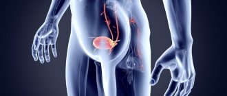
Cystoscopy of the bladder is the examination of the walls of the urethra, bladder, and ureteric outlets.
The study is carried out using special optics to diagnose various changes and diseases.
During the procedure, the doctor has the opportunity not only to identify the pathology, but also to perform a biopsy to take a biopsy sample and, if foci of the disease are identified, to carry out therapeutic measures in the form of administering medications.
The study is also unique because, thanks to it, the doctor examines the organ cavities, but also evaluates the condition of the kidneys based on the specific discharge from the ureters.
We can say that this opportunity becomes an auxiliary diagnostic method for identifying prostate pathologies.
But this plays a special role when preparing an operation to remove a kidney, when the condition of the remaining one will be decisive.
When is it carried out?
There is no age limit for conducting the study. Therefore, it is the main method for identifying certain diseases of the genitourinary system, due to its high information content.
Other methods, for example, ultrasound, although safer, do not provide the doctor with sufficient information.
In addition, as we noted above, during the course of this study, a biopsy can be performed for subsequent histological analysis.
If stones, tumors, or polyps are found during the examination, they can be destroyed and removed.
Having discovered ulcerative pathologies of the organ mucosa, the doctor cauterizes the damaged areas.
As for tumors in the prostate and inflammation in it, using research, the doctor determines how the process has spread to neighboring tissues, sees a picture of the specific involvement of the organ and urethra in the disease process.
Decoding the results
The results of the procedure will be issued in writing on average within 15 minutes after the study. The doctor will draw up a conclusion that reflects the condition, structure of the organ, characteristics and size, as well as the volume of residual urine. In general, such a study allows you to obtain the same information as a standard ultrasound - detect inflammatory diseases (signs of acute or chronic cystitis), suspect urolithiasis, see tumors and neoplasms. Additionally, the doctor will determine how much residual urine remains in the bladder after emptying.
If during the examination a residue of more than 30 ml is detected, the specialist will give recommendations on further actions. If prostate adenoma is suspected, additional diagnostic methods, consultation with a urologist, and laboratory tests may be required.
Indications and contraindications
The study is carried out for both diagnostic and therapeutic purposes. In the first option, it is carried out in a number of cases, including:
- Often worsening cystitis;
- Suspicion of a neoplasm;
- Overactive bladder;
- Chronic pain syndrome in the pelvic area;
- Detection of atypical cells in urine;
- Dysfunction when urinating - pain, difficulty, etc.
How is the treatment study indicated for:
- Excision of tumors;
- Neutralization of bleeding;
- Destruction and extraction of stones;
- Biopsies;
- Administration of medications.
In addition to the above, the procedure helps restore urinary tract patency.
The study is contraindicated if the patient has manifestations of inflammation of the genitourinary organs, as well as in case of damage to the urethra.
Cystoscopy of the bladder
When diagnosing bladder diseases, cystoscopy is often used. This procedure is similar to the well-known gastroscopy and allows you to examine the walls from the inside. It is carried out using a special instrument - a cystoscope, which is inserted through the urethra into the bladder. This equipment is equipped with a miniature camera and an LED flashlight, making the examination results as reliable as possible.
Main purposes of cystoscopy
Bladder cystoscopy can be performed not only as a diagnostic procedure. There are several types of procedure:
- Examination - simply an internal examination for the purpose of making a diagnosis;
- Operating room - intended not only for examination, but also for the treatment of various pathological conditions, for example, stopping bleeding inside the cavity, removing tumors, polyps, adhesions.
Also, in most cases, under the control of an optical instrument, it is possible to obtain a sample of the affected mucosal tissue for further histological analysis.
Indications for testing
Cystoscopy of the bladder is not performed just at the request of the patient.
It is usually prescribed when there is insufficient information obtained in other ways - using MRI, ultrasound, x-ray with contrast, etc. Cystoscopy is a very unpleasant procedure, but is usually performed under local anesthesia. Therefore, the urologist prescribes it only for the following indications:
- Urinary tract infections;
- Purulent, bloody or other uncharacteristic discharge;
- Long-lasting burning sensation during urination;
- Frequent urge to urinate when the bladder is already empty;
- The presence of atypical cells in urine analysis;
- Neoplasms on the inner wall of the bladder;
- Urinary stones.
The study cannot be carried out during menstruation in women. It is best, if possible, to schedule the procedure for 5-7 days after the end of regular bleeding.
Stages of cystoscopy
Visual examination of the internal cavity of the bladder does not require any special preparation.
It is important to perform thorough hygiene and also try to urinate no later than an hour before the procedure. The cystoscopy itself is performed as follows:
- Inserting an endoscopic device into the urethra is an unpleasant procedure. Therefore, the first thing the doctor does is provide high-quality local anesthesia. For men, local anesthesia is often not enough - the procedure is recommended to be performed under intravenous anesthesia while the patient is asleep;
- Next, a cystoscope is inserted through the urethra into the bladder. With its help, urine is drained and the inner cavity of the bladder is filled with saline solution;
- If there is blood, pus or other pathological secretions inside, the cavity is washed several times, and only after that the mucous membrane is visualized;
- The urologist examines the inner surface of the bladder, starting from the apex and front wall, then the right and left sides, and finally the back wall and bottom. Particular care should be taken to examine the so-called Lieto triangle, located between the right and left ureters and the internal opening of the urethra. It is here that pathological changes in the mucous membrane most often occur;
- If cystoscopy is performed to evaluate kidney function, a special dye is injected into the bladder. Next, the patient is observed, monitoring the rate of excretion of the dye in the urine.
Actions after cystoscopy
Upon completion of the diagnosis, the doctor will decipher the received data right on the spot and issue a conclusion.
If general anesthesia was not used, the patient can go home immediately. You can lead your usual lifestyle, with the only recommendation - drink more water. This will dilute it, which will make urination easier, eliminating the burning sensation in the urethra that will likely occur, no matter how delicately the procedure was performed. In rare cases, after diagnosis, especially when receiving material for histology, a small amount of blood may be present in the urine. This is completely normal and should go away completely within a day or two. But if you experience these symptoms, you should consult a doctor immediately:
- Increased body temperature, fever, chills;
- Acute urinary retention;
- Blood clots are observed in the urine;
- Aching pain in the lower back;
- Frequent urge to urinate, accompanied by severe cutting pain in the urethra.
Get diagnosed at our clinic
In our center of Modern Medical Technologies you can undergo a complete urological examination, including cystoscopy of the bladder.
The urology department is equipped with all the necessary equipment, operating rooms, and has a full-fledged laboratory for conducting tests. Experienced specialists regularly take advanced training courses abroad, gaining new knowledge. We keep up with the times, use the principles of evidence-based medicine in our work and treat our patients very sensitively. The prices of our services allow almost everyone to receive quality medical care. Make an initial appointment and you will see for yourself what modern medicine is without queues and with a guarantee of results.
How to prepare
Preparing for the study cannot be called difficult. The patient undergoes a urine test and some other tests. This is necessary to exclude contraindications.
Medicines that are responsible for reducing the rate of blood clotting taken by the patient are usually eliminated several days before the test.
There should be no sexual contact on the eve of the study.
Men are examined using, as a rule, spinal or general anesthesia, due to their anatomical characteristics.
For women, the procedure is usually performed under local anesthesia, which is applied to the tube of the device (endoscope).
Before the procedure, you need to have a bowel movement and thoroughly clean your genitals.
Everyone, without exception, must stop eating twelve hours before and drink three hours before.
How to prepare for the examination
Cystoscopy does not require special preparation. If it is performed under anesthesia, then you need to abstain from food 10-12 hours before the appointed time. 3-4 hours before the procedure, you should avoid consuming any liquids. Since you cannot drive after anesthesia, you should arrive at the clinic in another way. If the examination of the bladder walls is carried out without anesthesia, it is enough to simply appear on an empty stomach.
Attention! Before the procedure, external toilet of the genital organs should be performed.
Carrying out
The subject undresses and lies on his back, legs bent at the knees. The doctor should explain to him how the procedure is performed and what sensations may arise during it.
The external genitalia are treated with an antiseptic, and the device is lubricated with glycerin.
Men are given anesthesia into the urethra using a special syringe, and wait for the analgesic effect to begin. This continues for up to 10 minutes.
Depending on the chosen toolkit, there are options:
- Harsh intervention;
- The intervention is flexible.
Rigid involves the use of a rigid device that is mounted on a metal tube. Such a device has pros and cons. On the one hand, it straightens the tissue well, which is important for examination; on the other hand, it is more traumatic and causes significant discomfort to the patient.
Do not use a rigid device during pregnancy or with significant tumors localized in the pelvic organs.
A tube of the device is inserted into the urethra and a liquid is supplied to wash and straighten the folds of the organ membrane.
In order to supply and drain liquid, a special tap is connected to the device. For better visualization, the organ should be washed before the study. The water used to rinse the organ is collected for later study.
Flexible research involves the use of a flexible instrument. This is a movable polymer tube, at the end of which there is a lamp and optics.
The device easily passes into hard-to-reach places, which increases the information content of the study, and also significantly minimizes the possibility of injury and pain.
The advantages of the flexible option are obvious, so in modern medical practice it is replacing the hard option.
For children, a flexible version of the procedure using a pediatric endoscope is indicated.
Doctor's comment
Frequent urination or incontinence, lack of urge to urinate, weakened feeling of bladder fullness - are you tired of these symptoms? The impossibility of long walks, constant dependence on the availability of a toilet - urinary problems negatively affect your quality of life. To get rid of the disorders that have made your life uncomfortable, you must first of all make an appointment with a urologist who will prescribe examination and treatment. Our Center has the opportunity to undergo comprehensive diagnostics, which includes a variety of modern methods, including CUDI - a comprehensive urodynamic study of the lower urinary tract. During the examination, we will be able to determine the advisability of prescribing conservative therapy or verify the need for correction through surgery. By contacting us with such a delicate problem, you can count on an individual approach, and well-chosen treatment will forever relieve you of existing problems with urination.
Urologist Natalya Sumerova
Study in women
Typically, the test is easier for women than for men. The reason for this is the peculiarity of the urethra, short and straight.
General anesthesia is not required; a local option is used: an anesthetic is applied to the device.
Difficulties may arise if there are significant tumors in the uterus and pregnancy in its later stages, when the bladder is compressed by the uterus.
It must be understood that the procedure in the second case is performed only for health reasons: there is a high probability of provoking a spontaneous abortion.
Contraindications to the procedure
Ultrasound with determination of residual urine is carried out in one of three ways:
- transabdominal - through the anterior abdominal wall;
- transrectally - through the rectum;
- transvaginally - through the vagina.
The first method has no contraindications, with the exception of acute inflammatory diseases of the skin of the abdomen. Transrectal examination is not performed for anal fissures, exacerbation of hemorrhoids, proctitis, paraproctitis. The transvaginal method is not suitable for inflammatory diseases of the genital tract, and is also not used in women who have not begun to be sexually active.
Study in men
The anatomical features of a man (the length of the urethra can reach over twenty centimeters) cause certain difficulties in conducting the study.
The physician is required to be highly qualified and accurate. Mainly, this plays an important role at the stage when the device is inserted.
An anesthesiologist must be present during the examination, as there may be a risk of severe pain when it is necessary to administer an anesthetic drug.
Biopsy
We will separately talk about the procedure for taking a biopsy for subsequent histological examination.
The biopsy is taken in several ways:
- Biopsy is cold. The material is removed using special forceps inserted through the device.
- TUR biopsy. This is cutting off a piece of tissue or completely removing the tumor using an electrocoagulator, which is also carried out through an endoscope.
Both methods have pros and cons.
With the cold option there is minimal risk of damage to the organ wall, but there is little information about the spread of the tumor.
Cholangiography
TUR biopsy, on the contrary, provides comprehensive information about the development of the tumor and negates the risk of bleeding. The downside of this option is the high risk of injury.
Types of cystoscopes
To carry out the procedure, a cystoscope is used, which is a special design made of a thin tube with optics built into it, which transmits the resulting image to an external monitor. A cystoscope is inserted into the bladder through the urethral canal, sequentially examining all parts of the organ. To carry out the procedure, two types of structures have been developed:
- Rigid cystoscopes . The optical system of the device is designed to observe and assess the condition of the organ directly with the eyes without displaying the image on a large screen.
- Flexible cystoscopes. The image is transmitted to the monitor thanks to the special features of the optics.
Important. In modern medicine, rigid structures are extremely rarely used, since they have a number of negative features - their insertion and movement through the body causes pain, and the quality of visualization is not as accurate as that of flexible cystoscopes.
Complications
After the procedure, blood discharge may appear in the patient’s urine for several days.
Not excluded:
- Burning;
- Discomfort in the urethra;
- Pain when urinating.
To quickly resolve these symptoms, you need to consume plenty of fluids during the first few days.
Among the complications that may appear after the study are the following:
- Acute cystitis;
- Damage to the organ wall;
- Bleeding;
- Injury to the urethra, some others.
Possible complications
What happens after the end of cystoscopy?
If the subject has undergone anesthesia, he remains for several hours in the clinic under the supervision of an anesthesiologist. If the patient feels well, you can go home. He is preliminarily prescribed antibacterial drugs to prevent complications of cystoscopy in the form of exacerbation of cystitis.
For several days after the procedure, pink mucus will be released from the urethra, and pain will be felt during micturition. Drinking plenty of fluids will reduce it. This measure will reduce the concentration of urine and prevent irritation of the damaged urethral mucosa.
Attention! If after endoscopy you have a fever or blood begins to come out of your urethra, consult a doctor immediately.
You can conduct a cystoscopic examination at Medical. We perform it under anesthesia in the form of general anesthesia, spinal or local anesthesia. Therefore, all unpleasant sensations will be minimized.