Fungal infections (mycoses) are diseases that are caused by different types of microscopic fungi.
Mycoses of the skin, mucous membranes, and nails are the most common among all human fungal diseases. Systemic mycoses (affecting several body systems) are predominantly found in people with immunodeficiency. Most often these are HIV-infected patients, patients after organ transplantation, cancer patients, etc.
The main causative agents of skin mycoses: trichophyton fungi Trichophyton rubrum, Trichophyton mentagrophytes, var. interdigitale, Epidermophyton floccosum and candida Candida.
Infection with fungi occurs through direct contact with a patient, through shoes, clothing, when using general hygiene products (washcloths, manicure tools), when visiting gyms, baths, saunas, and swimming pools. The main factor of infection is the presence of abrasions and cracks in the skin, which occur with increased sweating, dryness, and poor circulation in the extremities. People with diabetes, immune disorders, blood diseases; Those taking antibiotics, glucocorticosteroids, and cytostatics for a long time are especially susceptible to the development of mycoses.
Most often, microscopic fungi cause damage to the skin of the feet and large skin folds (axillary, groin and others).
Risks of infection
- direct contact with the skin of an infected person;
- playing or caring for animals;
- using clothes and shoes of infected people, or personal hygiene items, such as combs, brushes or towels;
- exchanging sick children's toys, especially soft toys;
- touching contaminated surfaces.
Floors in bathrooms, showers, locker rooms, shower stalls, gym mats, and ship decks are at high risk, especially in schools or public swimming pools. In general, any surface that is used by a large number of people can harbor fungi, especially if the surface is wet or damp.
Researchers estimate that at least 20% of people suffer from some form of fungal infection of the skin, hair and nails. There are groups of people who are more likely to get sick than others:
- people living in warm, humid climates;
- athletes (especially swimmers);
- children attending kindergarten;
- people working with animals;
- obese people;
- people with reduced immunity due to illness (HIV infection, organ transplantation, some types of cancer) or taking medications.
Main symptoms and complaints
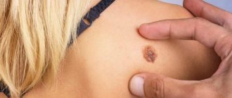
When infected with fungi, ring-shaped inflammatory foci appear on the skin and in the folds, with clear boundaries and a raised ridge along the edges. There may be small scales and bubbles on the surface. If large folds are affected, the surface may become weeping and macerated. Often observed in the groin areas, scrotum, armpits, and under the breasts.
Fungal disease of the feet is manifested by peeling of the skin in the spaces between the toes, soles, thickening of the stratum corneum of the soles and lateral surfaces of the feet, cracks.
With onychomycosis (“nail fungus”), yellowish and whitish stripes form in the thickness of the nail, the nail plate thickens, crumbles, and the edges of the nail become jagged. Sometimes thinning of the nails is observed, and detachment of the nail from the bed may occur.
Itching is the main complaint with fungal skin diseases. When the surface macerates (especially in folds), a burning sensation occurs. Fungal infections develop slowly and have a long course. Exacerbation usually occurs in the summer. It is important to remember: the use of ointments and creams containing hormones causes worsening of the condition and is strictly contraindicated for mycoses.
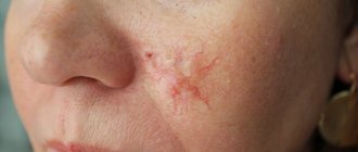
Among fungal diseases of the scalp, the most common is microsporia , which is caused by fungi of the genus Microsporum. Microsporia is a highly contagious disease. In Russia, Microsporum canis fungi are widespread and often infect animals (cats, dogs, rabbits, hamsters, guinea pigs). Human infection occurs through contact with a sick animal. Peaks of incidence are May-June and September-November. Children most often get sick, not only because of their ardent love for animals, but also because of the structural features of their skin - more delicate than that of adults. A symptom of microsporia is the appearance of round or oval lesions, with raised clear boundaries, covered with scales or crusts. When the scalp is affected, the hair in the lesion is broken off.
Yeast-like fungi are the cause of candidiasis. Urogenital candidiasis is an inflammatory disease of the genitourinary tract caused by fungi of the genus Candida. It is believed that 70-75% of women develop vulvovaginal candidiasis (“thrush”) at least once during their lives. The main symptoms in women are profuse white, cheesy or creamy discharge from the genital tract, itching and burning in the external genital area. Relapses of the disease often occur due to a decrease in immune defense, the use of antibiotics, or a change of sexual partner. In men, manifestations of urogenital candidiasis may include redness and swelling of the genitals, with a white coating on the surface of the rash.
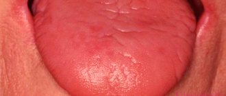
Candida can affect the mucous membranes of the oral cavity, most often in young children. Erosed elements with a white coating appear on the mucous membrane, accompanied by a burning sensation and discomfort when swallowing.
Treatment and prognosis
Treatment of fungal infections (mycoses) takes from several weeks in the case of skin mycoses to months in the case of nail mycoses. Smooth skin (without hair) is usually completely restored after treatment, scarring at the site of hair loss is rare and, as a rule, hair growth is restored. A complete cure for nails affected by fungi is not always possible, and nail restoration can take a long time.
Unfortunately, there is no immunity after a fungal infection, so infection can occur again.
The information contained in this publication should not be used as a substitute for medical care or consultation with a physician.
author of the publication Mikhail Sergeevich Betekhtin, PhD, dermatovenerologist, oncologist, cosmetologist.
Classification of mycoses
Mycoses are usually divided according to the degree of involvement of the skin, mucous membranes and appendages of the skin in the pathological process:
- Keratomycosis
. Fungal microorganisms multiply only in the stratum corneum of the skin; keratomycosis does not cause extensive inflammatory changes on the body. - Dermatophytes
. The changes affect the dermis and epidermis, as well as nails and hair follicles. - Candidiasis
. In addition to the skin, mucous membranes may be involved in the pathological process. - Deep mycoses
. Not only the skin is affected, but also a number of internal organs.
conclusions
Superficial fungal infections are most often caused by dermatophytes from the genera Trichophyton, Epidermophyton and Microsporum. These organisms metabolize keratin and cause a number of pathological clinical manifestations, including tinea pedis, dermatophytal dermatitis, dermatophytosis, etc. Based on clinical findings, the diagnosis of cutaneous dermatophyte infection can be strongly suspected. To confirm the diagnosis, potassium hydroxide should be used. Most dermatophyte infections can be managed with topical treatment. Examples of effective topical antifungal agents include azoles, allylamines, ciclopirox, butenafine, and tolnaftate. Oral antifungal therapy is used for widespread infections or infections that are resistant to topical therapy. Nystatin is not effective against dermatophyte infections.
List of literature / References
- Havlickova B, Czaika VA, Friedrich M. Epidemiological trends in skin mycoses worldwide // Mycoses. 2008 Sep;51 Suppl 4:2-15. doi: 10.1111/j.1439-0507.2008.01606.x. Erratum in: Mycoses. 2009 Jan;52(1):95. PMID: 18783559.
- Havlickova B, Czaika VA, Friedrich M. Epidemiological trends in skin mycoses worldwide // Mycoses 2008; 51 Suppl 4:2.
- Seebacher C, Bouchara JP, Mignon B. Updates on the epidemiology of dermatophyte infections // Mycopathologia 2008; 166:335.
- Ameen M. Epidemiology of superficial fungal infections // Clin Dermatol 2010; 28:197.
- El-Gohary M, van Zuuren EJ, Fedorowicz Z, et al. Topical antifungal treatments for tinea cruris and tinea corporis // Cochrane Database Syst Rev 2014; :CD009992.
- Alston SJ, Cohen BA, Braun M. Persistent and recurrent tinea corporis in children treated with combination antifungal/ corticosteroid agents // Pediatrics 2003; 111:201.
- Greenberg HL, Shwayder TA, Bieszk N, Fivenson DP. Clotrimazole/betamethasone diproprionate: a review of costs and complications in the treatment of common cutaneous fungal infections // Pediatr Dermatol 2002; 19:78.
- Rosen T, Elewski BE. Failure of clotrimazole-betamethasone dipropionate cream in treatment of Microsporum canis infections // J Am Acad Dermatol 1995; 32:1050.
- Hawkins DM, Smidt AC. Superficial fungal infections in children // Pediatr Clin North Am 2014; 61:443.
- Crawford F, Hollis S. Topical treatments for fungal infections of the skin and nails of the foot // Cochrane Database Syst Rev 2007; :CD001434.
- Korting HC, Tietz HJ, Brautigam M, et al. One week terbinafine 1% cream (Lamisil) once daily is effective in the treatment of interdigital tinea pedis: a vehicle controlled study. LAS-INT-06 Study Group // Med Mycol 2001; 39:335.
- Gupta AK, Cooper EA. Update in antifungal therapy of dermatophytosis // Mycopathologia 2008; 166:353.
- Bell-Syer SE, Khan SM, Torgerson DJ. Oral treatments for fungal infections of the skin of the foot // Cochrane Database Syst Rev 2012; 10:CD003584.
- Adams BB. Tinea corporis gladiatorum // J Am Acad Dermatol 2002; 47:286.
- van Zuuren EJ, Fedorowicz Z, El-Gohary M. Evidence-based topical treatments for tinea cruris and tinea corporis: a summary of a Cochrane systematic review // Br J Dermatol 2015; 172:616.6.
- Bourlond A, Lachapelle JM, Aussems J, et al. Double-blind comparison of itraconazole with griseofulvin in the treatment of tinea corporis and tinea cruris // Int J Dermatol 1989; 28:410.
- Cole GW, Stricklin G. A comparison of a new oral antifungal, terbinafine, with griseofulvin as therapy for tinea corporis // Arch Dermatol 1989; 125:1537.
- Panagiotidou D, Kousidou T, Chaidemenos G, et al. A comparison of itraconazole and griseofulvin in the treatment of tinea corporis and tinea cruris: a double-blind study // J Int Med Res 1992; 20:392.
- Faergemann J, Mörk NJ, Haglund A, Odegård T. A multicentre (double-blind) comparative study to assess the safety and efficacy of fluconazole and griseofulvin in the treatment of tinea corporis and tinea cruris // Br J Dermatol 1997; 136:575.
- Elewski BE, Hughey LC, Sobera JO. Fungal diseases. In: Dermatology, 3rd ed, Bolognia JL, Jorizzo JL, Schaffer JV (Eds), Elsevier Limited, Philadelphia; London 2012. Vol 2, p.1251.
- Voravutinon V. Oral treatment of tinea corporis and tinea cruris with terbinafine and griseofulvin: a randomized double blind comparative study // J Med Assoc Thai 1993; 76:388.
- Farag A, Taha M, Halim S. One-week therapy with oral terbinafine in cases of tinea cruris/corporis // Br J Dermatol 1994; 131:684.
- Smith KJ, Neafie RC, Skelton HG 3rd, et al. Majocchi's granuloma // J Cutan Pathol 1991; 18:28.
- Gill M, Sachdeva B, Gill PS, et al. Majocchi's granuloma of the face in an immunocompetent patient // J Dermatol 2007; 34:702.
- Cho HR, Lee MH, Haw CR. Majocchi's granuloma of the scrotum // Mycoses 2007; 50:520.
- Tse KC, Yeung CK, Tang S, et al. Majocchi's granuloma and posttransplant lymphoproliferative disease in a renal transplant recipient // Am J Kidney Dis 2001; 38:E38.
- Kim ST, Baek JW, Kim TK, et al. Majocchi's granuloma in a woman with iatrogenic Cushing's syndrome // J Dermatol 2008; 35:789.
- Akiba H, Motoki Y, Satoh M, et al. Recalcitrant trichophytic granuloma associated with NK-cell deficiency in a SLE patient treated with corticosteroid // Eur J Dermatol 2001; 11:58.
- Ilkit M, Durdu M, Karakaş M. Majocchi's granuloma: a symptom complex caused by fungal pathogens // Med Mycol 2012; 50:449.
- Novick NL, Tapia L, Bottone EJ. Invasive trichophyton rubrum infection in an immunocompromised host. Case report and review of the literature // Am J Med 1987; 82:321.
- Feng WW, Chen HC, Chen HC. Majocchi's granuloma in a 3-year-old boy // Pediatr Infect Dis J 2006; 25:658.
- Gupta AK, Prussick R, Sibbald RG, Knowles SR. Terbinafine in the treatment of Majocchi's granuloma // Int J Dermatol 1995; 34:489.
- McMichael A, Sanchez DG, Kelly P. Folliculitis and the follicular occlusion tetrad. In: Dermatology, 2nd ed, Bolognia JL, Jorizzo JL, Rapini RP (Eds), Elsevier Limited, St. Louis 2008.
- Gupta AK, Groen K, Woestenborghs R, De Doncker P. Itraconazole pulse therapy is effective in the treatment of Majocchi's granuloma: a clinical and pharmacokinetic evaluation and implications for possible effectiveness in tinea capitis // Clin Exp Dermatol 1998; 23:103.
- Burg M, Jaekel D, Kiss E, Kliem V. Majocchi's granuloma after kidney transplantation // Exp Clin Transplant 2006; 4:518.
- Liao YH, Chu SH, Hsiao GH, et al. Majocchi's granuloma caused by Trichophyton tonsurans in a cardiac transplant recipient // Br J Dermatol 1999; 140:1194.
- Bonifaz A, Vázquez-González D. Tinea imbricata in the Americas // Curr Opin Infect Dis 2011; 24:106.
- Cheng N, Rucker Wright D, Cohen BA. Dermatophytid in tinea capitis: rarely reported common phenomenon with clinical implications // Pediatrics 2011; 128:e453.
- Romano C, Rubegni P, Ghilardi A, Fimiani M. A case of bullous tinea pedis with dermatophytid reaction caused by Trichophyton violaceum // Mycoses 2006; 49:249.
- Al Aboud K, Al Hawsawi K, Alfadley A. Tinea incognito on the hand causing a facial dermatophytid reaction // Acta Derm Venereol 2003; 83:59.
- Veien NK, Hattel T, Laurberg G. Plantar Trichophyton rubrum infections may cause dermatophytids on the hands // Acta Derm Venereol 1994; 74:403.
Diagnostics
The diagnosis is confirmed by the detection of segmented hyphae in skin scrapings from the affected area with potassium hydroxide (KOH). When bubbles form, the top of the bubble can serve as an adequate sample. Alternative diagnostic procedures are dermatophyte culture test medium and culture method.
Patients who have significant erosion, ulceration, or malodor in the affected area should undergo Gram staining and culture to evaluate for secondary bacterial infection.
Differential diagnosis depends on clinical subtype:
Interdigital dermatitis of the foot
- Erythrasma
- Interdigital candidiasis
- Hyperkeratotic (moccasin) dermatitis of the foot
- Atopic dermatitis
- Chronic contact dermatitis
- Chronic palmoplantar (dyshidrotic) eczema
- Palmoplantar psoriasis
- Pitted keratolysis
- Juvenile plantar dermatosis
- Exfoliating keratolysis
- Keratoderm
Inflammatory dermatosis of the foot
- Acute palmoplantar (dyshidrotic) eczema
- Acute contact dermatitis
- Palmoplantar pustulosis
- Scabies
A positive KOH test demonstrating segmented hyphae distinguishes Tinea pedis from nonfungal diseases. With interdigital candidiasis, a test with the drug KOH will show budding yeast, pseudohyphae and hyphal septa.
Transmission routes
The main source of infection is humans
.
The main route of transmission is contact
. Activation of opportunistic flora is also possible when problems with the immune system occur. Fomites, that is, objects contaminated with pathogenic fungi, can be household items, shoes, clothing, and bedding of the sick person, if people who are sensitive to this fungus actively come into contact with them. Outbreaks are possible in organized groups, mainly in children's and army groups. Among public places, swimming pools, water parks and other structures where people can be barefoot inside are considered a common variant of fungal infection, plus a warm and humid microclimate is formed there, which contributes to the long existence of the pathogen outside the host.
In rare cases, for example, with microsporia, the source of infection can be a street or domestic animal, most often a cat. In common parlance, this disease is called “lichen.” “Ringworm” or, more precisely, “ringworm” in a child is microsporia. It is also possible to become infected with trichophytosis through contact in rural areas with hay or other substrates on which the secretions of sick rodents remain.
General symptoms of mycoses
During the incubation period, that is, during the proliferation of fungi, nothing bothers the infected person. The disease begins to manifest itself after the fungi begin to negatively affect the skin.
Depending on the type of fungal microorganism and the location of the infection, mycoses can manifest themselves in different ways, but there are several common symptoms:
- Itching of the skin.
- The appearance of red spots, rashes, blisters.
- Peeling of individual areas of the skin.
When localized on the feet and hands, peeling of the dermis between the fingers is possible.
Mycosis involving the nail plates is called onychomycosis. Nail fungus damage is indicated by a change in their color, delamination, the appearance of dark or yellowish spots, and thickening of the plate.