Preeclampsia
The patient is urgently hospitalized in the intensive care unit of the nearest hospital with a delivery room. The main therapeutic goal is to reduce reflex and central hyperreactivity, prevent seizures, stabilize vital functions, and correct multiple organ disorders. A pregnant woman with preeclampsia is prescribed a strict medical and protective regimen. The treatment regimen includes the following groups of drugs:
- Anticonvulsants
. The “gold standard” is the administration of magnesium sulfate through an infusion pump. The drug has a sedative, anticonvulsant, antispasmodic, hypotensive effect, and effectively reduces intracranial pressure. Simultaneously with the improvement of cerebral hemodynamics, the myometrium relaxes and increases the intensity of blood flow in the uterus. If necessary, tranquilizers are additionally used. - Antihypertensive drugs
. Imidazoline derivatives are preferred, which have a central α2-adrenomimetic effect, stimulate I1-imadazoline receptors in the nucleus of the solitary tract and thereby enhance the parasympathetic effect on the myocardium. Parenteral administration of peripheral vasodilators, hybrid β- and α1-blockers with a rapid antihypertensive effect is possible. - Infusion formulations
. To normalize oncotic and osmotic pressure, colloidal, protein, and balanced crystalloid solutions are administered intravenously. Infusion therapy can improve the rheological properties of blood, central and peripheral hemodynamics, tissue perfusion, reduce the severity of multiple organ disorders, and restore water and electrolyte balance.
According to indications, sedatives, direct anticoagulants, antioxidants, membrane stabilizers, and drugs to improve blood flow in tissues and prevent fetal respiratory distress syndrome are used. If intensive care is ineffective within 24 hours of hospitalization, emergency delivery by cesarean section is recommended. For patients with rapidly increasing symptoms of preeclampsia, surgery is performed within 2-4 hours. Natural childbirth with high-quality anesthesia (long-term epidural anesthesia), perineotomy or episiotomy is possible only with a significant improvement in the patient’s well-being, stable stabilization of blood pressure, and laboratory parameters.
National Institute of Health (NICE) guidelines
Aspirin 75 mg is recommended from 12 weeks of pregnancy if there is at least one high risk:
- arterial hypertension in a previous pregnancy;
- chronic renal pathology;
- autoimmune diseases (such as APS);
- diabetes;
- chronic arterial hypertension.
Aspirin 75 mg is recommended from 12 weeks if there are two or more moderate risks:
- first pregnancy;
- age over 40 years;
- the interval between pregnancies is more than 10 years;
- family history of preeclampsia;
- multiple pregnancy;
- BMI>35.
What is preeclampsia (PE)?
Without going into detail about various scientific formulations, it must be said that preeclampsia is a severe toxicosis in the second half of pregnancy, when the pregnant woman’s body ceases to cope with the increased load that occurs with increasing pregnancy. Preeclampsia is a leading cause of maternal and perinatal death or disability. In the structure of maternal losses, more than 50,000 cases per year occur due to PE.
There are two types of preeclampsia: moderate and severe.
Moderate occurs mainly after 37 weeks of pregnancy and is characterized by a favorable prognosis with early detection and correct treatment in a hospital setting. Severe preeclampsia begins after 20 weeks and up to 37 weeks, has a pronounced aggressive course with complications that can lead to death - eclampsia (convulsions or coma in women with PE), intracranial hemorrhage or stroke, disseminated intravascular coagulation and HELLP syndrome. Other serious complications include cerebral edema, blindness, kidney failure, liver failure, or pulmonary edema. In addition, a significant percentage of women with PE require premature delivery for maternal and/or fetal indications, which, in turn, increases the risks of developing complications associated with prematurity (in particular, neonatal mortality, intracerebral hemorrhage, seizures, respiratory and digestive disorders, jaundice, retinopathy) and leads to increased length of hospitalization. In the structure of perinatal mortality, about 25% of deaths are associated with PE and eclampsia, the consequences of which also include fetal growth retardation, which is detected in 15% of newborns. The risk of adverse maternal and fetal/newborn outcomes is significantly higher with early and premature (severe) PE than with full-term (moderate) PE. Long-term consequences: Women
who develop PE, compared with those who have an uncomplicated pregnancy, are at twice the risk of developing cardiovascular disease (CVD), including hypertension, coronary heart disease, stroke, and risk of death due to CVD.
It is currently unclear whether this relationship occurs due to PE-related vascular damage, which can potentially lead to the development of CVD, or, more likely, there is an inverse relationship - women with a predisposition to the development of CVD are exposed to specific stress during pregnancy, which leads to PE. Children
exposed to adverse effects of PE in utero, compared with those born during an uncomplicated pregnancy, have a 2-fold higher risk of cerebral palsy due to preterm birth, growth retardation, or both.
Preparing for a future pregnancy
If you had preeclampsia during pregnancy or postpartum preeclampsia, you are at high risk of complications in future pregnancies. But it is possible that you will not have complications during your next pregnancy. Your doctor will likely monitor you more closely throughout your future pregnancy for signs of complications.
Your doctor may also recommend preventative treatment to reduce the risk of complications
Translation by Svetlana Sheremeteva
Photos from free sources
Diagnostics
Differential diagnosis of preeclampsia and eclampsia must first be carried out with an epileptic seizure (“aura” before the attack, convulsions). Also, these complications should be distinguished from uremia and brain diseases (meningitis, encephalitis, hemorrhages, neoplasms).
The diagnosis of preeclampsia and eclampsia is established based on a combination of instrumental and laboratory data:
- Blood pressure measurement. Increasing blood pressure to 140/90 and maintaining these numbers for 6 hours, increasing systolic pressure by 30 units and diastolic by 15.
- Proteinuria. Detection of 3 or more grams of protein in the daily amount of urine.
- Blood chemistry. An increase in nitrogen, creatinine, urea (kidney damage), an increase in bilirubin (red blood cell breakdown and liver damage), an increase in liver enzymes (AST, ALT) - liver dysfunction.
- General blood analysis. An increase in hemoglobin (a decrease in the volume of fluid in the vascular bed, that is, blood thickening), an increase in hematocrit (viscous, “stringy” blood), a decrease in platelets.
- General urine analysis. Detection of protein in urine in large quantities (normally absent), detection of albumin (severe preeclampsia).
Preeclampsia (PE) is one of the most important problems in modern obstetrics, given its medical and socio-economic significance. Since the consequences of severe hypertensive disorders during pregnancy reduce the quality of life of women of reproductive age, and the frequency of disturbances in the physical and psychosomatic development of prematurely born children is quite high, this problem is significant both medically and socially [1-3].
For many years, PE remains one of the serious multisystem pathological complications of pregnancy, the frequency of which does not tend to decrease. Even Hippocrates in the 4th century BC. described a disease in pregnant women, the symptoms of which he compared with epilepsy. It was only in 1827 that R. Bright suggested that eclampsia (translated from Greek as “lightning”, “flash”) is a kidney disease based on the definition of albuminuria. In 1843, J. Lever described edema, albuminuria, pigment spots and headache in eclampsia. A. Delore in 1884 put forward an infectious theory and even isolated the so-called Bacillus eclampsae
. In 1886, E. Leyden introduced the term “nephropathy,” and in 1908, S.D. Mikhnov - “preeclampsia”. In 1905, J. De Lee suggested that eclampsia is the result of the action of toxins, and W. Freund coined the term “toxicosis of pregnancy.” Finally, in 1913, W. Zangemeister described the classic triad: arterial hypertension, edema, proteinuria, which is the basis for diagnosing and assessing the severity of P.E. Today, the diagnosis of PE is based on the occurrence of arterial hypertension, proteinuria and generalized edema, usually after 20 weeks of pregnancy, or on a sudden increase in blood pressure, the appearance or exacerbation of proteinuria in patients with chronic arterial hypertension (CAH) [4, 5].
Despite ongoing prevention and repeated attempts to select therapy, it was not possible to reduce the incidence of P.E. Data on the prevalence of PE in the world in the general population vary - from 2.3 to 23% [4, 6, 7]. According to O.V. Makarova et al. [4], in Russia P.E. occurs in 13-16% of the total number of births. Prevalence of P.E. among pregnant women in the Russian Federation in 2011 accounted for 17.4% of all those who completed pregnancy, and in 2012 - 16.7% [8]. When analyzing the frequency of this pathology during childbirth, an increase in the frequency of PE and eclampsia in parturient and postpartum women was revealed from 27.1% in 2011 to 30.3% in 2012 (by 11.6%) [9]. In some developed countries, in particular the USA, an increase in the incidence of PE has been noted in recent years [10]. Probably the reason for this is the increase in the incidence of conditions such as diabetes mellitus, obesity and CAH in the population [4, 11]. The leading risk factors for the development of PE are extragenital pathology (endocrinopathy, diseases of the cardiovascular system, kidneys), inflammatory processes of the genital organs and a burdened obstetric history [12]. B. Sibai [13] found that the frequency of PE in primiparous women varied from 4 to 11%, while the frequency of PE in women who gave birth was lower, although it also varied widely. At the same time, according to C. Ananth et al. [14], the probability of delivery at term is higher in multiparous women with hypertension than in primiparous women. The main risk factors for the development of PE, identified from anamnesis and clinically, are chronic kidney disease (58.6%), vascular pathology (51.7%), endocrine pathology (38.0%), metabolic syndrome (24.0%), heart disease (22.0%), gastrointestinal tract diseases (20.7%) [15]. According to our data [16], in 68% of cases, PE is of a combined nature and develops against the background of extragenital pathology, in particular against the background of CAH and kidney diseases. In pregnant women with CAH, the addition of PE was noted in 19% of cases. Other risk factors for PE include maternal age over 35 years, obesity, and multiple pregnancies [4].
According to the World Health Organization, PE is the main cause of premature birth, premature abruption of a normally located placenta, the development of placental insufficiency, intrauterine growth retardation, and the birth of low birth weight children. Complications of P.E. are hemorrhage and retinal detachment, acute renal failure, acute fatty hepatosis, HELLP syndrome, pulmonary edema and stroke [4, 5, 11, 17, 18].
In the global structure of maternal mortality, the share of PE is 12-15%, and in developing countries this figure reaches 30% [19]. According to Russian authors [20], this pathology remains one of the main causes of maternal mortality and accounts for about 20% of all cases of maternal deaths in the Russian Federation.
According to the Ministry of Health of the Russian Federation, hypertensive complications of pregnancy occupy 3-4th place in the list of causes of maternal mortality over the last decade and are its direct cause in 6.9-17.4% of cases [4, 8, 9, 18 , 20].
In Moscow, maternal mortality from various forms of PE and their complications ranks first among all causes of maternal mortality, ranging from 17 to 28% per year, i.e. the share of PE in Moscow turned out to be slightly higher than in Russia as a whole [ 9, 20]. PE requires close attention and monitoring of the patient not only during pregnancy and childbirth, but also in the postpartum period, especially in the early period. Up to 44% of cases of eclampsia occur in the postpartum period, especially during full-term pregnancy [21, 22]. Of the total number of women who died from PE, 96.8% died after childbirth [23].
For a long time, the prevailing idea was that this complication of pregnancy does not have a noticeable effect on the woman’s health in the long term after childbirth. However, today it is known that PE affects not only the course of pregnancy and childbirth, but also significantly affects the health of the mother and child and long-term prognosis.
The long-term prognosis of women who have undergone PE is characterized by an increased incidence of CAH, coronary heart disease, diabetes mellitus and strokes [24]. The results of recent studies indicate the presence of a high cardiovascular risk in this group of women in later life. According to G.T. Sukhikh [18], in the long term of the life cycle in women who have previously undergone PE (especially during the first birth), the risk of developing cardiovascular complications, including fatal ones, increases. Also, with the simultaneous combination of three cardiovascular risk factors in the obstetric history (preeclampsia, premature birth and low body weight for gestational age of the infant), a sevenfold increase in subsequent fatal outcomes is observed.
There is a tendency to earlier development of ischemic brain lesions in young women after undergoing P.E. The likelihood of developing an ischemic cerebral stroke in women who have suffered PE during childbirth is 60% higher than the likelihood of developing a stroke in those who have not suffered it [25]. PE is also a risk factor for the early development of atherosclerosis, as evidenced by the discovery of a greater thickness of the intima-media complex of the coronary vessels of the heart and femoral artery in women who underwent PE during childbirth than in women after a normal pregnancy [25]. G. Smith et al. [26] reported that 20% of women with PE exhibit residual microalbuminuria 1 year after birth, as well as increased blood pressure, total cholesterol, low-density lipoproteins, triglycerides, body mass index, fasting insulin, and creatinine.
In the UK, L. Bellamy et al. [27] conducted a meta-analysis of clinical studies to evaluate the relationship between PE and subsequent development of cardiovascular diseases. At the end of the meta-analysis, mortality from all causes was assessed among women who experienced PE during pregnancy. PE is characterized by an increased risk of developing various cardiovascular diseases and all-cause mortality in later life. There is no established relationship between PE and the development of malignant neoplasms, including breast cancer, which is the main cause of morbidity and mortality in older age.
Since women who have undergone PE often give birth early and by cesarean section, the frequency of disturbances in the physical and psychosomatic development of prematurely born children is quite high, and they subsequently suffer from various metabolic, hormonal, and cardiovascular diseases.
Children born to mothers with PE have higher blood pressure and body mass index, which must be taken into account in the primary prevention of cardiovascular diseases in such people [28].
Until now, the etiology and pathogenesis of PE are not fully known, scientists around the world are making attempts to connect all the links together, to determine the sequence and subordination of all changes that occur in the body of a pregnant woman, but so far all assumptions remain only assumptions and each previous one, often, is refuted by what follows. Currently, the highest priority is the placental theory of development, which explains the occurrence of PE as a result of pathological placentation, which in turn leads to placental ischemia with the further development of generalized vasospasm, endotheliosis and multiple organ failure. Even in the recent past, the issue of unified terminology and classification of various forms of arterial hypertension in pregnant women in the Russian Federation was extremely relevant.
In 2013, the Russian Society of Obstetricians and Gynecologists developed Federal clinical guidelines “Hypertensive disorders during pregnancy, childbirth and the postpartum period. Preeclampsia. Eclampsia”, where a clinical classification of hypertensive disorders during pregnancy was proposed [29, 30]:
- preeclampsia and eclampsia;
— preeclampsia and eclampsia against the background of chronic arterial hypertension;
- gestational (pregnancy-induced) arterial hypertension;
- chronic arterial hypertension (existing before pregnancy).
The use of a unified terminology and classification will allow for a correct assessment of statistical data on the prevalence of PE, will reduce the overdiagnosis of this condition, will facilitate the correct diagnosis, determination of the severity of the disease and the development of adequate management tactics both at the outpatient and inpatient stages and will ensure continuity between different medical institutions.
Among the many problems associated with PE and eclampsia, the problem of diagnosis, prognosis and assessment of severity occupies one of the most important places and is of great importance both for obstetricians and gynecologists and for doctors of related specialties. Reducing perinatal and maternal morbidity and mortality remains the main goal of obstetricians and gynecologists.
It is important to remember that even if PE is suspected, a pregnant woman must be hospitalized urgently for additional examination and clarification of the diagnosis. When the diagnosis is confirmed, the condition of the pregnant woman and the functional state of the fetus are assessed, and further management tactics depend on the gestational age and the severity of the disease.
The diagnosis of PE is established by a combination of arterial hypertension (systolic blood pressure - SBP ≥140 mm Hg and/or diastolic blood pressure - DBP ≥90 mm Hg [31]), proteinuria (the presence of protein in the urine ≥0.3 g/l in a daily sample (24 hours) or in two samples taken with an interval of 6 hours; when using a test strip (protein in urine) - indicator (≥"1+" [32]) and generalized edema [4]. Peripheral edema is observed in 70-80% of cases during physiological pregnancy, and therefore cannot be considered as a diagnostic criterion for PE [4, 33].However, edema of the face and hands often precedes the development of PE, and generalized, rapidly increasing edema, anasarca, fluid accumulation in cavities are considered one of the unfavorable prognostic criteria for severe PE [34].At the same time, PE occurring without generalized edema is recognized as more dangerous for the mother and fetus than PE with edema.
There is evidence that 10% of women with PE have no or minimal proteinuria in the urine [33], and 20% of women who develop eclampsia do not have proteinuria [33, 35]. In some women, atypical PE is reported, which is accompanied by multiple organ failure, generalized endotheliosis, but without arterial hypertension, without proteinuria, or without both of these symptoms [33].
The management of patients with PE depends on the gestational age and the severity of the disease.
In 2011, B. Sibai [36] described the following criteria for moderate PE: arterial hypertension: SBP ≥140 mmHg. or DBP ≥ 90 mmHg occurring at >20 weeks' gestation in a woman with a history of normal blood pressure, and proteinuria ≥0.3 g/L protein in a 24-hour urine sample.
Criteria for severe PE (presence of symptoms of moderate PE and ≥1 of the following criteria):
1) arterial hypertension: SBP ≥160 mm Hg. or DBP ≥110 mm Hg. with double measurements with an interval of 6 hours at rest;
2) proteinuria ≥5.0 g/l in a daily urine sample or >3 g/l in two portions of urine taken with an interval of 6 hours, or a value of 3+ on the test strip;
3) oliguria <500 ml in 24 hours;
4) cerebral or visual symptoms (headache, floaters, etc.);
5) pulmonary edema;
6) cyanosis;
7) pain in the epigastrium or right upper quadrant;
 liver dysfunction (increased activity of alanine and aspartate aminotransferases);
9) thrombocytopenia (<100·106/l);
10) intrauterine growth retardation.
The criteria for severe PE also include critical forms of PE (severe retinopathy, acute fatty hepatosis, HELLP syndrome, acute renal failure, pulmonary edema) and placental abruption [35].
In addition to the above criteria, severe PE is characterized by a progressive decrease in the blood content of total protein, albumin, platelets, and an increase in proteinuria and hematocrit. Currently, only a progressive deterioration of clinical and laboratory tests over time indicates the worsening of PE and its transition to a more severe form and requires early delivery, while the absolute values of clinical and laboratory indicators are not always reliable in identifying the true severity of the process [4]. For early diagnosis and verification of the severity of the process, it is possible to use 24-hour blood pressure monitoring (ABPM), determination of central hemodynamic parameters and microalbuminuria [4].
Despite the variety of proposed scales and methods, there is still no way to determine with a sufficient degree of reliability the degree of severity of PE and predict its further course: the severity of edema, proteinuria and arterial hypertension does not always determine the true severity of pathological processes in PE, due to which creates the need to search for new biochemical markers [4, 16, 37, 38]. Today, the role of angiogenic growth factors in the pathogenesis of PE and the possibility of using the determination of the imbalance of pro- and antiangiogenic growth factors in the prognosis, diagnosis and differential diagnosis of PE and identifying its severity are widely discussed [16, 37, 38]. The use of additional tests is extremely important nowadays due to the increasing frequency of PE and the registration of its atypical forms. For P.E. characterized by an increase in the level of antiangiogenic (soluble FMS-like tyrosine kinase-1-sFlt-1 and endoglin - sEng) and a decrease in the level of proangiogenic (PlGF - placental growth factor) factors compared to patients with physiological pregnancy [38, 39]. Since angiogenic processes in the body are regulated by a balance of pro- and antiangiogenic growth factors, an angiogenic coefficient (K) has been proposed, which is the ratio of sFlt-1/PlGF protein content. Such a coefficient is intended to quantitatively reflect the balance of angiogenic proteins and, consequently, the direction and/or intensity of angiogenic processes in the body. The higher the value of the K coefficient, the more pronounced is the imbalance between vascular growth factors towards the predominance of the antiangiogenic protein Flt-1 or a decrease in the level of the proangiogenic factor PlGF. The value of the angiogenic coefficient K in PE is 70 times higher than in healthy pregnant women [37, 38] and significantly increases with increasing severity of PE. A highly sensitive (62%) and highly specific (75%) criterion for severe PE is a decrease in proangiogenic placental growth factor (PlGF) to 14.5–42.8 pg/ml, which indicates progression of the pathological process in this category of patients [39].
The only treatment for PE today is delivery, removal of the fetus and placenta from the mother’s body. At a gestational age of more than 34 weeks, the tactics of managing patients with PE is reduced to preparing for delivery after stabilization of the pregnant woman’s condition. The method of delivery depends on the biological maturity of the birth canal, the obstetric situation, and the general condition of the pregnant woman and the fetus [4]. Caesarean section for PE is not the only method of choice for delivery. Spontaneous birth, when properly managed, is hemodynamically less stressful for the mother and reduces the incidence of respiratory complications in premature newborns. Caesarean section is the method of choice for the delivery of patients with PE at less than 32 weeks of gestation.
Currently, the main indication for delivery of pregnant women with PE at preterm gestation is deterioration in clinical and laboratory data and/or deterioration in the functional state of the fetus. In gestation periods of less than 34 weeks, management tactics are determined by the severity of P.E. With P.E. severe cases, the only correct treatment is delivery after stabilization of the patient’s condition, regardless of gestational age. The most controversial is the management tactics of pregnant women with moderate PE during premature pregnancy. When we talk about “prolonging” pregnancy during PE in order to stabilize the patient’s condition and prevent fetal RDS syndrome, in essence, we are delaying the transition from moderate to severe PE, which requires emergency delivery, and often resuscitation measures. In patients with moderately severe PE (moderate severity) up to 34 weeks of pregnancy, drug therapy can be used, which is carried out in a hospital setting, accompanied by careful monitoring of the condition of the mother and fetus and ends with preparation for labor and delivery. The use of such tactics in case of a favorable course of the disease in some cases helps to prolong pregnancy by 2 weeks [40]. At a gestational age of less than 34 weeks in pregnant women with moderate PE, according to some authors, the possibility of continuing pregnancy will depend on the effectiveness of therapy. In this situation, careful dynamic monitoring of the patient’s clinical and laboratory parameters is carried out for 24-48 hours [4]. The period of pregnancy prolongation in patients with PE averaged 8.12±1.27 days (from 0 to 19 days) [16], while the period of pregnancy prolongation in patients with moderate PE averaged 11.85±1. 61 days, which is significantly longer than in pregnant women with severe PE – 4.1±0.69 days ( p
<0,05) [16, 37, 39].
The criterion for starting antihypertensive therapy for PE is SBP ≥140 mmHg. or DBP ≥90 mm Hg. Antihypertensive therapy for PE must be carried out under Doppler monitoring, since a decrease in uteroplacental and fetal placental blood flow is possible [4, 40]. The main drugs currently used in the world for the treatment of arterial hypertension during pregnancy are methyldopa (first-line drug), the α-β-blocker labetalol, calcium antagonists (nifedipine) and β-blockers (the drug of choice is metoprolol), also possibly the use of hydrochlorothiazide, furosemide, verapamil, clonidine, prazosin. ACE inhibitors, angiotensin II receptor antagonists, spirolactone, calcium antagonists diltiazem and felodipine are contraindicated during pregnancy. It should be noted that some drugs that have proven themselves in practice and are widely used abroad (labetalol, diazoxide, hydralazine for parenteral administration) are not registered in our country [40].
It is necessary to ensure prolonged administration of a solution of magnesium sulfate before and during childbirth and in the postpartum period in order to prevent seizures [4, 40]. Magnesium sulfate is the drug of choice for the prevention and treatment of seizures, which is superior to benzodiazepines, phenytoin and nimodipine in the prevention of eclampsia and does not increase the incidence of cesarean section, bleeding, infectious diseases and neonatal depression [41]. Administration of magnesium sulfate should be administered before and during delivery, and should continue for at least 24 hours after delivery or 24 hours after the last episode of seizures [42, 43]. Magnesium sulfate is administered only intravenously, always using a device for continuous administration. The loading dose is 4-6 g of dry matter for 10-15 minutes, and the maintenance dose is 1-2 g of dry matter per 1 hour.
It is important to note that regional analgesia or anesthesia is the preferred method of pain management for patients with PE, both during vaginal delivery and during cesarean section (with a platelet level >75 109/L and in the absence of coagulopathy). Spinal and epidural, as well as combined spinal-epidural anesthesia are effective and equally safe.
Currently, the tactics of managing patients with PE in the Russian Federation are regulated by two documents: Order of the Ministry of Health of the Russian Federation No. 572n dated November 1, 2012 “On approval of the Procedure for the provision of medical care in the field of obstetrics and gynecology (except for the use of assisted reproductive technologies)” (as amended and additions) and Federal clinical guidelines “Hypertensive disorders during pregnancy, childbirth and the postpartum period. Preeclampsia. Eclampsia”, developed by the Russian Society of Obstetricians and Gynecologists in 2013.
From all of the above, it follows that the frequency of PE is not decreasing, a huge number of pregnant women are at risk for the development of this pathology, the consequences of PE are dangerous to the life and health of both the woman and the fetus, which leads to an increase in mortality and disability. Despite a large number of studies and publications, the root causes of PE and markers of the rate of progression of pathological changes in vital organs and systems remain unknown, which limits the possibilities of treating and preventing the development of PE. Thus, to reduce maternal mortality from PE and eclampsia, it is necessary to monitor risk factors for the development of this condition, ensure proper monitoring of pregnant women at high risk for PE, comply with the diagnostic criteria for PE when making a diagnosis, and develop adequate tactics for managing patients depending on the gestational age and the severity of the condition, ensure quick and gentle delivery, according to standards, ensure monitoring of patients after delivery in an intensive care unit. Research should be conducted to identify predictors of PE both in the high-risk group and in the general population to determine criteria for early and timely diagnosis and markers of process progression in order to optimize obstetric tactics and timing of delivery.
How to cope with postpartum preeclampsia
The time after childbirth can be challenging even without health problems. Recovering from childbirth and caring for a newborn can be stressful. While recovering from pregnancy, it is important to pay attention to your own health by monitoring symptoms and consulting with your doctor.
If you are diagnosed with postpartum preeclampsia while in hospital, you may need a longer hospital stay. Reach out to loved ones or talk to your healthcare provider to learn about options for additional support when you return home.
Symptoms of eclampsia and preeclampsia
Signs of preeclampsia
Preeclampsia is just a short interval between nephropathy and a seizure. Preeclampsia is a dysfunction of vital organs of the body, the leading syndrome of which is damage to the central nervous system:
- the appearance of spots before the eyes, flickering, blurriness of objects;
- tinnitus, headache, feeling of heaviness in the back of the head;
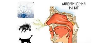
- nasal congestion;
- memory disorders, drowsiness or insomnia, irritability or apathy.
Preeclampsia is also characterized by pain in the upper abdomen (“in the pit of the stomach”), in the right hypochondrium, nausea, and vomiting.
An unfavorable prognostic sign is increased tendon reflexes (this symptom indicates convulsive readiness and a high probability of developing eclampsia).
With preeclampsia, swelling increases, sometimes for several hours, but the severity of edema does not matter in assessing the severity of the pregnant woman’s condition. The severity of preeclampsia is determined based on complaints, proteinuria and arterial hypertension (an increase in blood pressure for normotensive patients above 140/90 mm Hg should be alarming). If arterial hypertension is 160/110 or more, they speak of severe preeclampsia.
Kidney damage manifests itself in the form of a decrease in the amount of urine excreted (oliguria and anuria), as well as a high protein content in the urine (0.3 grams in the daily amount of urine).
Signs of eclampsia
Eclampsia is an attack of convulsions that consists of several phases:
- First phase. The duration of the first (introductory) phase is 30 seconds. At this stage, small contractions of the facial muscles appear.
- Second phase. Tonic cramps are a generalized spasm of all muscles of the body, including the respiratory muscles. The second phase lasts 10-20 seconds and is the most dangerous (the woman may die).
- Third phase. The third phase is the stage of clonic seizures. The motionless and tense patient (“like a string”) begins to beat in a convulsive seizure. The convulsions go from top to bottom. The woman is without a pulse or breathing. The third stage lasts 30-90 seconds and is resolved with a deep breath. Then breathing becomes rare and deep.
- Fourth phase. The seizure resolves. Characteristic is the release of foam mixed with blood from the mouth, a pulse appears, the face loses its cyanosis, returning to normal color. The patient either regains consciousness or falls into a coma.