In this article we will tell you:
- Causes of iridocyclitis
- Main types of iridocyclitis
- Symptoms and complications of iridocyclitis
- Diagnosis of iridocyclitis. Treatment
- Prevention of iridocyclitis
It happens that eye diseases concern one particular department, tissue or structure of an organ. So, with cataracts we talk about clouding of the lens, with keratitis - with inflammation of the cornea, with macular degeneration - with damage to the retina. Something else happens when several departments are involved in the pathological process at once. Such diseases are called combined, and it is to this group that the disease discussed in this article, iridocyclitis, belongs.
Iridocyclitis
is an inflammation of both the ciliary (ciliary) body and the iris of the eyeball.
The name of the disease comes from the Greek words iris - rainbow, and kykios circle, eye. Iridocyclitis is also called anterior uveitis. The disease is distinguished from uveitis proper by the localization of the inflammatory process: with ordinary uveitis, the choroid of the eye (uveal tract) is affected. The combination of lesions characteristic of ocular iridocyclitis occurs due to the anatomical similarity: the blood supply and innervation of the departments are closely related, and when one of them is affected, the inflammation quickly spreads to the other. Sometimes there are inflammatory processes in only one of the mentioned sections - the iris or the ciliary body. In these cases, we will talk about individual diseases: cyclitis and iritis.
You can get iridocyclitis at any age. Somewhat more often it affects young people 20-40 years old.
Causes of iridocyclitis
There are many reasons why iridocyclitis can develop. Among them are both exo- and endogenous. With the exogenous nature of the disease, the reason for its development lies in external factors. Endogenous iridocyclitis is a consequence of internal problems in the body and is more common.
The blood-ophthalmic barrier is normally responsible for the health of the eyes: it traps harmful bacteria, microbes, poisons - all those foreign objects that can cause diseases of the eye structures. When the permeability of this natural filter increases, harmful substances enter directly into the iris and then into the ciliary body, turning both sections into foci of inflammation.
The following are the main groups of causes of iridocyclitis:
- Infections: bacterial, viral, fungal.
- Helminthic infestations.
- Allergy.
Herpes, influenza, ARVI measles, tuberculosis, some sexually transmitted diseases (syphilis, gonorrhea, chlamydia), staphylococcal and streptococcal infections, malaria, brucellosis: all these ailments can affect the permeability of the blood-ophthalmic barrier and cause inflammation.
Seasonal hay fever or year-round allergies (to dust, food, household chemicals, medications, etc.) can also cause the disease.
- Chronic foci of inflammation in the oral or nasal cavities, otitis.
- Rheumatic diseases.
In persons suffering from rheumatism and syndromes associated with it, the incidence rate can reach up to 40%.
- Metabolic diseases (diabetes, sarcoidosis, gout, etc.).
- Injuries (contusions, wounds) and postoperative complications.
Risk factors for the occurrence of this disease may include:
- traumatic, stressful situations;
- hypothermia of the body;
- too intense physical activity;
- bad habits, unbalanced diet;
- immune failures.
It happens that even specialists fail to find out the cause of the disease: then they talk about iridocyclitis of unknown etiology.
Causes and provoking factors of iridocyclitis
As a rule, iridocyclitis develops against the background of chronic infections, both general and local (especially in the presence of lesions in the nasopharynx and oral cavity). Pathogenic pathogens can be viruses (herpes, cytomegalovirus, measles, influenza), pathogenic bacteria (brucellosis, syphilis, tuberculosis and many others), protozoan microorganisms and intracellular parasites (chlamydia, toxoplasmosis, malaria), fungal cultures (candidiasis, etc. ).
Systemic inflammation of the joints (rheumatoid arthritis, ankylosing spondylitis) often provokes the development of iridocyclitis. The listed forms of iridocyclitis are called endogenous (due to internal causes).
Exogenous (brought from outside) variants of iridocyclitis usually turn out to be complications of injuries, including ophthalmic surgery, and acute infectious and inflammatory processes in the cornea or sclera. Provoking factors in such cases are usually hypothermia, physical or emotional overload, and endocrine disorders.
Main types of iridocyclitis
According to the nature of the appearance and course of iridocyclitis, they are divided into:
- acute (begin abruptly. Symptoms of acute iridocyclitis of the eye are pronounced, easily diagnosed, duration – 3-6 weeks);
- chronic (they are more sluggish in nature compared to acute forms, symptoms are more difficult to relieve, duration is up to several months);
- recurrent - periods of attenuation of the disease are replaced by exacerbations (more often in winter).
According to the nature of inflammation, iridocyclitis is:
- serous. Iridocyclitis of this form is characterized by the accumulation of serous secretion in the chambers of the eye.
- hemorrhagic. Such iridocyclitis is characterized by severe damage to the vessels supplying blood to the ciliary body and iris.
- exudative. The main difference between this form is the accumulation of pus in the form of a crescent on the lower part of the iris.
- fibrinous-plastic. Iridocyclitis of this type occurs with the formation of fibrin threads. With fibrinous iridocyclitis, synechiae form in the anterior chamber of the eye, and fibrinous exudate accumulates.
Iridocyclitis
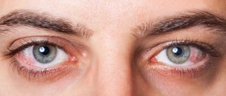
The severity and characteristics of the course of iridocyclitis depend on the nature and duration of exposure to the antigen, the level of permeability of the blood-ophthalmic barrier, the genotype and immune status of the body. With iridocyclitis, unilateral eye damage is usually observed. The first signs of acute iridocyclitis are general redness and pain in the eye, with a characteristic significant increase in pain when pressing on the eyeball. Patients with iridocyclitis experience photophobia, lacrimation, a slight (within 2-3 lines) decrease in visual acuity, and the appearance of “fog” before the eyes.
The course of iridocyclitis is characterized by a noticeable change in the color of the inflamed iris (greenish or rusty-red) and a decrease in the clarity of its pattern. The appearance of moderately severe corneal syndrome and pericorneal injection of the vessels of the eyeball is possible. Serous, fibrinous or purulent exudate may be detected in the anterior chamber of the eye. When purulent exudate settles at the bottom of the anterior chamber of the eye, a hypopyon is formed in the form of a gray or yellow-green stripe; When a vessel ruptures in the anterior chamber, an accumulation of blood is detected - hyphema.
The inflammatory process in the ciliary body, when exudate settles on the surface of the lens and the fibers of the vitreous body, can lead to clouding and a decrease in visual acuity.
On the posterior surface of the cornea, with iridocyclitis, grayish-white precipitates appear from pinpoint deposits of cells and exudate, during the resorption of which pigment lumps are observed for a long time. Swelling of the iris tissue and its close contact with the anterior capsule of the lens in the presence of exudate leads to the formation of posterior adhesions (synechias), causing irreversible narrowing (miosis) and deformation of the pupil, worsening its reaction to light. When the iris and the anterior surface of the lens fusion, a circular adhesion is formed along its entire length. If the course of iridocyclitis is unfavorable, synechia creates a risk of developing blindness due to complete occlusion of the pupil.
Often, intraocular pressure during iridocyclitis is below normal due to inhibition of the secretion of moisture in the anterior chamber. Sometimes, with acute onset iridocyclitis with severe exudation or fusion of the pupillary edge of the iris with the lens, an increase in intraocular pressure is observed.
Different types of iridocyclitis have their own clinical picture. Viral iridocyclitis is characterized by a torpid course, the formation of serous or serous-fibrinous exudate and light precipitates, and increased intraocular pressure.
Tuberculous iridocyclitis occurs with mild symptoms, manifested by the presence of large “greasy precipitates”, yellowish tubercles (tubercles) on the iris, opalescence of the anterior chamber moisture, the formation of powerful posterior stromal synechiae, blurred vision or complete occlusion of the pupil.
Autoimmune iridocyclitis is characterized by a severe recurrent course against the background of exacerbations of the underlying disease with frequent development of complications (cataracts, secondary glaucoma, keratitis, scleritis, atrophy of the eyeball). Each relapse is more severe than the previous one and often leads to blindness.
With traumatic iridocyclitis, sympathetic inflammation of the healthy eye (sympathetic ophthalmia) may develop. Iridocyclitis in Reiter's syndrome, caused by chlamydial infection, is accompanied by conjunctivitis, urethritis and joint damage with minor manifestations of inflammation of the choroid.
Symptoms and complications of iridocyclitis
The main symptoms of iridocyclitis are nonspecific: they are inherent in many ophthalmological (and not only) diseases. Patients with inflammation of the ciliary body and iris complain of:
- headache;
- blurred vision;
- photophobia;
- lacrimation;
- blurred vision;
- hyperemia.
Signs characteristic of acute iridocyclitis are:
- sharp pain in the eyeball, radiating to the head;
- increased pain when touching the eye in the projection area of the ciliary body;
- thickening of the iris, narrowing and immobility of the pupil;
- increased pain at night;
- swelling of the eyelids;
- the vessels visible along the outer edge of the iris are first bright red, later violet.
In chronic iridocyclitis, smoothing of the iris pattern, pronounced exudation, and the formation of protein tubercles, adhesions, and synechiae are observed. The pain is less pronounced than in the acute form.
Untimely or ineffective treatment of the disease often leads to complications. The inflammatory process can spread to other parts - then the patient develops chorioretinitis, scleritis, keratitis, vasculitis, endo- or panophthalmitis. In addition, complications of iridocyclitis include:
- the appearance of cataracts, glaucoma;
- retinal disinsertion;
- adhesions, pupil fusion;
- corneal dystrophy.
In difficult cases, we can talk about irreparable loss of vision and even removal of the eyeball.
Iridocyclitis: treatment methods
Drops that dilate the pupil are prescribed as first aid. The specialist prescribes medications that act on the causative agent of infection, increase immunity, and eliminate inflammation. Usually these are drugs that contain antibiotics. Corticosteroids, cytostatics, immunomodulators, enzyme agents, and absorbable exudates can be used.
With timely initiation of therapy, the prognosis is favorable. If treatment is prescribed late or the disease is severe, then various consequences are possible: sympathetic inflammation of the second eye, glaucoma, atrophy of the eyeball, blindness.
Diagnosis of iridocyclitis. Treatment
Patients with any form of the disease should undergo treatment under the supervision of a specialist. Self-diagnosis and self-medication in this case are unacceptable: after all, the consequences can be too severe.
To clarify the diagnosis, the ophthalmologist will prescribe general tests: blood tests for various parameters, X-ray and ultrasound examinations, and, if necessary, tests for allergens and rheumatism. Local examination will consist of examination with a slit lamp, palpation, measurement of intraocular pressure, and biomicroscopy of the eye.
The doctor will develop a treatment plan. Most likely, it will be carried out in a hospital. The set of drugs will depend on the cause of the disease. In addition to medications, a specialist may prescribe a course of physiotherapeutic procedures, for example, electrophoresis.
What it is?
Iridocyclitis is an intraocular inflammation that affects the iris and the anterior part of the ciliary body.1 It is a fairly rare disease - about 0.3 cases per 1000 population. In the structure of eye pathology they occupy 5-12%.2
If iridocyclitis is not chronic, it can be cured in about 1-3 months. If we talk about all forms of intraocular inflammation, this pathology is one of the most common forms. Quite often, acute iridocyclitis develops into chronic recurrent forms.
Complications may also occur, including glaucoma and cataracts. To avoid such a scenario, it is important to diagnose iridocyclitis early and treat it.
Treatment
Before starting treatment, the patient must undergo urine and blood tests (general clinical tests), and be examined by an allergist and phthisiatrician in order to identify the causes of the disease iridocyclitis. Treatment of this disease must be comprehensive; this process is long. To reduce the risk of fusion, treatment should be carried out from the very beginning of the disease.
The ultimate goal of treating iridocyclitis is not only to prevent adhesions, but also to accelerate resorption, remove exudate from the eye chamber, and reduce inflammation. For this, antiviral agents, various antibiotics, hormones in the form of drops or ointments, as well as means for dilating the pupils can be used.
Physiotherapeutic methods of treatment are also effective in most cases - heating, the use of special UHF devices, electrophoresis, autohemotherapy. If the symptoms of iridocyclitis include increased intraocular pressure, diuretics and antiglaucomatous drops are additionally used as treatment.
Prevention
To prevent iridocyclitis, it is necessary to monitor the immune system, strengthen it, treat even non-dangerous common diseases (flu, colds), prevent the spread of foci of infection, and not make them chronic.
If there is any suspicion of visual impairment, especially if it has been going on for a long time, you should consult a doctor. The consequences and complications of advanced chronic iridocyclitis can be cataracts, glaucoma, optic nerve atrophy and a number of other eye diseases that can lead to complete or partial loss of vision.
Treatment of cyclitis
Treatment of cyclitis is carried out on an outpatient basis, prescribed by an ophthalmologist. The list of drugs is as follows:
- Patients are prescribed non-steroidal anti-inflammatory drugs - Nimesulide, Dexketoprofen, Ketonal. They are taken in tablets for 5-7 days. Non-steroidal anti-inflammatory drugs are also used in the form of intramuscular injections. The tactics of administration depend on the severity of the condition and are determined by the attending physician.
- In case of severe inflammation, hormonal drugs are prescribed - Dexamethasone, Prednisolone. They are used both as injection solutions and as eye drops.
- For bacterial etiology of cyclitis, antibiotics are prescribed for treatment. The choice is made based on the results of determining the sensitivity of pathogens to drugs. Antibiotics are taken orally, and are also used in the form of ointments and drops (Tetracycline, Ofloxacin).
- If the disease is caused by viruses, then pathogenetic therapy is prescribed (non-steroidal anti-inflammatory drugs, mydriatics, hormonal agents, vitamins are used). If cyclitis is caused by a herpes virus, then specific medications are prescribed (for example, Acyclovir).
- If cyclitis is caused by pathogenic fungi, then specific drugs are also prescribed - antimycotics (Fluconazole, Amphotericin B, Ketoconazole, Intraconazole). They are prescribed both in tablets and in the form of topical preparations.
- To dilate the pupil, mydriatics are used - phenylephrine, cyclopentolate. They not only dilate the pupil, but also improve metabolism in the organ of vision.
Vitamin eye drops (for example, Taufon) and artificial tear preparations can be used as additional means. During the treatment period, doctors recommend wearing sunglasses, since patients are concerned about photophobia.
Possible complications
If iridocyclitis is not treated properly, the following complications may occur: 8
- Cataract. Most often develops with a long course of the disease.
- Irregular pupil shape. Scar tissue can cause the iris to fuse with the lens.
- Glaucoma. Recurrent iridocyclitis can lead to this incurable disease.
- Calcium deposits on the cornea. They provoke corneal degeneration, which impairs vision.
- Swelling inside the retina. Fluid-filled cysts in the back of the eye can reduce your central vision.
Risk factors
The risk of developing iritis increases if: 6
- There is a specific genetic change. This change is referred to as HLA-B27 and causes the immune system to not function fully.
- There is a sexually transmitted infection. Syphilis and HIV are associated with a significant risk of iridotskilitis.
- I have an autoimmune disease. Iridocyclitis often develops in people with ankylosing spondylitis and rheumatoid arthritis. About 40% of cases of iridocyclitis occur against the background of a systemic disease.
- There are pockets of chronic infection of the ear, throat, nose or mouth.
- There are bad habits. Tobacco smoke has a negative impact on eye health.
Causes
Not in every case it is possible to accurately determine the cause that initiated the development of iridocyclitis.
The initiators may be: disruptions in the functioning of the endocrine systems; overwork of both mental and physical types; a sharp and prolonged drop in body temperature due to the environment. Most often, iridocyclitis occurs as a consequence of general diseases of the human body; in this case, it is endogenous iridocyclitis. The reasons for its development may be:
- chronic infections: cysts on the roots of teeth, caries, sinusitis, sinusitis, infections in the nasopharynx;
- sarcoidosis, gout, rheumatism and other systemic connective tissue diseases;
- bacterial and fungal infectious diseases: influenza, tuberculosis, measles.
In a smaller number of cases, iridocyclitis of the eye appears as a consequence of a penetrating type of injury to the eyeball, inflammatory diseases of other parts of the eye - the sclera, cornea.
Iridocyclitis can belong to one of the types, depending on the characteristics of the disease; there are the following types: chronic, acute or subacute. Depending on the nature of the inflammation, there are hemorrhagic, serous, exudative, and fibrinous-plastic iridocyclitis.
For reasons leading to the disease, iridocyclitis can be infectious and non-infectious, allergic, postoperative, post-traumatic, iridocyclitis of uncertain origin.