Make an appointment by phone: +7 (343) 355-56-57
+7
- About the disease
- How to cure
- Prices
- Sign up
- Specialists
- About the disease
- Prices
- Sign up
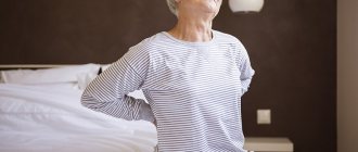
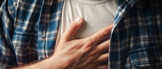
Spinal diseases affect 70% of the world's population. Pain reduces social activity and leads to temporary disability. The process can occur in different parts of the spine. Often the cause of ailments is destruction of the lower back, which characterizes lumbosacral radiculitis.
Causes
- degenerative changes: osteochondrosis (protrusion, disc herniation), spondyloarthrosis, vertebral displacement (spondylolisthesis, spondylolysis)
- vertebral compression fractures due to osteoporosis or trauma
- problems with posture (scoliosis, kyphosis, lordosis);
- inflammatory process (spondylodiscitis, spondyloarthritis, rheumatoid arthritis, abscess)
- tumors;
The process can be triggered by: physical activity, hypothermia, lifting (carrying) heavy objects, a sedentary lifestyle and excess weight, pregnancy.
Which doctor should I contact?
Neurologists are involved in the diagnosis and treatment of radiculitis; if necessary, other specialized specialists are involved - vertebrologists, orthopedic traumatologists, and neurosurgeons. Complex therapy can be carried out with the participation of a chiropractor, physical therapist and reflexologist.
Symptoms
The main place among the symptoms of lumbosacral radiculitis is pain. Classification by type of pain syndrome:
- 1. Lumbago. This is a reflex spasm of the paravertebral muscles. Characterized by acute pain in the lower back. Often occurs during (after) physical activity.
- 2. Lumboischialgia. This is a reflex spasm of the paravertebral muscles and piriformis muscle (usually on one side) irradiating to the gluteal region.
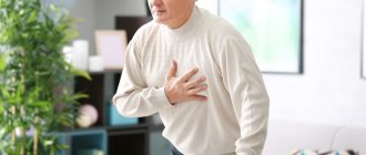
- 3. Lumbar radiculopathy: pain in the lumbar region with irradiation along the affected radicular nerve, sensory and motor disturbances in the area of nerve intervention. The upper segments 1-3 are the perineum, genitals, 4-5 and 1 cross are the lower limbs. In the chronic course of the disease, sensory and motor impairments may no longer recover, a chronic pain syndrome may form, the most dangerous complication is spinal canal stenosis with pressure on the spinal cord or spinal cord roots (cauda equina). Paresis of the lower extremities and pelvic dysfunction develop. This complication requires an emergency consultation with a neurosurgeon and a solution to the issue of surgical intervention and decompression of the spinal canal.
Radiculitis lumbosacral
12024 June 29
IMPORTANT!
The information in this section cannot be used for self-diagnosis and self-treatment.
In case of pain or other exacerbation of the disease, diagnostic tests should be prescribed only by the attending physician. To make a diagnosis and properly prescribe treatment, you should contact your doctor. Lumbosacral radiculitis: causes of occurrence, what diseases it occurs with, diagnosis and treatment methods.
Definition
Lumbosacral radiculitis, or radiculopathy, is a symptom complex that occurs as a result of pinching and inflammation of the spinal nerve roots in the lumbar and/or sacral spine.
Typically, this condition develops in people over 45, but in recent years, young people have also begun to complain of sciatica.
People whose work involves heavy physical labor or prolonged stay in one position, professional athletes are susceptible to the development of radiculopathy; patients with autoimmune diseases; those who are overweight and/or have metabolic diseases.
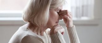
Radiculitis is characterized by lower back pain combined with shooting or pulling pain along the side or back of the leg (sometimes down to the foot), numbness and paresthesia, muscle weakness, increased pain when raising a straight leg.
Varieties of radiculitis
Radiculopathy can result from mechanical damage or an infectious process. Damage can develop both in the intervertebral discs and directly in the vertebral body or its processes.
Pain with radiculitis can be sharp (lumbago), shooting or stabbing (sciatica), burning or aching (lumboischialgia).
- Lumbago is an acute pain that appears suddenly as a result of sudden lifting of weights, sneezing, coughing, due to hypothermia of the lower back (for example, under air conditioning) or spasms of the lower back muscles, or intervertebral hernias.
- Sciatica is a shooting or stabbing pain that radiates to the buttocks, thighs, and calf muscles, as a result of which the patient may complain of muscle weakness (“legs can’t hold up”) and even numbness of the limbs.
- Lumboischialgia is a burning or aching pain that is localized in the gluteal muscles and thighs. It is characterized by a gradual increase.
Possible causes of sciatica
- Hernia or protrusion (bulging) of the intervertebral disc.
- Spondylosis is a chronic degenerative lesion of the spine that develops as a result of aging, overload or injury.
- Spinal stenosis.
- Osteophytes are growths of bone tissue that resemble hooks or spines in shape.
- Sacralization or lumbarization of the sacrum.
- Spondyloarthrosis is a chronic disease of the intervertebral joints, manifested by degenerative changes in cartilaginous tissues and leading to a decrease in the height of the intervertebral discs, tightening of the ligaments, and deformation of the intervertebral joints.
- Ankylosing spondylitis (Bechterew's disease).
- Vertebral fractures (including compression fractures due to osteoporosis).
- Hormonal changes that reduce the strength of cartilage and bone tissue.
Which doctors should you contact if you have lower back pain
? If you have signs of radiculitis, you should consult a doctor for examination and clarification of the diagnosis. In the future, he is engaged in the treatment of radiculitis.
Diagnostics and examinations for radiculitis
Before prescribing instrumental examinations, the doctor collects anamnesis and conducts physiological tests to identify symptoms of “tension” - specific signs of pathology of the nerve roots.
To diagnose spinal diseases the following is prescribed:
- X-ray of the lumbosacral spine with functional tests;
Diagnostics
To establish the level of damage, topical diagnosis is of great importance. The main radicular syndromes are presented in the table: [6]
| Spine | Sensory impairments | Movement disorders | Reflexes |
| C3, C4 | Shoulder girdle | Diaphragm | |
| C5 | Anterior shoulder, deltoid region | Deltoid muscle and partially biceps brachii muscle | Decreased reflex from the biceps brachii muscle |
| C6 | Radial surface of the shoulder and forearm, thumb | Triceps brachii, pronator teres, pectoralis major, often pollicis eminence muscles | Decreased or absent reflex from the biceps brachii muscle |
| C7 | Middle and index fingers | Small muscles of the hand, especially the eminence of the little finger | Decreased or absent reflex from the triceps brachii muscle |
| C8 | Little finger | Quadriceps femoris | Decreased reflex from the triceps brachii muscle |
| L3 | Anterior thigh | Quadriceps femoris, tibialis anterior | Decreased knee reflex |
| L4 | Medial surface of the leg | Extensors of the big toe | |
| L5 | Medial surface of the foot, big toe | Foot flexors | Decreased or lost Achilles reflex |
| S1 | Lateral surface of the foot, little toe |
Radiculopathy requires a CT or MRI scan of the affected level of the spine. In order to assess the level of research, it is necessary to find out the symptoms of the disease, neurological status during admission. If the level of the lesion cannot be determined, electromyography is prescribed, which helps to target the affected root, but does not allow the cause to be determined.
If neuroimaging does not reveal atomic changes, it is necessary to study the cerebrospinal fluid to exclude infectious and inflammatory causes, as well as determine the level of glucose in the blood to exclude diabetes.
FAQ
Is it possible to warm the lower back with a heating pad if you have radiculitis?
During an exacerbation of radiculopathy, it is recommended to keep the lower back warm. However, warming with a heating pad, a “blue lamp”, heated salt or sand is not recommended. This may increase swelling of the roots and lead to increased pain. You can use special belts, including those made of dog hair, etc.
I have a stomach ulcer. What painkillers can I take for sciatica?
Many drugs used for exacerbation of radiculopathy are not recommended for patients with a history of gastric and duodenal ulcers. Only a doctor can prescribe adequate treatment after examination. Don't self-medicate!
An MRI of my spine revealed a herniated disc. Should I delete it?
Indications for surgery to remove a herniated disc can be determined by a doctor during a consultation. Hernias most often do not require surgical intervention. It is necessary to observe a special protective regime, carry out preventive treatment and engage in physical therapy.
III. Examination for radiculopathies
It is carried out both to clarify the diagnosis and to exclude diseases that are potentially life-threatening for the patient (for example, tumors, metastases, spinal fractures and others) and to select the optimal treatment tactics.
Most often prescribed:
- radiography (spondylography) with functional tests,
- electroneuromyography
- computed tomography (CT)
- magnetic resonance imaging (MRI),
- Ultrasound diagnostics
- osteoscintigraphy,
- Dopplerography of blood vessels (for symptoms of the cervical spine),
- densitometry
- additional types of examination (blood tests, serological tests, abdominal organs, radiography of the skull, pelvic bones, etc.)
What is an intervertebral hernia
If a tear forms on the spinal disc, and it itself shifts away from the spinal column, resulting in inflammation and swelling, this is an intervertebral hernia. It is often a consequence of osteochondrosis. It can be identified by the following symptoms:
- the pain is pinpoint, the location of the lesion is clearly defined, and is poorly relieved by painkillers;
- movements are constrained (for example, a turn cannot be performed in the usual volume);
- with physical activity the pain increases sharply;
- atypical reflexes when trying to stand up.
There are several stages of this disease: disc prolapse, protrusion, extrusion, sequestration.
Pathogenesis
The onset of the development of the disease is two factors that are related to each other: mechanical irritation of the root and/or spinal ganglion and inflammatory changes in the perineural tissue that occur as a result of penetration of the disc into the epidural space.
In this case, factors of root compression can be both disc herniations and bone growths (uncovertebral, spondyloarthritic). Compression can also be caused by hypertrophied ligaments and periarticular tissues, vascular structures (epi- and subdural hematomas, arteriovenous malformations, epidural hemangiomas).
Until now, “blank spots” remain in the pathophysiological concept of radicular pain. It is assumed that the basis of radicular pain is axonal dysfunction caused by various etiological factors, including neural compression, ischemia, damage by inflammatory and other biologically active substances. Spinal roots (unlike peripheral nerves) have a weak blood-neural barrier, making the axon more susceptible to compression injury.
Increased vascular permeability due to mechanical compression of the root leads to endoneurial edema. As a result, a precedent arises that prevents full capillary blood supply and the formation of interneural fibrosis. The spinal root receives up to 58% of its nutrition from the surrounding cerebrospinal fluid (CSF). Perineural fibrosis prevents the complete supply of axonal tissue with nutrients due to diffusion from the CSF, which also contributes to increased sensitivity of the fiber to pressure.
Studies using experimental root compression have shown that even at minimal pressure (5–10 mm Hg) venous blood flow ceases.
The occlusion pressure of the radicular arterioles is significantly higher (approximately corresponds to mean blood pressure), but depends on the potential for venous stasis. Ischemia of nerve fibers or venous congestion leads to biochemical changes that can maintain pain sensations.
Work with experimental root compression demonstrates that compensatory diffusion of nutrients from the CSF is impaired in the setting of epidural inflammation or in the presence of fibrosis.
Recent studies have shown that degenerative changes in the nucleus pulposus and annulus fibrosus can lead to local neural changes and the synthesis of algogenic agents such as metalloproteinases, tumor necrosis factor (TNF), interleukin (IL)-6 and prostaglandin E2. Pathogenetically, the pain syndrome in radiculopathy is of a mixed nature, including nociceptive and neuropathic components.
Preventive measures:
- active lifestyle, regular sports and swimming;
- avoiding hypothermia and excessive stress;
- correct load distribution when lifting and carrying heavy objects;
- balanced healthy diet;
- proper organization of the workplace, purchasing ergonomic furniture;
- timely seeking help for diseases of the musculoskeletal system.
At the CELT clinic they know how to return you to normal life! If you are tired of enduring pain, contact us!
Make an appointment through the application or by calling +7 +7 We work every day:
- Monday—Friday: 8.00—20.00
- Saturday: 8.00–18.00
- Sunday is a day off
The nearest metro and MCC stations to the clinic:
- Highway of Enthusiasts or Perovo
- Partisan
- Enthusiast Highway
Driving directions
Non-drug treatment
Non-drug methods are used after the acute symptoms have subsided, when there is no longer severe pain. Their task is to strengthen the muscle corset, improve blood supply to the lumbosacral region and stimulate recovery processes. Help speed up recovery:
- physiotherapy – laser, ultraviolet, magnet, electrophoresis with painkillers, hormones, therapeutic mud;
- massage sessions using soft techniques to avoid re-pinching the nerve;
- special exercises that the physical therapy instructor will show help relieve excess stress from the spine and improve posture;
- reflexology;
- manual therapy;
- traction traction using dry or underwater methods.
Drug treatment
Non-steroidal anti-inflammatory drugs (NSAIDs) are prescribed to relieve acute pain, inflammation and muscle spasms. These are drugs based on diclofenac, nimesulide, ibuprofen, meloxicam. The choice of dosage form depends on the intensity of the symptoms: severe pain is eliminated with intramuscular injections, moderate pain can be relieved with tablets. In the acute period, local agents - ointments, gels and creams - can additionally be used. The second choice drugs are steroids - Hydrocortisone, Dexamethasone, Diprospan. They have a powerful anti-inflammatory effect. If NSAIDs with hormones are ineffective, then therapeutic blockades with Lidocaine and Novocaine are performed.
Antispasmodics and muscle relaxants (Mydocalm) help relax spasmodic muscles. If swelling is present, diuretics and magnesium sulfate are prescribed. Swollen tissues put additional pressure on the nerve, so removing excess fluid greatly alleviates the condition. To enhance the effect of analgesics and normalize the functioning of the nervous system, sedatives are added. Treatment of lumbar radiculitis also includes B vitamins, which improve nerve conduction. The most commonly used are Milgamma and Neuromultivit. If the disease is of non-infectious origin, then ointments with a warming effect can be prescribed - based on turpentine, snake and bee venoms, camphor. They relieve pain due to local irritant action.
Prevention
Preventive measures to prevent radiculitis are simple:
- competent organization of sleeping space. A comfortable bed is an elastic mattress of medium hardness and a small pillow of medium thickness. The ideal choice is orthopedic bedding;
- a complete and balanced diet will not only provide the body with healthy vitamins and minerals, but will also help maintain normal weight;
- adequate physical activity. Hard work is bad for your back, so if possible, you should reduce the load. Physical inactivity also threatens with negative consequences for the spine, and you should exercise at least once a week.
Doctors at our center have been working with patients suffering from radiculitis for many years. With timely treatment, the prognosis is always favorable. With us you will receive complete treatment, starting from examination and diagnosis, and ending with advice on lifestyle correction.
Surgical intervention
Operations for diseases of the spine
- Cost: 100,000 - 250,000 rubles.
- Duration: 40-60 minutes
- Hospitalization: 2-3 days in hospital
More details
Surgical intervention is carried out exclusively according to indications:
- lack of effect from conservative treatment;
- inhibition of tendon reflexes;
- feeling of weakness in the lower limb;
- decreased sensitivity of the lower limb.
Neurosurgeons at the CELT multidisciplinary clinic perform gentle operations through micro-incisions or punctures while preserving all supporting structures of the spine. Three-dimensional modeling of the operation and planning of each stage are carried out in advance. The process uses microsurgical and endoscopic techniques, laser and ultrasound, which allows our patients to quickly recover and return to work within two to three weeks after surgery.
Modern treatment of radiculitis at RAMI
If surgical methods of assistance are not required, then modern outpatient treatment of radiculitis is based on the causes of damage to the roots. If the causes are infectious, then appropriate antibiotics or antivirals, anti-inflammatory drugs, immunomodulators, and then vitamins and metabolites are used. If the cause of radiculitis is injury or impaired blood flow (ischemia), then the basis of treatment will be rest, immobilization (wearing corsets and reclinators), anti-inflammatory drugs, vascular agents and vitamins.
When radiculitis occurs from compression of the root by a herniated disc, then decompression is carried out (relieving pressure on the root), removing swelling from the nerve, optimizing the blood supply to the root, and also strengthening the structure of the discs. All these effects are provided by complex drugs.
The therapeutic procedure of introducing “Heel” drugs into the area of the damaged root is called biopuncture. In this case, a “cocktail” of 2-3 drugs is injected into the area of the damaged root with a special thin needle. A course of treatment requires no more than 6 procedures with an interval of 1-2 days at the beginning, and 1 week at the end of treatment.
The effectiveness of biopuncture is so high that patients often forget about their symptoms after just a couple of days. After this, rehabilitation methods are used - physiotherapy (MILT, TENS, BRT), manual methods (manual therapy, massage, osteopathy), reflexology (acupuncture, micro-acupuncture, moxa therapy, laser puncture), as well as a course of chondroprotectors and restorative drugs. Practice shows the greatest effectiveness of this set of methods in the treatment of radiculitis.
If you are really looking for your doctor...
Reviews of doctors providing the service – Lumbar radiculitis
In 2000, Andrei Arkadyevich performed spinal surgery on me.
Four days in the clinic and I have been living a full life for 20 years without restrictions on movement and I remember with gratitude Dr. A.A. Khodnevich. God bless him. And in 2000 he could walk no more than 10 meters. Read full review Viktor Alexandrovich
20.05.2020
Low bow to Alexander Semenovich Bronstein and Andrei Arkadyevich Khodnevich. I arrived at CELT on July 2, 2021 with extreme pain that I endured for 10 days. Hernia C6-C-7. I was given two blockades in Ivanovo, about 9 complex IVs, I lost 6 kg in a week and was in a panic, I didn’t see a way out and nothing happened to me... Read full review
Elena Nikolaevna L.
20.10.2019