The term “urticaria” covers a heterogeneous group of diseases and conditions, united by similar symptoms and one primary skin element - a blister.
In 40% of cases, urticaria is accompanied by the development of angioedema - swelling of the deep layers of the dermis, subcutaneous tissue and submucosal layer, with virtually no involvement of the superficial layers of the skin. Some patients may develop isolated angioedema without urticaria.
Epidemiology
Some types of urticaria affect 15–25% of the population, with acute urticaria (lasting less than 6 weeks) accounting for more than 60% of all cases. In 30% of cases, urticaria becomes chronic and recurrent. Acute urticaria develops more often in children and adolescents, chronic urticaria in adults, more often in women 20–40 years old. In 20–35% of patients with chronic urticaria, symptoms persist for the next ten years. In 30–50% of cases, chronic urticaria is caused by some kind of autoimmune disease.
In most cases, the cause of chronic recurrent urticaria in adults remains unknown.
Classification, types of urticaria and pathogenesis
A wheal is a local swelling of the dermis, usually characterized by itching, limited swelling of the skin, and hyperemia.
The blister completely resolves within a relatively short time. In addition to dividing urticaria into acute and chronic, there is a division into types depending on the factors that provoke exacerbation. Different forms of urticaria can occur simultaneously in the same patient. In addition, a number of conditions have been described that were once classified as urticaria, but are not such, or contain urticaria/angioedema as one of the symptoms of the clinical picture:
- Urticaria pigmentosa (mastocytosis) is a disease caused by the accumulation and proliferation of mast cells in tissues.
- Polymorphic skin rashes, including urticarial ones, are characteristic.
- Urticarial vasculitis - vasculitis with skin rashes - nodules and blisters, angioedema. After resolution of the elements, hyperpigmentation may be observed.
- Nonhistaminergic angioedema, such as hereditary angioedema, often associated with defects in the complement or kinin system.
- Exercise-induced anaphylaxis/urticaria.
- Cryopyrin-associated periodic syndrome (in the clinic there is an urticarial rash, fever, arthralgia or arthritis, increased fatigue and headaches. Includes familial cold autoinflammatory syndrome (Muckle-Wells syndrome (with the development of sensorineural hearing loss and renal amyloidosis) and infantile multisystem inflammatory disease .
- Schnitzler syndrome is a chronic urticaria with monoclonal IgM gammopathy.
- Gleich syndrome is episodic angioedema with eosinophilia.
- Wells syndrome is a granulomatous dermatitis with eosinophilia.
The key point in the pathogenesis of urticaria is the release of mast cell mediators (histamine, serotonin, chemotactic factors, arachidonic acid metabolites) and the development of their effects: vasodilation, increased permeability of the vascular wall with the appearance of edema and hyperemia; smooth muscle spasm; phenomena of itching and burning.
Classification of the disease
There are two forms of urticaria:
- acute (duration is about 6 weeks),
- chronic (over 6 weeks).
- There are different types of immunological urticaria:
- Urticarial vasculitis.
- Allergic
- Compliment addict.
- Autoimmune.
- Physical is divided into:
- mechanical: vibration, dermographic;
- temperature: thermal or cold contact;
- allergies that develop under the influence of other extraneous factors.
- There are also the following forms of urticaria:
- Contact.
- Aquagenic.
- Adrenergic.
- Idiopathic.
- Urticaria caused by non-IgE-mediated mast cell degranulation.
- Cholinergic.
- Drug-induced urticaria with development mechanisms different from those previously described.
Clinic: course and symptoms of urticaria
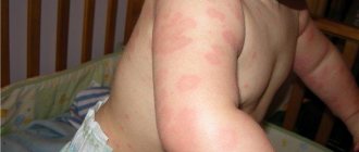
Usually, the development of urticaria begins with itching, followed by the appearance of characteristic rashes - blisters, clearly limited, rising above the surface of the skin, of various shapes and sizes, sometimes confluent, with a color ranging from pale pink to intense hyperemia (sometimes with clearing in the center). The lifetime of the rash is from 30 minutes to 1–2 days, the resolution is complete. In most cases, the rashes come and go - they can last for several hours in one place and then disappear and reappear in another. When urticaria occurs in a chronic form, rashes often occur in the evening, which should be taken into account when planning a visit to the doctor. The general condition, as a rule, does not suffer, but prolonged itching certainly reduces the quality of life.
Angioedema is characterized by diffuse swelling of loose subcutaneous connective tissue in the area of the dorsal surface of the hands and feet, eyelids, lips, genitals, and mucous membranes; asymmetrical swelling and slight itching (usually a burning sensation or pain, bloating), the superficial skin may appear unchanged. Swelling of the neck, pharynx and larynx can lead to breathing problems and dysphagia; swelling of the intestinal wall - cause vomiting, diarrhea, abdominal pain. Resolution of the elements takes longer - up to 72 hours.
In the case of allergic urticaria associated with a reaction to physical factors, there is usually an association between the exposure and the appearance of the rash. The localization and type of elements will have their own characteristics.
So, with cold urticaria, a rash appears in those places that have been in contact with cold objects or cold air. Urticaria remain relatively short-lived and disappear when warmed.
With dermographic urticaria, the elements often look like linear blisters along the course of scratching. Rashes with aquagenic urticaria occur after contact with water of any temperature and look like small urticaria against the background of erythematous spots. Fortunately, this is a rare form of the disease.
Causes
Often, rashes are the result of an allergic reaction of the body to unusual products or common allergens: dust, animal hair, cosmetics, medications, insect bites, cold or heat, chemical components.
The rash can also appear against the background of other diseases. Urticaria occurs in people with problems of the gastrointestinal tract, kidney and liver pathologies, hormonal disorders, and tumor processes. The immune system reacts with nettle fever to infection with bacterial, viral and fungal infections, as well as parasites.
Anamnesis
Given the ephemeral nature of urticaria rashes, it is extremely important to properly collect anamnesis. The doctor must understand whether it is urticaria, what provokes it and what mechanisms support it. Therefore, you should pay attention to the following points:
- description of the elements and associated sensations (itching, burning, pain);
- life expectancy of elements and the presence of secondary changes - hyperpigmentation, erosions, crusts, etc.;
- the cyclicity of the appearance of elements, the duration and frequency of the disease. For women, it is worth paying attention to the connection with the phases of the menstrual cycle;
- triggers, including medications, food intake, insect bites/stings, as well as physical factors and stress;
- previous therapy and its effectiveness;
- presence of allergic diseases in personal and family history;
- the presence of concomitant somatic pathology, including those associated with chronic urticaria, infectious diseases, autoimmune diseases, non-infectious chronic inflammatory diseases, especially involving the gastrointestinal tract, paraneoplasia;
- professional activity/hobby.
Why do sun allergy symptoms occur?
The appearance of photodermatitis is promoted by internal and external causes.
Internal causes include chronic diseases of the liver and adrenal glands, some metabolic disorders, and malfunctions of the immune system. It should also be noted that people with fair skin and children are prone to photodermatitis.
External causes are the effects on the body of substances that can be divided into three large groups:
- Furocoumarins are special substances secreted by plants such as hogweed, parsnip, ash, parsley, dill, St. John's wort, and can cause allergies. Bergamot, lavender, musk, and sandalwood oils also have an allergenic effect.
- Components of cosmetics - eosin, para-aminobenzoic acid, retinoids, boric and salicylic acids, phenol, mercury compounds.
- Medicines - sulfonamides, barbiturates, aminazine, some antibiotics (doxycycline, tetracycline), some cardiovascular drugs (amiodarone, Trazicor), cytostatics, oral contraceptives, some non-steroidal anti-inflammatory drugs (indomethacin, ketoprofen, naproxen, meloxicam, diclofenac, lornoxicam) , both in the form of tablets and in the form of ointments, creams and gels.
Cosmetic peeling, permanent make-up or tattoos, even temporary ones, can also trigger photodermatitis.
Inspection
If a patient comes to an appointment with a rash, then it is necessary to understand how similar what he sees is to a urticarial rash. The presence of elements other than a blister, as well as atypical secondary elements - crusting, erosion, persistent hyperpigmentation - casts doubt on the diagnosis.
Also, during the examination, you should pay attention to the signs of atopic diseases and other somatic pathologies. If you suspect a connection between physical factors and the development of urticaria, provocative tests are informative. The doctor can apply streak stimulation with a spatula, apply heated or cooled objects, apply vibration sources, irradiate with light of different wavelengths, offer physical exercises and hang weights from the patient’s limbs, followed by assessment of the skin.
Additional research:
- skin test when using autologous serum;
- rheumatological tests;
- nasal swab for the presence of eosinophils;
- TSH, T3, T4, antibodies to thyroid peroxidase and thyroglobulin;
- culture of throat flora, throat culture for sensitivity to antibiotics and bacteriophages;
- stool analysis for sensitivity to bacteriophages and dysbacteriosis;
- antibodies to helminths, as well as roundworms and lamblia;
- coprogram;
- HBsAg, AT to hepatitis C, RW, AT to HIV;
- ultrasound examination of the thyroid gland;
- Ultrasound of the abdominal cavity;
- consultation with a gastroenterologist to identify H. pylori, endoscopy, consultation with a neurologist, ENT specialist (VSD);
- ECG;
- if urticarial vasculitis is suspected, a dermatovenerologist performs a skin biopsy.
Laboratory and instrumental diagnosis of urticaria
In addition to general clinical studies, tests should be performed to look for concomitant somatic pathology. Often, patients in this category have diseases of the gastrointestinal tract and hepatobiliary system of infectious and non-infectious origin, autoimmune diseases, including autoimmune thyroiditis, which is widespread today. At the same time, examination results often lie within the reference values, and it is difficult to associate the appearance of urticaria with any concomitant pathology.
Differential diagnosis is carried out with a number of diseases, such as urticarial vasculitis, Dühring's dermatitis herpetiformis, erythema multiforme exudative, contact urticaria, as well as blood-sucking insect bites.
Diagnosis of sun allergy
If you notice an unusual reaction to the sun, I recommend contacting an allergist or dermatologist, who will prescribe you a certain range of analyzes and tests that can be used to confirm the presence of an allergy or a predisposition to it.
It is impossible to fight a disease without eliminating its causes, so be prepared to be offered a comprehensive examination:
- skin tests (scarification tests, prick tests, patch tests). Skin allergy tests are the fastest, safest and most reliable method of examination in immunology. Testing is carried out for patients from 5 to 50 years old;
- determination of the level of total immunoglobulin E;
- determination of the level of specific immunoglobulin E to household, epidermal, pollen, fungal, food, medicinal and bacterial allergens, as well as insect poison;
- determination of the level of specific immunoglobulins to food antigens - diagnosis of food intolerance;
- examination of sputum and secretions from the nasal cavity.
Traditional therapy
How to treat hives? The first step is to eliminate the causes and triggers of the disease if possible. Refusal of medications that provoke rashes, an attempt to avoid the influence of provoking physical factors, as well as adherence to a hypoallergenic diet can reduce the frequency of relapses of the disease, but all this has rather low effectiveness without drug therapy.
An important task is the rehabilitation of foci of chronic infection and correction of concomitant somatic pathology. Sometimes these measures are enough to prevent relapses.
The main drugs for urticaria, with which drug therapy begins, are, of course, H1-histamine receptor blockers. According to the recommendations of the World Allergy Organization, therapy should begin with second-generation antihistamines.
If symptoms persist for 2 weeks, then it is permissible to increase the dose to four times and continue taking it for up to 4 weeks. A sedating H1 blocker may be prescribed at night. If ineffective, it is recommended to change the drug or add antileukotriene drugs. During exacerbations, a short course of systemic glucocorticosteroids (3–7 days) is acceptable.
If manifestations of urticaria/angioedema persist, the issue of switching to 2nd line drugs must be resolved - glucocorticosteroids, immunosuppressants (cyclosporine), monoclonal antibody drugs (omalizumab). In the treatment of urticaria pigmentosa, as well as urticaria caused by physical factors, the stabilizer ketotifen is used mast cell membranes.
Angioedema (with the exception of hereditary forms associated with defects in the complement system or kinin system) is treated according to the same principles. In case of life-threatening conditions associated with the risk of asphyxia (swelling of the larynx, tongue), emergency treatment with the administration of adrenaline is required; emergency intubation or tracheostomy may also be necessary.
Photodermatoses
Ending. Read the beginning of the article in No. 5.
Xeroderma pigmentosum (atrophoderma pigmentosum)
A rare, hereditary disease characterized by increased photosensitivity, the development of pigmentation and skin atrophy, photophobia, neurological symptoms, a progressive course and a very high risk of developing skin tumors.
Increased sensitivity to ultraviolet radiation is caused by impaired repair of DNA damaged by UV radiation.
The type of inheritance is autosomal recessive, possibly autosomal dominant.
The disease develops in newborns or early childhood.
The first symptoms are photodermatitis, photophobia, conjunctivitis, lacrimation. Later, pigment spots such as freckles and lentigines, telangiectasia, hyperkeratosis, areas of depigmentation, and dry skin appear on open areas of the body. Atrophic changes with foci of sclerosis lead to the formation of microstoma, narrowing of the nasal openings, thinning of the ears and the tip of the nose. Already in childhood, actinic keratosis, keratoacanthosis, basal cell skin cancer, melanoma, angio- and fibrosarcoma, etc. develop.
The prognosis is unfavorable. No treatment has been developed.
Phototoxic (photodynamic) reactions are caused by the presence in the body of substances that have a photosensitizing effect.
These substances may be of endogenous or exogenous origin. Often, topical and systemic drugs act as substances with photodynamic action (
).
A photosensitizing effect can also be caused by products of coal tar, shale, and oil; the juice of some plants, for example, the umbrella plant - hogweed, bergamot oil, etc.
After discontinuation of the drug that caused photosensitivity, increased sensitivity of the skin to UV radiation may persist for several months, which may be important for making a diagnosis of the disease.
Depending on the concentration of the photosensitizer, exposure to sunlight and the intensity of irradiation, the manifestations and severity of the phototoxic reaction may vary.
There are three types of phototoxic reactions:
1) immediate erythema and urticaria; 2) delayed burn-type reaction, developing after 16–24 hours and later; 3) delayed melanin hyperpigmentation, after 72–96 hours.
The rashes are localized on the skin of open areas of the body: on the forehead, cheekbones, nose, ears, side and back surfaces of the neck, the front surface of the chest, and the extensor surface of the limbs. In this case, there is a sharp boundary between healthy and affected skin, corresponding to the edge of clothing, a watch strap, etc.
However, in some cases there may be atypical localization of the rash. So, with good local immunity and the presence of a tan, not all open areas of the body may be affected. On the other hand, rashes can also be localized in closed areas, if the protective effect of clothing is insufficient.
When in contact with plants that cause photosensitization of the skin (phytodermatitis, meadow dermatitis), the boundaries of the erythema clearly follow the contours of the leaves in the contact areas.
A typical example of endogenous sensitization is porphyria (translated from Greek as purple dye).
This is a group of diseases that are hereditary or with a hereditary predisposition, varying in severity, clinical manifestations and prognosis, having a common feature - a violation of porphyrin metabolism and accompanied by an increased content of porphyrins or their precursors in the body.
Porphyrins are pigments that are derivatives of porphyrin and perform an important function in the life processes of animals and plants. Porphyrins are aromatic compounds that easily form complexes with metals and have intense colors.
In the body of a healthy person, intermediate products of porphyrin synthesis are contained in small quantities. Clinical forms of porphyrin metabolism disorder (porphyria) are accompanied by changes in tissue content and increased excretion of free porphyrins. Genetically determined porphyrins are characterized from a biochemical point of view by a deficiency of the enzyme that catalyzes one of the porphyrin metabolism reactions. Determination of the content of porphyrins in the blood, urine, feces and enzyme activity is a reliable diagnostic test for porphyria. Porphyrins are determined by fluorimetric and spectrophotometric methods.
In addition to genetically determined causes, impaired porphyrin metabolism is observed in liver diseases (cirrhosis), some poisoning (lead), as well as in patients with malignant tumors. Such conditions do not apply to porphyrias.
Erythropoietic protoporphyria is caused by a defect in the ferrochelotase enzyme, resulting in overproduction of protoporphyrin and its accumulation in the bone marrow, red blood cells, and increased excretion in bile and feces. It was first isolated from the group of porphyrias in 1961 by J. Magnus et al. Inherited in an autosomal dominant manner. It is the most common of the erythropoietic porphyrias and in most cases is benign. Skin lesions are caused by the photodynamic effect of protoporphyrin. The toxic effect of protoporphyrin is less pronounced than that of uroporphyrin I. The disease usually develops in early childhood, often in the first year of life. The clinical picture is characterized by increased sensitivity to solar radiation with a wavelength of 400 nm or more. A few minutes after exposure to the sun, a phototoxic reaction similar to a sunburn develops in exposed areas of the skin, burning, stabbing and burning pain, severe swelling and erythema appear. The photoresponse decreases 24–36 hours after exposure. With prolonged exposure to the sun, hemorrhagic rashes are observed. Occasionally, blisters and bullous rashes with hemorrhagic contents form at the burn sites, which ulcerate and leave small superficial scars. A burn in such patients can even occur through window glass and synthetic underwear, since rays of 400 nm or more pass through them freely.
Some patients develop a chronic eczema-like reaction. Over time, the skin of exposed skin thickens, becomes rough, and shagreen-like.
Developing in early childhood, the disease for a long time can manifest itself only as subjective disorders. Complications include hypochromic anemia with a high iron content in the blood serum, a tendency to form stones in the gall bladder, and in rare cases, the deposition of a huge amount of porphyrins in the liver with the development of liver failure and cirrhosis.
The prognosis is generally favorable. Over the years, photosensitivity becomes less pronounced. Liver cirrhosis may develop, but death is rare.
Treatment involves reducing the skin's sensitivity to sunlight. Carotenoids, vitamin E, nicotinic and lipoic acids, Riboxin, ATP, methionine are prescribed. Sunlight should be avoided. Photoprotective drugs are ineffective, so clothing that protects from the sun is recommended.
Urocoproporphyria (porphyria cutanea tarda). The disease was first identified as an independent clinical form in 1937 by J. Waldenstrom. The pathogenesis is based on deficiency of the enzyme uroporphyrinogen decarboxylase, inherited in an autosomal dominant manner and manifested under the influence of unfavorable factors, primarily hepatotoxic (alcohol, lead, heavy metals, arsenic, previous hepatitis, malaria, long-term use of estrogens, barbiturates, griseofulvin , tetracyclines, antidiabetic, antituberculosis and sulfa drugs).
The disease is characterized by severe skin symptoms, which develop in people over 40 years of age. The main signs are increased sensitivity to mechanical injury and solar radiation with the formation of blisters and scars on exposed skin. Hypertrichosis in the temporomygomatic region, increased growth of eyelashes and eyebrows, and darkening of hair are often observed. Continuous hyperpigmentation or hyperpigmented spots of dirty gray or brown color can be combined with scleroderma-like skin changes resembling diffuse scleroderma, as well as with foci of dystrophy.
Microcysts in the form of milia-like elements are found in long-term sufferers of porphyria cutanea tarda on the skin of the back of the hands and fingers, face, ears, and back of the head. Sometimes there may be nail lesions in the form of subungual hyperkeratosis, deformation and complete photoonycholysis.
Biochemically, an increased amount of iron in the blood serum, saturation of the liver parenchyma with iron, siderosis of hepatocytes and Kupffer cells, as well as liver damage of varying severity are determined. Increased lipid peroxidation under the influence of UV irradiation leads to the formation of free radicals and the occurrence of a free radical chain reaction. To neutralize free radicals, an enzymatic (superoxide dismutase, catalase, peroxidase, glutathione reductase) and non-enzymatic (tocopherol, sulfide groups) antioxidant defense system is activated. A decrease in the activity of these enzymes and tocopherol correlates with the duration and severity of cutaneous porphyria tarda and leads to an increase in the final product of lipid peroxidation - malondialdehyde, an increase in iron levels, and destruction of membrane lipids.
In urine and blood plasma, the concentration of uroporphyrin and coproporphyrin increases, with more uroporphyrin than coproporphyrin. Uro- and coproporphyrin also increases in feces.
Treatment consists primarily of stopping the consumption of alcoholic beverages and eliminating other provoking factors (estrogens, barbiturates, other drugs that have a porphyrinogenic effect).
Good results are observed with treatment with deferoxamine (Desferal), which forms complex compounds with iron. Intramuscular administration of 0.5–1.0 g of the drug in the form of a 10% solution once a day for 15 days promotes the excretion of iron and iron-containing proteins.
Chloroquine and hydroxychloroquine (Plaquenil) allow you to achieve long-term remission, but side effects such as retinopathy, agranulocytosis, nausea, vomiting, etc. are possible.
The use of Delagil and its analogues is not recommended.
Treatment with antioxidants is effective - Beta-carotene in combination with contaxanthin (Fenoro) or alpha-Tocopherol acetate 100 mg in the form of an oil solution in capsules every other day, alternating with methionine (0.5–0.75 g/day).
It is advisable to treat with Unithiol in the form of a 5% solution intramuscularly, 5 mg 2 times a day for 10 days, Carbolen, activated carbon orally.
Hereditary coproporphyria
An autosomal dominant disease caused by a defect in coproporphyrinogen oxidase. Its clinical manifestations are similar to variegated porphyria, but its course is milder. The most common symptom is abdominal pain. Neurological and mental disorders are less common, photosensitivity is low, increases during attacks, often in the form of bullous photodermatosis and mild skin vulnerability.
During an exacerbation, an increased amount of delta-aminolevulinic acid and porphobilinogen is found in the urine (lower than in acute intermittent porphyria). The amount of coproporphyrin in urine and feces is sharply increased. During the period of remission, the amount of delta-aminolevulinic acid and porphobilinogen may be normal with an increased content of coproporphyrin in the urine and feces.
The disease is rare and often occurs latently.
Treatment is the same as for variegated porphyria. It is necessary to avoid provoking medications and hepatotoxic agents. The prognosis is favorable.
Photoallergic reactions
The mechanism of antigen formation and the initial stages of the development of photoallergic reactions are not clear enough. In this regard, two theories are discussed.
According to the first (Willis J., Kligman AM, 1968), a chemical substance, by adsorbing light energy, forms a stable photoproduct, which, when combined with a protein, forms an antigen.
The second theory (Harber LC et al., 1966) suggests that irradiation generates free radicals, the high reactivity of which allows them to conjugate with epidermal proteins and form a complete antigen.
Considering the tropism of many medications for the skin, the possibility of the formation of conjugates with proteins under the influence of light is quite likely under any route of administration of the substance into the body. The free radical theory of antigen formation under the influence of sunlight is supported by a decrease in the activity of antiradical defense, an increase in lipid peroxidation products, a change in the spectrum of membrane lipids, a decrease in the natural antioxidant tocopherol, etc.
A number of signs make it possible to distinguish phototoxic reactions from photochemical ones.
Diseases whose pathogenesis involves photoallergic processes include the type of diseases described below.
Photodermatosis polymorphic
In the domestic literature, it is customary to distinguish two independent pathologies - solar prurigo and solar eczema. In foreign literature, it is customary to consider these diseases as a variant of polymorphic photodermatosis.
The pathogenesis of this disease is not fully understood, but the involvement of the immune system is undoubted. There is a significant increase in the total number of B lymphocytes in the peripheral blood and an increase in IgA in more than 50% of cases.
Photodermatosis can appear in people of any age.
Polymorphic photodermatosis can appear in people of any age, most often in 10-30 year old women.
The onset of the disease usually occurs suddenly. Between sun exposure and clinical manifestations, 7–10 days may pass if there is no repeated exposure to the sun. The course is wavy - it worsens 1 day after being in the air on a sunny day, and in subsequent days it subsides.
Clinic. Clinical signs of the disease are pruriginous or vesicular rashes located on open areas of the skin. There may be the development of conjunctivitis and cheilitis. The course of the disease is seasonal: rashes appear in the spring and stop with the onset of autumn. With exacerbation, the rash may spread to closed areas of the skin.
A characteristic feature is a pronounced polymorphism of the rashes - small lichenoid papules, large ones - of the prurigo type, can resemble various granulomas - including annular, can occur of the eczematous type, urticarial rashes.
The most characteristic type of rash is pink-red papules, 0.2–1.0 cm in diameter, located on an erythematous background. When papules coalesce, plaques are formed that resemble lesions of discoid lupus erythematosus. Due to severe itching - a large number of excoriations and hemorrhagic crusts. In some patients, papulovesicular rashes predominate on open areas of the skin, accompanied by eczema-type weeping.
There may be clinical manifestations of a mixed nature with a predominance of either papules or vesicles.
The diagnosis is made on the basis of the clinic, anamnesis, and laboratory data (relationship with solar radiation, localization on open areas of the skin).
Treatment:
1. Photoprotection. 2. Antioxidants - carotenoids, alpha-Tocopherol acetate 100 mg orally every 1 day, methionine 0.25 g 3 times a day, every other day, Teonicol 0.15 g 3 times a day.
In the acute period - external corticosteroid ointments.
Erythema solar persistent
Rare photodermatosis. The pathogenesis of the disease is associated with photoallergy to drugs, plants and other substances.
The clinical picture is characterized by persistent erythema of a bluish-red color, with clear boundaries in open areas of the skin with rounded or irregular outlines.
The diagnosis is made on the basis of the clinic and anamnesis - a characteristic picture of the lesion, itching, burning, connection with solar radiation, seasonal dynamics in the form of weakening or disappearance of the process in winter, resumption - in the spring, after insolation.
Differential diagnosis: lupus erythematosus, actinic reticuloid.
Treatment. Resistance to treatment is noted. Positive dynamics are known under the influence of Aevit (1 capsule/day), Presotsil (start with 6 tablets per day, followed by a dose reduction to 1 tablet per day). Externally - steroid ointments with Unna cream (1:1).
Lightpox
The disease was first described in 1862 as a bullous skin lesion that occurs on exposed skin and is associated with sun exposure. At first, the diseases were associated with photosensitivity to porphyrins. However, further clinical observations showed that the typical picture, as a rule, is not accompanied by porphyrinuria.
Currently, the view on the pathogenesis of this disease is controversial. According to some authors, some clinical forms are associated with impaired porphyrin metabolism, others are not. There is evidence of a decrease in urocanic acid, a protective factor, in the epidermis.
The clinical picture of the disease begins after sun exposure in the first years of life and is characterized by seasonality. There are cases of morbidity among relatives, as well as cases of spontaneous recovery.
Characteristic rashes are small and medium-sized blisters, dense, with transparent contents, an umbilical depression in the center (the contents can be purulent or hemorrhagic).
The blisters are covered with a brown crust, after which, when rejected, a smallpox-like scar is formed. The rash is accompanied by burning, itching, and there may be malaise and increased body temperature.
Localization of rashes on the ears and cornea can lead to deformation of the ears and clouding of the cornea. Extensive erosions filled with pus are possible on the lips. In some cases, the rash may be weakened and leave no scars.
Differential diagnosis should be made with porphyrias (congenital, late), solar prurigo, necrotic acne.
1. Detection of porphyrins in blood and urine. 2. Sensitivity to UV rays for smallpox is characteristic of long wavelengths - 290–320 nm, porphyria - 400 nm. 3. Acne is not related to sun exposure.
Treatment:
1. Photoprotection. 2. Complamin (Teonikol) and Lipamide. 3. Aevit 1 capsule per day. 4. Beta-carotene.
Actinic reticuloid
The disease is a chronic dermatosis, combined with severe photosensitivity and histologically resembling lymphoma. Only men over 50 years of age are affected. The pathogenesis is unknown. A very rare disease.
Clinic . The outbreaks are localized in open areas and have clear boundaries. On an erythematous background there are papular elements of a pink-red color, merging with each other, forming edematous plaques with fine-plate peeling. There may be follicular papules up to 0.5 cm in diameter, pink-red in color with a dense consistency, and itching. Lymph nodes are not enlarged. Mucous membranes, hair, and nails are not affected.
Histologically, massive acanthosis is determined. In the stratum corneum there is parakeratosis, in the spinous layer there is edema, exocytosis, single microabscesses.
In the dermis there is an infiltrate of lymphocytes and histiocytes. There are large cells with hyperchromic nuclei, reminiscent of “mycosis”.
The diagnosis is made based on a combination of clinical and morphological changes. The disease is benign and usually does not transform into lymphoma.
Treatment . A short-term effect is observed when using corticosteroids, cytostatics, and antimalarial drugs.
The best results come from:
1) use of photochemotherapy; 2) a constant increase in UV exposure produced a clinical effect; 3) a positive result was observed from the combined use of prednisolone + hydroxychloroquine + azatnocrine (Haynes HK, 1984).
Solar urticaria
A relatively rare photodermatosis, significantly different from other photodermatoses. Pathogenesis has not been studied enough. However, the possibility of passive transfer with serum of hypersensitivity to light served as the basis for the assumption of the participation of immunoglobulins in the mechanisms of the occurrence of rashes.
Clinic . Immediately after irradiation, a reaction appears in the form of rashes, which depend on the exposure: short-term - small, pink-red itchy urticarial elements, with prolonged insolation - large blisters of pale pink color with a red border along the periphery.
Urticaria disappear within 15–30 minutes, erythema after 2–3 hours.
Gradually, in open areas, rashes stop appearing with repeated irradiation, but in closed areas, exposure to sunlight is immediately accompanied by a response.
Treatment . Antihistamines are selected individually. Antimalarial drugs. Dosed UV irradiation in a gradual increase in dose to create resistance to sunlight.
Literature
- Skripkin Yu. K. Skin and venereal diseases, M., 2000.
- Fitzpatrick T., Johnson R. et al. Dermatology, atlas-reference book. M., 1999.
- Brawan J., Andersen W., Lee J., Labadie R., Solares G. Photoaging versus intrinsic aging: a morphologic assessment of facial skin // J Cutan Pathol. 1995; 22 (2): 154–159.
- Dalle Carbonare M., Pathak MA Skin photosensitizing agents and the role of reactve oxygen species in photoaging // J Photochem Photobiol. 1992; thirty; 14 (1–2): 105–124.
- Daynes RA Phenotypic and physical characteristics of lymphoid cells involved in then immunity to sunergeneic UV-induced tumor // J Immunol. 1979; 122(6):2458–2464.
- Egbert Ch. Die acuten Lichtschaden // Kosmetik Journal. 1986.
- Egbert Ch. Die chronischen Lichtschaden der Haut // Kosmetik Journal. 1986.
- Finkel P. Sun protection factor and UVA protection labeling in Europe // Euro Cosmetics. 1999; 4:34–38.
- Goldfarb MT, Ellis CN, Weiss J. S, Voorhees JJ Topical tretinoin for photoaged skin // Cutis. 1989; 43 (5): 476–82.
- Kligman AM Current status of topical tretinoin in the treatment of photoaged skin // Drugs Aging. 1992; 2 (1): 7–13.
- Kligman AM, Grove GL, Hirose R., Leyden JJ Topical tretinoin for photoaged skin // J Am Acad Dermatol. 1986, Oct; 15 (4 Pt 2): 836–59.
- Kligman LH Photoaging. Manifestation, prevention ion and treatment // Clin Geriatr Med. 1989; 5 (1): 235–251.
- Krutmann J. UVA-Strahlung auch Kanzerogen // Dermaforum. 1999: 6; 6.
- Ley RD Ultraviolet radiation-induced malignant melanoma in Monodelphis domestica // Photochem Photobiol. 1989; 50 (1): 1–5.
- Stuzin JM, Baker TJ, Gordon HL Treatment of photoaging. Facial chemical peeling (phenol and trichloroacetic acid) and dermabrasion // Clin Plast Surg. 1993; 20 (1): 9–25.
- Thiele JJ, Schroeter C., Hsieh SN, Podda M., Packer L. The antioxidant network of the stratum corneum // Curr Probl Dermatol. 2001; 29: 26–42.
N. F. Yarovaya , Candidate of Medical Sciences, Associate Professor
GOU DPO RMAPO , Moscow
Table 2.
New trends
The effectiveness of treating urticaria with alternative groups of drugs is currently being studied. Among them: antidepressants, androgens, slow calcium channel blockers (nifedipine), M-anticholinergic blockers, sulfasalazine, methotrexate, colchicine.
The use of plasmapheresis and phototherapy in patients with solar urticaria is actively discussed. Separately, it is worth dwelling on the issue of the use of biological agents. The idea of binding and inactivating molecules involved in immune responses has always seemed extremely attractive in the search for a cure for hives. The appearance in practice of monoclonal antibodies (MAbs), which make it possible to solve this problem with high specificity and selectivity for target molecules, gave rise to a number of biomedical studies that studied the possibility of using MAbs for the treatment of immune-mediated diseases, including allergological diseases.
Currently, mAbs against immunoglobulin E, the drug omalizumab, are registered in Russia. The drug binds to IgE, blocks its interaction with receptors on mast cells, which prevents their subsequent degranulation; there is also a decrease in the number of IgE receptors on the surface of basophils. Omalizumab, originally used to treat severe atopic asthma, later showed its effectiveness in the treatment of chronic urticaria. However, scientists do not rest on this and continue to search for new biological agents to treat different types of urticaria.
Symptoms of urticaria
- Blisters can appear as pinpoint or large-focal, sometimes they merge with each other.
- A blister, the primary element in the manifestation of a skin rash, is a local swelling of the papillary dermis, which occurs as a result of accelerated blood flow, vasodilation, and increased vascular permeability.
- Blisters may also appear as cavityless exudative ephemeral elements. Dense, swollen, bright pink in color, they can rise above the skin level and have different shapes (large scalloped, rounded, etc.), different sizes (diameter - from 0.5 to 10-15 cm) and often a pale area in the center. Blisters may disappear without a trace (even after a few minutes).
- Sometimes there may be a burning sensation or a blister may be accompanied by unpleasant itching of the skin.
- Inflammation can occur anywhere. But most of all skin rashes appear in places where skin and clothing come into close contact: under straps, belts, and suspenders.
- The duration of the visual presence of the rash can range from several minutes to a day.
- In half of the patients, urticaria is accompanied by angioedema (Quincke's edema).
If swelling and rash have spread to the surface of the mucous membranes of the gastrointestinal tract, nausea and vomiting have appeared, this indicates a possible threat to human life and requires immediate intervention by a specialist.
Very often, urticaria is not treated and the disease is not given due importance. However, it is important to know that hives can be a signal of the appearance of diseases of the internal organs or some other serious infection in the body.
Do not forget about the predisposition to urticaria. This will be indicated by increased sensitivity of the skin and white stripes that remain after light scratching (white dermographism). If hidden infections are detected in time, the immune system will be able to function normally. It is important to assess the condition of the internal organs, being aware that if their functioning is disrupted, skin diseases may occur.
Forecast
Often, an episode of acute urticaria may be the only one in a patient's life. If the patient is less fortunate and the allergy becomes chronic, then spontaneous remission can be expected in 50% of cases.
Potentially life-threatening are laryngeal edema with urticaria and the acute form of the disease with anaphylactic reactions. For patients with cold urticaria, swimming in cold water can be fatal due to asphyxia or systemic reactions with a drop in blood pressure.
Treatment of the disease at the Mama Papa Ya clinic
Clinic "Mama Papa Ya" is located in Lyubertsy. Here we see qualified doctors of various specialties who will provide assistance to patients with urticaria:
- in the clinic you can do the necessary tests and undergo the necessary examination;
- can be treated for both adults and children;
- consultations with specialists in various fields provide the most complete treatment for urticaria;
- We offer affordable prices for medical services and convenient work hours.
To make an appointment, call the phone number listed on the website or through the form provided.
Reviews
Good clinic, good doctor!
Raisa Vasilievna can clearly and clearly explain what the problem is. If something is wrong, she speaks about everything directly, not in a veiled way, as other doctors sometimes do. I don’t regret that I ended up with her. Anna
I would like to express my gratitude to the staff of the clinic: Mom, Dad, and me. The clinic has a very friendly atmosphere, a very friendly and cheerful team and highly qualified specialists. Thank you very much! I wish your clinic prosperity.
Anonymous user
Today I had a mole removed on my face from dermatologist I.A. Kodareva. The doctor is very neat! Correct! Thanks a lot! Administrator Yulia Borshchevskaya is friendly and accurately fulfills her duties.
Belova E.M.
Today I was treated at the clinic, I was satisfied with the staff, as well as the gynecologist. Everyone treats patients with respect and attention. Many thanks to them and continued prosperity.
Anonymous user
The Mama Papa Ya clinic in Lyubertsy is very good. The team is friendly and responsive. I recommend this clinic to all my friends. Thanks to all doctors and administrators. I wish the clinic prosperity and many adequate clients.
Iratyev V.V.
We visited the “Mama Papa Ya” Clinic with our child. A consultation with a pediatric cardiologist was needed. I liked the clinic. Good service, doctors. There was no queue, everything was the same price.
Evgeniya
I liked the first visit. They examined me carefully, prescribed additional examinations, and gave me good recommendations. I will continue treatment further; I liked the conditions at the clinic.
Christina
The doctor carefully examined my husband, prescribed an ECG and made a preliminary diagnosis. She gave recommendations on our situation and ordered additional examination. No comments so far. Financial agreements have been met.
Marina Petrovna
I really liked the clinic. Helpful staff. I had an appointment with gynecologist E.A. Mikhailova. I was satisfied, there are more such doctors. Thank you!!!
Olga
{loadmodule formrec}
How is allergic urticaria treated?
First of all, when allergic urticaria develops, the effect of the allergen should be eliminated. In case of contact allergies, remove the irritating substance from the surface of the skin; in case of an insect bite, remove the sting, stop eating or taking medication, take enterosorbent (activated carbon, Enterosgel), and, if possible, rinse the stomach.
Before seeking medical help, you can take an antihistamine and use an ointment that does not contain hormones. There is no need to try to treat yourself; further prescriptions should be made by a doctor, since uncontrolled use of hormonal and other potent drugs can lead to negative consequences.
Medical care consists of a more precise selection of allergy medications that have minimal side effects, the prescription (in severe cases, with chronic urticaria) of systemic corticosteroids, and in some cases, immunosuppressants. Source: Modern principles of treatment of acute and chronic urticaria. Skorokhodkina O.V., Klyucharova A.R. Practical medicine No. 7, 2012. p. 45-49.
Emergency care for a patient with allergic urticaria is required when Quincke's edema develops. If it occurs, you should immediately call an ambulance. In order to alleviate the patient’s condition before the ambulance arrives, you need to calm him down, provide an influx of fresh air, and unfasten clothing that is restricting breathing. To relieve swelling, doctors provide the necessary assistance on site, including adrenaline injections and tracheal intubation, then the patient is taken to the hospital, where further treatment is carried out.