In this article we will tell you:
- Causes of viral conjunctivitis
- Classification of viral conjunctivitis
- Adenoviral conjunctivitis
- Herpetic conjunctivitis
- Epidemic keratoconjunctivitis
- Epidemic hemorrhagic conjunctivitis
- Differences between viral conjunctivitis and bacterial and allergic
- Treatment and prevention of viral conjunctivitis
More than 45 different viruses can cause conjunctivitis, researchers say. This disease causes severe discomfort, most often affects children, but often occurs in adult patients.
In terms of prevalence, viral conjunctivitis is inferior to bacterial conjunctivitis. However, due to its high contagiousness, outbreaks of the disease are extremely difficult to control: isolated cases of viral conjunctivitis quickly develop into epidemic ones, entailing various socio-economic consequences - from the introduction of quarantines to a sharp increase in temporary disability.
From this article you will learn about what viral conjunctivitis is, how to distinguish it and react correctly if suspicious symptoms appear.
Viral conjunctivitis
is an inflammatory disease of the conjunctiva, the membrane lining the outer surface of the eyeball and the inner surface of the eyelids, caused by a virus.
Symptoms of conjunctivitis
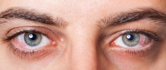
The main signs of inflammation of the conjunctiva include:
- Redness and swelling of the affected eye;
- Itching (especially in cases where conjunctivitis is allergic in nature);
- Burning sensation in the eye, pain, feeling of a foreign object;
- Fear of bright light;
- Tearing;
- Purulent or watery discharge from the eye (typical of conjunctivitis caused by a viral infection).
At the first signs of illness, you should consult a doctor!
Development of the disease
The clinical picture of conjunctivitis occurs in 4 stages:
- the initial signs of the disease resemble the symptoms of ARVI (weakness, headache, fever, loss of appetite, lethargy, sleep disturbance, cough, runny nose, sore throat);
- unilateral damage to the conjunctiva of an inflammatory nature, accompanied by its redness and increased lacrimation;
- the eyelids swell, mucous discharge appears, and there is a sensation of a foreign body in the orbit;
- Photophobia develops, lymph nodes enlarge, blisters and follicles form, which cause burning, itching and pain.
It is at the last stage that there is a high probability of determining the type of conjunctivitis and making the correct diagnosis.
Causes of conjunctivitis
- Bacteria. The most common “culprits” of bacterial conjunctivitis are staphylococci, gonococci, streptococci and chlamydia.
- Viruses. Inflammation of the mucous membrane of the eye is most often caused by adenoviruses and herpes viruses.
- Allergies. Almost everyone who suffers from seasonal allergies, that is, hay fever, has encountered allergic conjunctivitis. In this case, rhinitis joins conjunctivitis.
- Mushrooms. The cause of the inflammatory process is pathogenic fungi, which can be obtained mainly from the external environment - from food, soil, plants and animals.
- Physical and chemical factors. Exposure to radiation or irritating chemicals can also cause conjunctivitis.
Most often, conjunctivitis is allergic or infectious (viral), but to prescribe adequate treatment, the ophthalmologist needs to be sure of the nature of the disease.
To treat conjunctivitis, an ophthalmologist needs to identify the cause of its occurrence.
Differences between viral conjunctivitis and bacterial and allergic
Diagnosis of viral conjunctivitis is usually based on laboratory results. However, there are characteristic signs of each of the forms that allow the specialist to make an assumption about the nature of the disease after examination.
Differences between viral conjunctivitis
So, viral conjunctivitis differs from the bacterial form:
- The nature of the discharge.
- The degree of swelling of the eyelids: with the viral form the swelling is less.
- Changes in the lymph nodes.
With viral conjunctivitis, the discharge only sometimes contains small patches of pus, while the discharge with bacterial conjunctivitis is mostly purulent.
Typically, bacterial conjunctivitis does not affect them. The lymph nodes react to the viral form by becoming enlarged and painful.
Allergic conjunctivitis is also different from viral conjunctivitis. With allergies, the discharge is more abundant, viscous, resembling mucus. The lymph nodes are not tense, but the swelling is much stronger. In addition, allergic conjunctivitis is accompanied, unlike viral conjunctivitis, by severe itching.
Diagnostics
To diagnose conjunctivitis, it is enough for an experienced specialist to conduct an external examination of the eyes. It is much more difficult to determine the nature of the inflammation, so additional research may be required to determine the cause of the disease:
- Blood and urine analysis;
- Collection and examination of eye secretions;
- Culture of material obtained from a smear;
- Analysis of antibodies in blood serum;
- Allergy diagnosis – contact with the suspected allergen and analysis of the body’s reaction.
Survey
Diagnosis is carried out by an ophthalmologist and includes the following steps:
- Collection of complaints. The specialist finds out what symptoms bother the patient - discomfort, itching, burning, lacrimation, decreased vision.
- History of the disease. Duration of discomfort, contact with infectious diseases or allergens, existing diseases, connection with the change of season, reaction to exposure to sunlight, etc.
- Eyesight check. The results are compared with those obtained previously and help track the dynamics of the disease.
- Objective examination. Biomicroscopy is performed using a slit lamp and reveals hyperemia and swelling of the conjunctiva, injection of the eyeball, and the presence of discharge.
- Lab tests. They are not always prescribed by a doctor to identify the causative agent of conjunctivitis in case of ineffective treatment, frequent relapses, or drug intolerance.
- Special ophthalmological tests The Shimmer test is performed if keratoconjunctivitis sicca is suspected to clarify the intensity of tear production. The end of a strip of filter paper is placed behind the edge of the lower eyelid and after 5 minutes, see how wet it is.
- The fluorescein drop test is used to identify associated corneal damage that temporarily changes color when exposed to the dye.
The ophthalmologist takes a smear from the conjunctiva with a cotton swab from the fold between the back surface of the eyelid and the eye.
If demodicosis is suspected, the eyelashes are also examined under high magnification to detect microscopic insects.
In exceptional cases, with allergic conjunctivitis, allergic skin, nasal, conjunctival or sublingual tests are performed. Various allergens are introduced into the body one by one and they see which one reacts to it.
If a specific cause of the disease is suspected, consultation with other specialists may be recommended.
What can be confused with
Differential diagnosis of conjunctivitis is carried out with the following diseases:
- Keratitis. Characteristic symptoms are photophobia, pain and the desire to keep the eyelids closed. Due to swelling and clouding of the cornea, vision is blurred.
- Episcleritis and scleritis. Painful sensations are more pronounced, especially when moving the eyeball.
- Uveitis (iritis, iridocyclitis, choroiditis). Characterized by pain in the eye, photophobia, constriction of the pupil and blurred vision. There is no separation.
- Acute attack of angle-closure glaucoma. Swelling cornea, pain in the eye and in half of the head on the same side, dilated pupil, sharp deterioration of vision. There are no allocations.
- Foreign body of the eye. Usually the patient remembers the moment of injury. There is pain and discomfort, a desire to blink, rub the eye, and lacrimation. Without treatment, an infection may develop.
- Canalicular obstruction with dacryocystitis. Redness is not expressed, profuse lacrimation, when pressing with a finger just below the inner corner of the eye, pus may be released.
- Barley. Limited redness, itching and swelling of the eyelid, an increase in its thickness in the projection of inflammation. Before the abscess breaks, there is no discharge.
In each described case, the nature of the redness is different and is determined by the ophthalmologist during eye biomicroscopy.
Treatment of conjunctivitis
There is no universal treatment; each type of conjunctivitis is treated differently.
The drugs for viral therapy are selected by an ophthalmologist. To relieve symptoms and prevent complications, the doctor will also recommend special eye hygiene during illness and medicinal compresses.
For bacterial conjunctivitis, antibiotic drops will be prescribed. On average, bacterial conjunctivitis resolves within a week.
If conjunctivitis occurs due to allergies, antihistamines will be prescribed first. If possible, it is necessary to reduce contact with the allergen. At the discretion of the ophthalmologist, topical corticosteroids may be prescribed to help reduce inflammation. To combat symptoms, your doctor may recommend cold compresses on the eyes.
Find your nearest optical store
If you use contact lenses, you will have to give them up for a while.
What forms of conjunctivitis occur in adults?
Both adults and children are characterized by the same forms of the disease. Optometrists classify conjunctivitis based on different symptoms. They can manifest themselves in different ways, but they all have one thing in common: inflammation of the visual organs. Other symptoms of conjunctivitis “accompany” the main symptom and allow one to distinguish one form of the disease from another.
Conjunctivitis is classified based on what symptoms appear in adults. First of all, the ophthalmologist needs to determine the type of disease. Based on the symptoms, doctors distinguish between acute and chronic forms of the disease. In the first case, signs of conjunctivitis appear almost immediately after a person is infected and develop rapidly. Most often, the cause of acute conjunctivitis is infectious pathogens. It is not always possible to begin timely treatment for this form of the disease. Symptoms of conjunctivitis appear very quickly, and it is not always easy for a person who has contracted this disease to notice the preconditions that precede the pathology. Chronic conjunctivitis does not manifest itself so quickly. Its occurrence is preceded by various symptoms, on the basis of which it is not easy to make a diagnosis. It may take a long time before chronic conjunctivitis in adults manifests itself. Typically, this form of the disease occurs due to exposure of the visual organs to dust, computer radiation and other factors that can lead to irritation of the mucous membrane. The chronic form of conjunctivitis is often a consequence of other ophthalmological pathologies.
Complications of conjunctivitis
Although at first glance conjunctivitis seems to be a fairly mild disease, complications can lead to serious consequences, including blindness.
Without the necessary treatment, conjunctivitis can become chronic. If the mucous membrane of the eye is constantly inflamed, sooner or later the process will spread to the cornea, and this may result in complete loss of vision.
The sooner you receive treatment, the easier it will be to deal with conjunctivitis. Therefore, at the first signs of illness, you should consult an ophthalmologist.
Prevention
To avoid infection in the eyes, it is enough to follow these rules:
- Do not touch your face and eyes with dirty hands.
- Wash your hands with soap and use a disinfectant before applying cream or cosmetics around the eyes or plucking eyebrows.
- Maintain a work-rest regime during intense visual work, and regularly do eye exercises.
- If foreign particles get into your eyes, they must be removed with a clean bandage or paper towel.
- When using contact lenses, follow the rules of hygiene, and at the first sign of inflammation, stop wearing them.
- Promptly treat dry eye syndrome, teeth, diseases of the nasopharynx, and facial skin.
Sources:
- Renee Solomon, MD. COVID-19 patients with conjunctivitis may shed viral particles in tears and conjunctival secretions. — American academy of ophthalmology, 2021.
- Melvin I. Roat, MD, FACS, Sidney Kimmel Medical College at Thomas Jefferson University. Overview of conjunctivitis. - MSD reference book, professional Russian version, 2021.
- Michael A Silverman, MD. Acute Conjunctivitis (Pink Eye). - English-language database of current medical data Medscape, 2021.
How is conjunctivitis transmitted?
Conjunctivitis is highly contagious unless it is allergic in nature. Infectious inflammation can be transmitted from person to person through coughing, sneezing, touching, or using common household items, for example, the same towel.
Conjunctivitis is transmitted by airborne droplets, touch and objects
The most dangerous period occurs in the first 48 hours, it is at this time that the spread of infection is maximum. Children of preschool and school age are especially susceptible to this. If your child gets conjunctivitis, you will have to miss at least two days of school, otherwise other children will also become infected.
The spread of the disease within a family also occurs very quickly, so it is important to take precautions and pay special attention to hygiene.
Physiotherapy
Physiotherapy for ptosis is a mandatory component of non-surgical treatment. It includes massage, special gymnastics and exercises to tone up.
Self-massage
The peculiarity of the impact of such a massage is the ability to work directly on the desired area, as well as the fact that you can do it yourself without outside help. Self-massage technique:
- Preparatory stage. Disinfect your hands.
- Working stage. Apply massage oil to the upper eyelid in the direction from the inner corner of the eye to the outer and in the opposite direction when treating the lower eyelid. Execution time: 2 minutes.
- Leading stage. Lightly tap with your fingertips around the eyes in the same direction. And so on for 3 minutes.
- Climax stage. Continuously apply light pressure with the cones of your fingers on the upper eyelid for 2 minutes.
- The final stage. Place cotton pads soaked in chamomile infusion on your eyelids. The exposure time of chamomile infusion is not limited.
It is recommended to perform massage in a calm environment, without being distracted by various external stimuli. It is advisable to be in a separate room without strangers.
Eyelid massage is one of the components of non-surgical treatment of ptosis.
Physiotherapy
The exercises included in this complex are especially useful for congenital ptosis. Standard treatment of this myogymnastics in some cases allows one to avoid surgery. A set of therapeutic exercises:
- With your eyes wide open, perform 5 circular movements, close your eyes, and do not open your eyes for about a minute. Repeat 4 times.
- Open your eyes as much as possible and hold them in this position for 10 seconds. Afterwards, close your eyes as sweatily as possible for 10 seconds. Do it 7 times.
- Use your index finger to stroke your eyebrows using massaging movements, gradually increasing the pressure. Continue for up to 5 minutes.
This exercise should be repeated daily in the morning and evening. It is better to do this after water procedures, when the muscles have already reached an active state.
Exercises to tone the muscles of the eyelids
Exercising to tone will not take much time, but it stimulates muscle function. Its advantage: you can perform the complex right at your workplace. All exercises are best done while sitting:
- Describe a circle with your eyes. The head remains motionless. Having completed the circle, look up, then down and in both directions. Repeat 6 times.
- Open your eyes wide for 10 seconds, then close your eyes as tightly as possible for 10 seconds. Perform 5-6 times.
- Tilt your head back and look at your nose for 15 seconds. And so 6-7 times.
It is advisable to exercise 4 times a day at equal intervals. For example, at 7.00 - in the morning at home, at 11.00 - at work, at 15.00 - at the end of the lunch break, at 19.00 - in the evening at home.
Hygiene for conjunctivitis
To reduce the risk, the patient must have his own household items - dishes, towels, bedding, cosmetics. After complete recovery, bedding, clothes and towels must be washed, and cosmetics used by the infected person will have to be thrown away (at least eye makeup products). Your ophthalmologist will tell you more about hygiene for conjunctivitis.
During treatment you will have to stop using contact lenses. If you have been wearing contact lenses since contracting conjunctivitis, be sure to replace the pair with a new pair.
All family members should wash their hands frequently with warm water and soap, even if you do not leave the apartment.
Instillations - how to instill drops correctly
For a speedy recovery, not only the correct selection of medications is important, but also their correct use. To safely instill drops, you must follow these instructions:
- Wash your hands thoroughly with soap and dry with a paper towel.
- Wash or dry your eyes before the procedure
- Warm the bottle with the drug in your hands, especially if you took it out of the refrigerator
- Prepare a small mirror to avoid hurting yourself.
- Take a comfortable position - sitting or lying on the bed.
- Tilt your head back.
- Pull down your lower eyelid with your finger to create a “pocket” that allows fluid to enter more easily.
- Take the bottle, turn it over and bring the tip to the eye without touching the eyelids, mucous membranes or eyelashes.
- During instillation, it is recommended to look up, but the tip should not go out of sight.
- Apply 1-2 drops of the medicine, trying to get as close to the outer corner of the eye as possible.
- Then close your eyes for 2-3 minutes to distribute the drug evenly.
- Dry your eye with a clean tissue.
Photo: correct instillation: lower eyelid retracted, look up, drip from the outer corner.
If there is a large amount of pus, the eyes should be rinsed first. If the doctor has prescribed several drugs for instillation, it is necessary to maintain an interval of 10-30 minutes between their use. Ointment is always used last.