Classification
The classification of foot and mouth disease is based on clinical manifestations, severity of symptoms and severity of the infectious process. A detailed classification of the disease with explanations is presented in the table.
| Sign classification | Form of foot and mouth disease | Comments |
| Clinical manifestations | cutaneous | characterized by damage to the skin, mucous membranes remain clean |
| mucous membrane | the main manifestation of the disease is aphthous stomatitis, when ulcerative rashes are observed only on the oral mucosa | |
| mucocutaneous | the most common form of infection, vesicular-ulcerative elements cover the mucous membranes of the mouth and nose, as well as the skin (mainly in the area of the fingers) | |
| Symptom severity | typical | occurs with pronounced characteristic symptoms |
| erased | manifests itself as slight stomatitis and mild malaise | |
| asymptomatic | There are no clinical manifestations of the disease | |
| Heaviness | acute | the severity depends on the patient’s age, the state of the immune system, and concomitant diseases; criteria for assessing the severity of the disease are the severity of general intoxication and the prevalence of rashes on the skin and mucous membranes |
| moderate severity | ||
| heavy |
Causes and sources of the disease
The disease develops due to the entry into the body of a virus of the genus Aphtovirus. The pathogen is stable in the external environment, remains viable on animal fur for up to 4 weeks, and on clothing for up to 3.5 weeks. Withstands exposure to low temperatures and drying, dies when boiled, ultraviolet irradiation, or treated with antiseptic solutions.
The source and reservoir of infection are wild and domestic ungulates, and some rodents. The pathogen is excreted by infected animals through milk, saliva, and feces. The disease is transmitted through contact and ingestion when caring for sick animals and eating contaminated foods. No cases of transmission of infection from a sick person to a healthy person have been identified.
Etiology
The causative agent of the disease is an RNA-containing virus that accumulates in the epithelium of ulcers on the skin, oral mucosa and in the lymph circulation system. In the first two days, at the earliest stages of the development of the disease, the pathogen can be detected in significant quantities on the surface of aphthae, and in smaller quantities in saliva, blood, and feces. There are seven types of virus in total, but the most common are:
- Strain O
- Strain, A
- Asia-1;SAT-2.
The situation is also aggravated by the fact that an animal that has been ill with one strain of the virus can again become ill with any other.
The virus lives outside the body for quite a long time. The virus survives in contaminated areas of soil for up to 150 days, on clothing and wool for more than a month, in feces for up to 170 days, and in water for up to three and a half months. The virus survives in cold milk for up to six weeks, but when heated to 40°C it dies within 12 hours; the virus does not survive in sour milk media.
When heated to 60°C, the virus dies in 15 minutes; when boiled, it dies instantly. The presence of caustic soda or formaldehyde in disinfectant solutions also has a detrimental effect on the virus.
Pathogenesis
The entry points for infection are the mucous membranes of the oral cavity, the digestive tract, or damaged skin. At the site of entry of the pathogen, a primary focus of infection is formed - a small bubble where the virus multiplies and accumulates. Then the pathogen penetrates the blood, causing a response from the body, manifested by symptoms of general intoxication (fever, headache and muscle pain, weakness). As the infection develops, new foci form - multiple blisters (aphthae) on the mucous membranes of the mouth, nose, urethra and skin.
How dangerous is the foot and mouth virus for humans?
Fortunately, foot and mouth disease does not have serious consequences for the human body. Only in rare cases may the following occur:
- myocarditis – inflammation of the muscular lining of the heart. Manifested by the occurrence of life-threatening rhythm disturbances and heart failure;
- sepsis is massive blood infection with the foot-and-mouth disease virus. Characterized by the appearance of fever, shock;
- pneumonia – inflammation of the lung tissue due to activation of secondary microbial flora (Haemophilus influenzae, pneumococcus, mycoplasma, chlamydia, etc.);
- meningitis is a severe lesion of the meninges.
Symptoms of foot and mouth disease
The incubation period for foot and mouth disease lasts from 2 to 12 days, on average 3-4 days. The onset of the disease is acute, often sudden. The body temperature rises sharply to 39-40 ºC, headaches and aches throughout the body appear. On the first day of illness, patients complain of a burning sensation in the mouth, increased salivation, and if the urethra is affected, pain when urinating. Subsequently, when examining the patient, the following signs of the disease are revealed:
- redness of the conjunctiva (the outer layer of the eyes);
- enlargement and tenderness of nearby lymph nodes;
- swelling and redness of the mouth;
- multiple small blisters with clear or cloudy contents on the mucous membranes of the mouth and the tip of the tongue;
- rashes in the folds between the fingers, near the nails.
After a day, the blisters open, forming ulcers that dry out and disappear without a trace 7-10 days after the onset of the disease. Full recovery occurs after 1-2 weeks, but with extensive damage to the mucous membranes and skin, the disease can drag on for up to a month.
Note! Foot and mouth disease is especially severe in children.
Often the main symptoms of the disease are accompanied by indigestion (abdominal pain, vomiting, diarrhea). In some cases, with aphthous fever in children, blisters may reappear.
Foot and mouth disease, features of the disease in humans
A. E. Kudryavtsev, Candidate of Medical Sciences
G. K. Alikeeva, Candidate of Medical Sciences
MGMSU, Moscow
Foot and mouth disease is a typical zoonosis, an acute infectious disease caused by a filterable virus, transmitted to humans from sick animals, characterized by the appearance of blisters and erosions on the oral mucosa (aphthous stomatitis), as well as on the skin of the hands and feet.
The causative agent of foot-and-mouth disease is a filterable ultramicroscopic virus that belongs to the Picornoviridae family. The foot and mouth disease virus is characterized by high variability both in laboratory and in natural conditions.
Foot and mouth disease has long been found in all parts of the globe, mainly in agricultural countries. The first scientific reports of foot-and-mouth disease in humans were published in Norway in 1764 by Sagar, who observed more than 1,500 cases of foot-and-mouth disease (aphthous foot-and-mouth disease).
The virus is stable in the external environment. In milk it remains for up to 25–30 hours (in refrigerator conditions - up to 10 days), in butter - up to 2 months, in sausages and corned beef - up to 50 days, in bran - from 2 to 5 months, in dried saliva ( on animal fur, on clothing) - from 1 to 3 months. The virus persists in frozen carcasses for up to 687 days. Ultraviolet (sun) rays and boiling (for 5 minutes) inactivate the virus.
The reservoir and source of infection are sick animals, especially cattle, as well as pigs, sheep and goats. Wild animals have also been noted to be susceptible to foot and mouth disease: moose, roe deer, reindeer, saigas, etc.
Young animals are especially seriously ill, among which there is a high mortality rate. In addition to the animals listed above, camels are susceptible to foot and mouth disease. Horses, dogs, and cats occasionally suffer from foot-and-mouth disease. Carriers can be rodents susceptible to this disease (gophers, rats, mice), as well as some birds (which themselves are not sick, but excrete the virus that came with food through the intestines) and the gadfly. Animals become infected through mutual contact on pastures, in barns and barns. Excreted from the body of sick animals with saliva, milk, manure, urine, and the contents of vesicles, the foot-and-mouth disease virus ends up on bedding, feed, and overalls of service personnel, where it can persist for a long time.
Animals with foot-and-mouth disease usually have characteristic aphthous rashes of vesicles and blisters (later ulcerating) on the mucous membranes of the mouth, nose, gums, tongue, lips, in the spaces between the hooves, and less often near the horns (“snout-hoof” disease). The udder of cows becomes covered with a rash, which is not without some epidemiological significance, although the milk itself becomes infectious before the appearance of canker sores and contains the virus for 10–12 days from the onset of the disease. Animals that have recovered from the disease exhibit virus carriage, which may not be accompanied by clinical manifestations. In the vast majority of cases, from the 10th to 12th day of illness, virus shedding stops. In cattle, the so-called malignant form of foot and mouth disease can be observed, which in approximately 60% of cases ends in death within 1–1.5 days from the onset of the disease. Animals that have recovered from foot-and-mouth disease acquire immunity for up to 1.5 years.
A person becomes infected with foot and mouth disease by consuming raw milk (more than 60% of cases) from sick animals (cows, goats), and less often by consuming meat from forcedly slaughtered sick animals. When contacting sick animals, people become infected in 30% of cases. Persons in certain professions are at risk of infection: milkmaids, cowgirls, calf workers, shepherds, workers in meat processing plants and slaughterhouses, veterinarians and livestock specialists.
The foot-and-mouth disease virus enters the human body through tiny lesions in the skin, as well as through the mucous membranes of the eyes, nose, mouth and gastrointestinal tract. Infection can also occur aerogenously by inhaling air containing virus particles. Children are most susceptible to foot and mouth disease.
Mechanical transmission of the foot-and-mouth disease virus by service and veterinary technical personnel (on shoes, clothing, hands) and infection of healthy animals is possible.
The possibility of infection of healthy animals from a sick person has been proven.
Major outbreaks of this disease in our country were observed in 1941–1943, 1952–1953 and 1965. In 2000–2001 An outbreak of foot-and-mouth disease emerged in England and the Netherlands with the threat of infection spreading to other European countries.
Outbreaks of foot and mouth disease among domestic animals have been registered in Kazakhstan (Mirnoye village, Karaganda region), China, North Korea, South Korea, and Vietnam.
A constantly unfavorable epidemiological situation regarding foot and mouth disease is observed in countries such as Iran, Turkey, and Afghanistan.
Having penetrated the human body, the foot and mouth disease virus begins to multiply in the epithelial cells of the mucous membrane or epidermal cells of the skin. In this case, an inflammatory reaction occurs in the form of vesicles filled with serous contents (primary affect), which made it possible for some authors to define foot and mouth disease as ectodermatosis (epitheliosis).
Subsequently, the virus from the sites of primary reproduction enters the blood, spreading throughout the body (generalization of the process), which is clinically manifested by symptoms of intoxication (fever, headache, etc.).
Dissemination of the virus is accompanied by damage to the mucous membranes of the mouth, tongue, nose, urethra and the skin of the hands, especially around the nails, in the interdigital folds. So-called secondary aphthae and skin lesions are formed in the form of bubbles - vesicles. Specific aphthae can also occur on the mucous membranes of the stomach, intestines and genitals. Generalization of infection is rarely accompanied by myocardial damage.
The transferred disease leaves a strong, but not long-lasting (1–1.5 years) species-specific immunity.
The incubation period lasts from 2 to 12 days (usually 3–8 days). In the vast majority of cases, foot and mouth disease begins acutely, suddenly, although gradual development of the disease is also possible. Severe chills, headache, malaise, and muscle pain appear, especially in the lumbar region. Appetite decreases sharply. Body temperature quickly rises to 38–40°C and remains at a fairly high level for 5–6 days. 1–2 days after the onset of the disease, inflammatory changes in the oral mucosa are observed (burning sensation in the mouth, pain when chewing, salivation). The process involves the mucous membranes of the lips, gums, cheeks, and larynx. The tongue is swollen. At the same time, conjunctivitis develops (sometimes one-sided), and pain appears when urinating (due to the development of urethritis). 1-2 days after this, when examining the oral cavity on the mucous membranes and especially on the edges and tip of the tongue, small bubbles (the size of wheat to lentil grains) with transparent and then cloudy yellowish contents can be detected. Sometimes there are so many bubbles that they are densely located on the oral mucosa, including the gums and palate. Within a day or two, the blisters burst on their own, and in their place are painful, irregularly shaped, bright red superficial ulcerations (aphthae), sometimes merging with each other. After opening the vesicles, the temperature, as a rule, decreases somewhat, but the patient’s well-being worsens. Speech and swallowing are difficult, salivation increases sharply, lips and tongue are swollen, covered with plaque and crusts. Regional lymph nodes swell and become painful. Bubbles and then ulcers may appear on the mucous membrane of the nose, the back wall of the pharynx, vagina, urethra, and conjunctiva. The patient is characterized by a bent posture and the need to resort to a towel due to literally flowing streams of saliva, as well as a pained facial expression and irritability.
In addition to damage to the mucous membranes, in most patients, vesicles appear on the skin, and for foot and mouth disease their most typical location is between the fingers and toes, as well as at the base of the nails. The bubbles are polymorphic, some of them can reach the size of a bulla. In addition, erythematous spots are sometimes observed not only on the extremities, but also on the torso. In some cases, the hands and feet become swollen, and patients feel a burning sensation, goosebumps, and itching in their area. Patients may also be in a forced position: fingers splayed, arms and neck bent, legs pressed together. In some patients, the nails subsequently fall out.
The rash period lasts about a week. Aphthae on the mucous membrane of the mouth, lips, and tongue usually heal quickly, within 3–5 days, without leaving scars. The temperature drops to normal levels, and a recovery phase begins, lasting an average of 10–15 days. However, in a number of patients, repeated eruption of blisters on the mucous membranes and skin may occur, delaying treatment of the disease for several months. In severe cases, a hemorrhagic rash may appear on the neck, chest, and back.
Changes in internal organs are usually not observed in uncomplicated foot and mouth disease. In children, the disease may be accompanied by the development of gastroenteritis.
The hemogram of foot-and-mouth disease at the height of the disease is characterized by eosinophilia, and in some patients leukopenia is noted.
In addition to clinically pronounced forms of foot and mouth disease, there are also erased forms without clinical manifestations, which can manifest themselves in the form of uncharacteristic stomatitis and general malaise, or be characterized by a moderate headache, malaise, accompanied by the appearance of characteristic vesicles in the interdigital folds, which open after 1–2 days and heal quickly. Asymptomatic forms occur without any clinical manifestations.
In the chronic, protracted course of the disease, skin rashes do not look like blisters, but tubercles, which resolve, and the skin above them does not peel off.
There is no generally accepted clinical classification of foot and mouth disease. The following clinical forms of foot and mouth disease are usually distinguished:
- cutaneous, in which a few aphthae are found only on the skin, usually in the area of primary penetration of the virus (this form is more common with occupational infection);
- mucous membrane, the main manifestation of which is aphthous stomatitis;
- mucocutaneous (most common), characterized by damage to the mucous membrane (oral cavity and eyes) and skin (mainly in the finger area).
In general, foot and mouth disease progresses favorably. The disease usually ends with recovery occurring in 2–3 weeks.
Very rarely, the disease is complicated by myocarditis, and due to the addition of secondary bacterial microflora - pneumonia and even sepsis. Virus carriage in foot and mouth disease can last up to 120–150 days after the illness, posing the threat of a new outbreak among animals.
The diagnosis of foot and mouth disease is based on the following data:
- epidemiological anamnesis, aimed at identifying the presence of a sick animal in a given area, cases of contact with sick animals, consumption of animal products (raw milk), as well as sour cream, cream, cottage cheese prepared from unsterilized milk of animals affected by foot-and-mouth disease;
- the presence of typical clinical manifestations of the disease (a combination of sudden onset, fever, intoxication, as well as characteristic changes in the oral mucosa - aphthous stomatitis - and skin in the area of interdigital folds, nail damage);
- laboratory data, for which virological and serological methods are used, as well as a bioassay.
Among the serological methods for diagnosing foot and mouth disease, RSK and RPGA are used, with the help of which antibodies to the foot and mouth disease virus are detected in the blood serum of patients. Recently, PCR has been used to quickly diagnose the disease.
The bioassay is carried out on guinea pigs, which are infected by scarification or by intradermal injection of the test material into the plantar surface of the hind legs. In the presence of the virus, after 24–36 hours, primary vesicles are formed at the site of infection, which open and turn into erosions. After another 1–3 days, secondary vesicles develop on the tongue and on the soles of the forelegs.
It should be remembered that there are also atypical forms of foot and mouth disease, which were mentioned earlier.
The differential diagnosis of foot and mouth disease should be carried out primarily with aphthous stomatitis, which is more common in young children. With aphthous stomatitis, there is no fever, symptoms of intoxication, excessive salivation and skin lesions. Only the oral mucosa is affected, the ulcers are deeper and never merge with each other, and their bottom is covered with a whitish coating. There is no eosinophilia in the blood.
Chickenpox, with which in some cases it is necessary to differentiate foot-and-mouth disease, is characterized by:
- autumn-winter seasonality;
- contact with patients with chickenpox or herpes zoster;
- a specific chickenpox rash is vesicular, involving the scalp and mucous membranes, the simultaneous presence of elements at different stages of development. With chickenpox, the rash is never located on the hands and feet. Vesicles on the oral mucosa and in the pharynx never ulcerate. Severe salivation is also not typical for chickenpox.
Foot and mouth disease also needs to be differentiated from enteroviral infections - herpangina and enteroviral exanthema (Boston or epidemic exanthema).
With herpangina, from the very first days of the disease, first small red papules appear on the mucous membrane of the soft palate, tongue and especially the arches, then characteristic vesicles (their size reaches the head of a pin), filled with transparent contents and not merging with each other even with a profuse rash (from 5– 6 to 30). The blisters open after 1–2 days, leaving behind small, quickly healing erosions. The patient's well-being changes little.
With enteroviral exanthema, the febrile state is complemented by the appearance of a rash on the skin of the face, torso and limbs. The rash is rubella-like, but can be maculopapular, petechial, ulcerous, or bullous. Sometimes the rash appears at the peak of the fever, but more often during its decline (fever lasts 1–8 days) and disappears after 1–2 days without peeling or pigmentation. Spotted exanthema may also appear on the oral mucosa. Disappears in the same time frame as the skin rash. During the acute period of the disease, pharyngitis and conjunctivitis often occur.
For the differential diagnosis of foot and mouth disease and the above-described forms of enterovirus infection, epidemiological history and laboratory tests are important.
Foot and mouth disease sometimes has to be differentiated from Lassa hemorrhagic fever. The disease, unlike foot and mouth disease, begins gradually - with chills, fever, and moderate headaches. The temperature reaches high numbers (39–40°C) by the 3rd–5th day of illness. Characteristic damage to the pharynx is ulcerative-necrotic changes on the tonsils and soft palate. On the 5th day, cough, chest and abdominal pain, nausea, vomiting and profuse watery stools appear. Flushing of the face, neck and generalized lymphadenopathy are noted. At the end of the 1st week, a mixed rash (petechiae, roseola, macules, papules) appears on the skin of the face, trunk and limbs. The liver is moderately enlarged. Patients develop hemorrhagic syndrome: hemoptysis, hemorrhages on the skin and mucous membranes, gastric and intestinal bleeding. Blood pressure drops and diuresis decreases. Infectious-toxic shock may develop.
Establishing a diagnosis of Lassa fever is helped by a correctly collected epidemiological history (the disease is epidemic in the countries of Western and Central Africa), blood tests (leukopenia, then leukocytosis, neutrophilia, increased ESR), urine (proteinuria, microhematuria, cylindruria, hyperazotemia), as well as virus isolation by serological methods (RSK, RNIF), PCR.
Patients with foot and mouth disease, regardless of the severity of the disease, must undergo treatment in a hospital, where they must remain for at least 14 days from the onset of the disease until complete clinical recovery, healing of ulcers on the mucous membranes and skin.
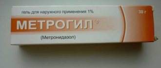
Antibiotics are ineffective for this disease. They are accepted only in case of secondary bacterial infection. Some effect was obtained when using interferon drugs and interferon inducers that have antiviral activity for the treatment of foot and mouth disease.
Proteolysis inhibitors (contrical, gordox, trasylol) and antioxidants (emoxipin, mexidol) have a beneficial effect on the body of a patient with foot-and-mouth disease, especially when combined into the lymphatic vessels or lymph nodes.
To reduce the degree of toxemia, patients with foot and mouth disease require detoxification therapy (hemodesis, rheopolyglucin, crystalloids).
Careful patient care and nutritional management (fractional meals with liquid or semi-liquid food) are important. The patient is given milk, cream, kefir, liquid porridge, slimy soups, etc. Patients need to be fed in small doses 6–7 times a day. In severe cases, when the patient is unable to eat, it is necessary to resort to tube feeding. Before eating, to reduce the intensity of pain due to the presence of aphthae in the oral cavity, a patient with foot and mouth disease is prescribed anesthesin powder (0.1 g) 20–30 minutes before meals. To rinse the mouth, use a 3% solution of hydrogen peroxide, a 0.01–0.1% solution of potassium permanganate, and chamomile infusion. To reduce pain, ointments containing anesthesin and novocaine are also used. During the healing period of aphthae, it is recommended to lubricate them with rosehip oil, sea buckthorn oil or carotoline. For eye damage, use a 30% solution of albucid.
It is also necessary to prescribe vitamins, and in severe cases, cardiovascular drugs.
To prevent the spread of foot and mouth disease among people, it is necessary to eliminate it among animals, which is achieved by establishing strict quarantine measures (fencing, disinfection of vehicles traveling outside the outbreak, etc.). Persons in contact with sick animals must work in special clothing. Milk must be boiled for at least 5 minutes before consumption, and meat from sick animals can only be used after thorough heat treatment. Premises are equipped for milk disinfection, processing and temporary storage. People working with animals sick with foot-and-mouth disease are prohibited from drinking water, eating or smoking in the area of infection. Pregnant women, teenagers and people with microtrauma to their hands are not allowed to work in farms unfavorable for foot-and-mouth disease.
Healthy animals are vaccinated. In some cases, if FMD is suspected, a herd of animals must be completely destroyed.
The article was published in the journal The Attending Physician
Diagnosis of the disease
The leading place in the diagnosis of the disease is given to laboratory methods. If FMD is suspected, patients are prescribed the following tests:
- a general blood test shows an increase in the level of eosinophils (a type of white blood cell) and ESR (erythrocyte sedimentation rate);
- bacteriological analysis of the contents of aphthae, saliva, feces reveals the presence of a virus in the biomaterial;
- Serological blood tests (complement fixation reaction, indirect hemagglutination reaction) show antibodies to the virus in the blood serum.
In some cases, to diagnose foot and mouth disease, a biological test is used by rubbing the contents of aphthae into the pads of the paws of guinea pigs. When the virus is present, animals develop rashes typical of infection.
Treatment of foot and mouth disease
Patients with suspected FMD are hospitalized in the infectious diseases department of the hospital. During the period of fever they are prescribed bed rest. The main therapeutic measures for foot and mouth disease are aimed at caring for the oral cavity and skin, relieving symptoms of intoxication.
With extensive damage to the oral cavity, patients eat semi-liquid, non-irritating food; in some cases, nutrition is provided through a tube or parenterally (intravenously). Anti-inflammatory ointments and physiotherapeutic methods (Ural irradiation, laser irradiation) are used locally. Depending on the symptoms, patients are prescribed antipyretic, analgesic and antiallergic drugs, vitamins and immunomodulators.
Complications of foot and mouth disease
Complications from foot and mouth disease develop quite rarely, mainly in children and people with weakened immune systems. As a rule, the cause of complications is the penetration of the virus into internal organs. Foot and mouth disease can lead to the following secondary infections:
- pneumonia - pneumonia;
- myocarditis – inflammation of the heart muscle;
- purulent diseases of the skin and mucous membranes;
- sepsis - general blood infection.
Note! To prevent complications, especially in children under three years of age, if even minor rashes appear on the skin or mucous membranes of the mouth, you should immediately consult a doctor.
Epizootic data
Artiodactyl animals are susceptible to the disease; cattle, camels and deer are more often affected. To a lesser extent, pigs and small animals are infected. In rare cases, domestic carnivores become infected (usually by consuming infected milk). Horses and birds do not suffer from foot and mouth disease. In laboratory conditions, rodents get sick. Animals of any age can become infected and become ill, but the disease is most severe in young animals.
The source of the disease is animals with clinical signs of foot and mouth disease and virus carriers. Infection occurs by eating contaminated feed or aerogenously. In large farms, the virus appears when sick and recovered animals are imported, or through contact with wild carriers. Pigs most often become infected by eating unprocessed slaughter products, contaminated feed or milk. The disease can also be caused by untreated transport for transporting animals or feed.
Prevention of foot and mouth disease
The main preventive measures to prevent the spread of foot and mouth disease are veterinary monitoring of the health of farm animals, their routine vaccination with inactivated foot and mouth vaccines, and compliance with quarantine measures when infected animals are identified.
Much attention is paid to sanitary and hygienic control over the health and working conditions of workers in livestock enterprises, preventive examinations are organized, and explanatory work is carried out with residents of rural areas.
Individual prevention consists of observing personal safety measures when working with pets, processing raw materials, as well as thoroughly processing dairy and meat products before consumption.
How to avoid getting foot and mouth disease?
People whose work is in any way connected with animals should be vaccinated with an inactivated foot-and-mouth disease virus vaccine, which significantly reduces the risk of the disease. Sick animals must be treated, and dead animals must be burned. When working, it is important to observe the rules of personal hygiene and not allow persons with even minor skin injuries to work. Do not eat raw dairy products, boil milk for at least 5 minutes, and heat-treat meat products.