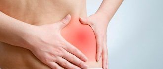
Lower back pain occurs quite often. Patients say “my lower back hurts”, “my lower back is pinched”, “shot in the lower back”. If the pain is not acute, they may say “lower back hurts,” “lower back pulls,” “lower back ache.” Sometimes the pain is described as a burning sensation in the lower back.
Lower back
called the lower back - from the place where the ribs end to the tailbone. Perhaps a separate word for the lower back was needed just to indicate the place where it hurts. After all, if your back hurts, then in most cases it is your lower back that hurts.
What can low back pain look like?
Most often, lower back pain occurs suddenly, sharply and is acute. In this case they talk about lumbago
(outdated popular name -
lumbago
). The pain is described as sharp, “shooting.” Movements are constrained, sometimes it is even impossible to straighten your back. With any movement the pain intensifies.
An attack of pain can last a couple of minutes, or it can last for a longer period of time (up to several days). It may be that the attack will pass and the pain will no longer remind itself, but often the pain returns and the person gets used to the fact that his lower back can hurt.
Lower back pain can not only be acute (sharp), it can be nagging and chronic. Mild but constant pain in the lower back, sometimes worsening, for example, during physical activity, an infectious disease, hypothermia, etc., is called lumbodynia
. Sometimes there is no direct pain, but stiffness remains in the lower back, and the patient experiences discomfort.
Symptoms
Pain in the lumbosacral region is the main symptom of lumbago, lumbodynia, and lumbar ischialgia.
- The pain may radiate down the front, side or back of the leg (lumbar ischialgia), or it may be localized only to the lower back (lumbago, lumbodynia).
- The feeling of lower back pain may intensify after exercise.
- Sometimes the pain may be worse at night or when sitting for long periods of time, such as during a long car ride.
- There may be numbness and weakness in the part of the leg that is in the area of innervation of the compressed nerve.
For timely diagnosis and treatment, a number of criteria (symptoms) deserve special attention:
- Recent history of trauma, such as a fall from a height, a traffic accident, or similar incidents.
- The presence of minor injuries in patients over 50 years of age (for example, a fall from a small height as a result of slipping and landing on the buttocks).
- A history of long-term use of steroids (for example, these are patients with bronchial asthma or rheumatological diseases).
- Any patient with osteoporosis (mostly elderly women).
- Any patient over 70 years of age: at this age there is a high risk of cancer, infections and diseases of the abdominal organs, which can cause lower back pain.
- History of oncology
- Presence of infectious diseases in the recent past
- Temperature over 100F (37.7 C)
- Drug use: Drug use increases the risk of infectious diseases.
- Lower back pain intensifies at rest: as a rule, this type of pain is associated with oncology or infections, and such pain can also occur with ankylosing spondylitis (Bechterew's disease).
- Significant weight loss (without obvious reasons).
- The presence of any acute dysfunction of the nerve is a signal for urgent medical attention. For example, gait disturbances and foot dysfunction are typically symptoms of acute nerve injury or compression. Under certain circumstances, such symptoms may require emergency neurosurgery.
- Bowel or bladder dysfunction (both incontinence and urinary retention) may be a sign of an emergency that requires emergency treatment.
- Failure to respond to recommended treatment or increased pain may also require seeking medical help.
The presence of any of the above factors (symptoms) is a signal to seek medical help within 24 hours.
Causes of lower back pain
Lower back pain can be caused by various reasons, but the statistics are as follows:
- in 90% of cases the pain is caused by problems with the spine and back muscles;
- in 6% the cause of pain is kidney disease;
- 4% - diseases of other internal organs (genitourinary system, intestines).
The spine accounts for the majority of all cases of low back pain, and this is no coincidence. In humans, the center of gravity of the body is located exactly at the level of the lower back, and when walking, the entire load falls almost entirely on the lumbar spine (animals that move on four legs do not have this problem). And when a person sits down, the vertebrae of the lower back and sacrum experience the same force of pressure with which a 170-meter layer of water presses on a diver. Naturally, this area is particularly vulnerable.
Treatment at the Energy of Health clinic
If you are experiencing back pain, welcome to the Health Energy clinic! Here you will find experienced doctors of various specialties, as well as modern diagnostic equipment that will help you accurately determine the cause of pain. When choosing treatment, we are guided by the principle of an integrated approach and use:
- modern drug regimens, selected individually;
- drug blockades to quickly relieve pain and restore mobility;
- a variety of physiotherapy courses for the treatment and prevention of diseases;
- your own exercise therapy room, where they will select a set of therapeutic exercises for you, teach you how to perform the exercises correctly, and help you organize daily exercises at home;
- massage room, where restorative and therapeutic massage of the lumbar region and the whole body is available;
- acupuncture and manual therapy sessions.
Together we will find an approach to treating any pathology. You will be monitored by specialized specialists throughout the course of therapy.
Diseases of the musculoskeletal system that cause lower back pain:
- pinched sciatic nerve. The nerve roots extending from the spinal cord are compressed by neighboring vertebrae. In this case, a sharp, shooting pain occurs. As a rule, pinched roots become possible due to degenerative changes in the spine (osteochondrosis): the intervertebral discs separating the vertebrae from each other are destroyed, the gap between the vertebrae narrows and sudden movement (tilting, turning) can lead to pinching of the nerve branch;
- sciatica (lumbosacral radiculitis). Pinched nerve roots can become inflamed. Inflammation of the nerve roots is called radiculitis (from the Latin radicula - “root”); To indicate inflammation of the sciatic nerve, a special name is sometimes used - sciatica. If the sciatic nerve is damaged, lumbar ischialgia may be observed - pain in the lower back, also spreading to the buttock and leg along the sciatic nerve;
- intervertebral disc herniation – protrusion of a fragment of the intervertebral disc into the spinal canal. Occurs as a result of injury or degenerative changes in the spine (osteochondrosis);
- myositis of the lumbar muscles. Myositis is an inflammation of skeletal muscles. The cause of myositis of the lumbar muscles can be hypothermia or sudden tension.
Also, lower back pain can be caused by diseases such as multiple sclerosis, degenerative sacroiliitis, osteoporosis.
Treatment
Treatment for low back pain depends on its cause. A neurologist, a urologist, a gynecologist, and a surgeon can deal with pathology. When it comes to diseases of the musculoskeletal system, doctors use medicinal, non-medicinal and surgical methods to improve the patient’s condition.
Drug treatment
The most common treatments for low back pain are nonsteroidal anti-inflammatory drugs (NSAIDs). These are drugs based on diclofenac, nimesulide, ibuprofen, meloxicam and their derivatives. They are prescribed in the form of tablets, intravenous and intramuscular injections, rectal suppositories, as well as creams, ointments and patches for topical use. The decision on the dosage of the drug, as well as the duration of the course, is made by the doctor, since uncontrolled use of these drugs can cause unpleasant side effects.
If NSAIDs are ineffective, doctors prescribe hormonal drugs (corticosteroids). They also stop the inflammatory process and help reduce pain.
The third group of drugs that improve the patient’s condition are antispasmodics (mydocalm, sirdalud). They relieve muscle spasms in the lumbar region.
Additionally, the following may be assigned:
- decongestants to reduce swelling of the pinched root;
- B vitamins to improve nerve conduction;
- sedatives.
Non-drug methods
Non-drug treatments complement drug regimens. Depending on the clinical situation, this may include:
- physiotherapeutic procedures (magnetic therapy, laser exposure, electrophoresis, etc.);
- physical therapy: a course of exercises is developed individually in accordance with the underlying and concomitant diseases; gymnastics should be performed regularly, not only in the clinic office, but also at home, only in this case it has an effect;
- restorative and therapeutic massage (performed outside of exacerbations);
- acupuncture;
- manual therapy and osteopathic assistance.
Surgery
The help of surgeons is necessary if the attending physician, based on the general picture, identifies one of the indications for surgical treatment. The presence of a herniated intervertebral disc in itself is not an indication for surgical treatment, regardless of its size. Depending on the indications, doctors can remove a herniated disc, eliminate compression of the spinal cord root, remove a tumor, etc. The decision to carry out a particular operation is made on an individual basis.
Prevention of low back pain
The occurrence of lower back pain is often provoked by a careless attitude towards one’s own health. Pain may be caused by:
- staying in the same position for a long time (for example, during sedentary work);
- incorrect posture;
- low mobility;
- excessive physical activity.
All these factors contribute to the development of diseases manifested by lower back pain. The risk of pain can be reduced by following the following advice from doctors:
- watch your posture;
- Avoid uncomfortable postures when working while sitting. It is advisable that the knees are slightly higher than the hip joints. To do this, use a low chair or footrest. Place a small pillow between your lower back and the back of the seat;
- When working sedentarily, you need to get up from time to time to move around. Take five-minute breaks every hour; how to lift weights correctly
- It is advisable to sleep on an orthopedic mattress (elastic and quite hard);
- You need to lift weights by bending your knee joints, not your back. That is, you need to squat down, bending your knees, and then straighten them, while maintaining a straight line of your back;
- When carrying a load, it must be evenly distributed between both hands; you cannot carry the entire load in one hand (one heavy bag);
- Every day you should do a set of exercises aimed at strengthening the abdominal and back muscles.
LUMBAR PAIN
Lumbar pain (LP), like headaches, is one of the most common complaints with which patients turn to both a local (family) general practitioner and a neurologist. According to WHO experts, almost 90% of people have experienced lower back pain at least once in their lives.
Among the most common causes of LBP are diseases of the spine, primarily degenerative-dystrophic (osteochondrosis, spondylosis deformans) and overstrain of the lumbar muscles. However, we should not forget that various diseases of the pelvic and abdominal organs, including tumors, can cause the same symptom complex as a herniated disc that compresses the spinal root.
The 20th century made serious adjustments to the understanding of the etiology and pathogenesis of BE. Initially, the main cause of their occurrence was considered to be inflammation of the nerve roots and trunks, which was reflected in such terms as lumbosacral radiculitis, radiculoneuritis, funiculitis, etc. Back in the 40-50s, “radiculitis” was often treated with massive doses of antibiotics. But subsequently, the infectious-inflammatory theory of the pathogenesis of BE began to be replaced by a vertebrogenic one, which was greatly facilitated by successful operations for disc herniation. They began to look for the cause of all BE in degenerative-dystrophic changes in the spine, in compression of a nerve root disc by a herniated disc. This period also corresponds to certain terminology: discogenic radicular compression syndrome, vertebrogenic radiculopathy, vertebrogenic reflex syndrome. In the 80-90s, the theory of the predominantly muscular origin of BE began to prevail among neurologists. Many researchers believe that in almost 90% of cases the cause of BE is myofascial syndromes, and vertebrogenic disorders account for no more than 10%. This is reflected in the corresponding terminology: dorsalgia, lumbodynia, myofascial syndrome.
- Etiology and pathogenesis of lumbar pain
The most common causes of BE are: pathological changes in the spine (primarily degenerative-dystrophic); pathological changes in muscles (most often myofascial syndrome); pathological changes in the pelvic and abdominal organs; diseases of the nervous system. The following are considered risk factors for the development of BE: heavy physical activity; physical stress; uncomfortable working posture; injury; cooling, drafts; alcohol abuse; depression and stress; consequences of “harmful” professions (exposure to high temperatures in hot shops and radiant energy, sudden temperature fluctuations, vibration, etc.).
The pathogenesis of PB can be reduced to the following simplified scheme. Painful impulses, regardless of the source (spine, overstrained muscle, “sick” internal organ) enter the spinal cord, from where it goes to special organs of muscle sensitivity - muscle spindles, overexcitation of which causes muscle spasm, leading to a change in body posture and increases it, increasing pain . This creates a vicious circle of maintaining pain. Muscle spasms can be aggravated by depression and chronic stress, which lower the threshold for pain perception, as well as alcohol, which softens the control of maintaining posture.
Among the vertebrogenic causes of BE there are: root ischemia (discogenic radicular syndrome, discogenic radiculopathy), resulting from compression of the root by a disc herniation; reflex muscle syndromes, the cause of which may be various degenerative changes in the spine.
Various functional disorders of the lumbar spine may have a certain significance in the development of LBP, when, due to incorrect posture, blocks of the intervertebral joints occur and their mobility is impaired. In the joints located above and below the block, compensatory hypermobility develops, leading to muscle spasm.
- Lumbar pain in various diseases of the spine
In addition to degenerative-dystrophic changes, relatively rare spinal pathology may also be important in the development of BE.
Spondylolisthesis - literally translated (from the Greek spondylos - vertebra and olisthesis - slipping) - a sliding vertebra that is displaced from the underlying one (most often the 4th or 5th, rarely the 3rd lumbar vertebra). Spondylolisthesis is observed in 2-4% of the population and in 7-10% of cases causes lumbosacral pain. The disease does not always manifest itself clinically and can be detected accidentally during an X-ray examination of the spine. It all depends on the nature of the displacement, i.e., on whether it is partial or complete. Depending on the angle of displacement of the sliding vertebra, stable and unstable spondylolisthesis are distinguished. Its stable form is characterized by the fact that when the spine bends and turns, the relationship between the protruding and underlying vertebrae is not disturbed. With unstable spondylolisthesis (more severe), these relationships are periodically disrupted.
Low back pain in childhood and adolescence is most often caused by abnormalities in the development of the spine. Spinal bifida (spina bifida) occurs in 20% of adults. Neurological symptoms occur in the cystic form of this pathology, when the dura mater protrudes and the epidural tissue grows. Upon examination, attention is drawn to hyperpigmentation, birthmarks, multiple scars, funnels and hyperkeratosis of the skin of the lumbar region. Sometimes urinary incontinence, trophic disorders, and weakness in the legs are noted. In this case, it is necessary to exclude rigid filament terminalis syndrome, which is characterized by thickening and shortening of the fila terminalis, which leads to overstretching of the spinal cord.
BE can cause lumbarization - the transition of the S-1 vertebra in relation to the lumbar spine and sacralization - the attachment of the L-5 vertebra to the sacrum. These anomalies are formed due to individual characteristics of the development of the transverse processes of the vertebrae.
Ankylosing spondylitis. In 1882, this disease was first described by the outstanding Russian neurologist V.M. Bekhterev under the name “stiffness of the spine with curvature.” It is currently referred to as “ankylosing spondylitis.” Almost all patients complain of lower back pain. According to various authors, the disease occurs in 0.08-2.6% of the population, and on average its prevalence is one case per 100. The vast majority of patients (up to 90%) are men aged 20-40 years. Rarely, the disease occurs in children and adults over 50 years of age.
Ankylosing spondylitis is manifested by inflammatory lesions primarily of low-moving joints (intervertebral, costovertebral, lumbosacral joints) and spinal ligaments. Gradually, ossification develops in them, the spine loses elasticity and functional mobility, becomes like a bamboo stick, fragile and easily injured. In the stage of pronounced clinical manifestations of the disease, the mobility of the chest during breathing and the vital capacity of the lungs are significantly reduced.
Tuberculous spondylitis. Chronic inflammation of the spine caused by tuberculosis. As a rule, one of the vertebrae is initially affected, in which a tuberculous granuloma develops, gradually destroying bone tissue. The strength of the outer layers of the vertebra prevents its deformation and the process is asymptomatic for a long time. This is the so-called prespondylytic phase, which in adults can last for many years. Clinically, tuberculous spondylitis begins to manifest itself when the process spreads to tissues adjacent to the vertebra or the affected vertebra is deformed. Often (up to 30% of cases), bruises contribute to the manifestation of the disease, and in children, in addition, various infections.
In children, the disease is more violent and the process often spreads to the vertebrae above and below. Gradually, the pain intensifies, local pain occurs when pressing over the lesion, gait becomes difficult, the mobility of the spine is sharply limited and its configuration changes, which leads to the formation of a hump.
When one or more vertebrae are affected by the tuberculosis process, purulent-necrotic masses can form an abscess that sometimes breaks out, forming a fistula.
Diagnosis of tuberculous spondylitis is initially based on X-ray data of the spine and other organs. The first radiological sign is a narrowing of the intervertebral disc. Then local osteoporosis, bone cavity, marginal destruction, wedge-shaped deformity and, finally, edema abscesses appear in the vertebral body. Breakthrough of caseous masses under the posterior longitudinal ligament into the epidural space is usually accompanied by compression of one or more roots, sometimes the spinal cord with the development of lower paraparesis.
Luetic spondylitis. It is a complication of secondary or tertiary syphilis. Differential radiological signs include severe osteosclerosis, defects of adjacent plates of the affected vertebrae, osteophytes (without ankylosis). The luetic nature of the process should be assumed in the case of recurrent meningitis, meningoencephalitis, repeated strokes (especially at a young age), and spontaneous subarachnoid hemorrhages. To confirm the diagnosis, blood and cerebrospinal fluid tests are performed - the Wassermann reaction and immobilization of treponema pallidum (TIPT).
Brucellous spondylitis. The etiology of the disease is associated with undulating fever with widespread rises in temperature (patients tolerate them relatively easily), profuse sweating, arthralgia and myalgia, lymphadenitis with a predominant enlargement of the cervical and, less commonly, inguinal lymph nodes. The diagnosis is confirmed by serological reactions of Wright and Heddelson.
Typhoid spondylitis. It occurs against the background of a long period of imaginary recovery. The diagnosis is confirmed by the Widal serological reaction.
Dysenteric spondylitis. The diagnosis is confirmed by the results of culture of intestinal contents in the acute period of dysentery.
Rheumatic spondylitis. Sometimes it complicates the course of rheumatism, which is typical for young people and is accompanied by relapses, changes in the heart, polyarthritis with damage to large joints. If bacterial endocarditis is added, then in this case there is fever, leukocytosis, a shift in the blood count to the left, and a sharp increase in ESR. To confirm the diagnosis, rheumatic tests, repeated blood cultures, ECG, and echocardiography are performed.
Nonspecific spondylitis. They can complicate any infection, but most often the intestinal and urinary tract.
Spinal osteomyelitis. Inflammatory damage to the bone marrow with subsequent spread of the process to all elements of bone tissue. Accounts for about 2% of all cases of bone osteomyelitis. The lumbar vertebrae are affected more often than the cervical and thoracic ones. There are nonspecific and specific osteomyelitis. The first is caused by pyogenic pathogens (mainly staphylo- and streptococci); the second - may be tuberculosis, syphilitic and other etiologies. With nonspecific osteomyelitis of the spine, the focus of purulent infection can be located in any part of the body in the form of boils, carbuncles, infected skin wounds, eczema, etc. and is transmitted hematogenously. However, the spine is often affected from nearby purulent foci, for example, with open infected fractures and gunshot wounds. Spinal osteomyelitis can occur acutely, subacutely and chronically. With an acute onset, fever, pronounced blood changes, and sharp, shooting pain in the lower back occur quickly. In the subacute and chronic course of the disease, which occurs more often, the pain is less pronounced. Characteristic changes on radiographs are revealed only after 1.5-2 months. after the onset of the disease.
Epidurit. There is a purulent focus: in 80% of cases, pyoderma, septic changes in the blood - leukocytosis, increased ESR, shift of the formula to the left; rapid increase in pain, fever, symptoms of intoxication. At the beginning of the disease, the pain is local, then signs of compression of one or more roots appear: symptoms of tension, sensory and motor disorders; meningeal symptoms. In the advanced stage of the disease, after an average of two days, gradually increasing pelvic and conduction disorders join these symptoms.
Additional studies include magnetic resonance imaging, cultures of blood, urine and discharge from purulent lesions. Lumbar puncture is contraindicated due to the risk of infection in the subarachnoid space.
Intramedullary abscess. In the initial stage, it is clinically similar to epiduritis. The main differential feature is the type of distribution of conduction disorders: ascending with epiduritis and descending with intramedullary abscess.
Multiple myeloma. It manifests itself as local pain in the thoracic or lumbar spine, which occurs gradually against the background of progressive weight loss, sweating, undulating fever and proteinuria. X-rays reveal diffuse osteoporosis, osteosclerosis, and later secondary spinal deformity. When pathological fractures occur, the pain increases sharply, symptoms of tension, radicular disorders, and lower paraparesis appear. In 70% of cases, ESR increases and normochromic anemia is detected.
| In the 80-90s, the theory of the predominantly muscular origin of BE began to prevail among neurologists. Many researchers believe that in almost 90% of cases the cause of BE is myofascial syndromes, and vertebrogenic disorders account for no more than 10%. This is reflected in the corresponding terminology: dorsalgia, lumbodynia, myofascial syndrome |
Electrophoresis of blood proteins reveals paraproteinemia, hypogammaglobulinemia, and Bence Jones protein in the urine. Hypocalcemia is detected in the blood. The diagnosis is confirmed by examining the puncture of the sternum. In this case, myeloma cell proliferation is determined in 90% of cases. In addition, an X-ray of the skull, chest, and pelvic bones should be performed—favorite sites for myeloma.
Spinal tumors. They can be benign or malignant, originating primarily from the spine or metastatic. Benign tumors of the spine (osteochondroma, chondroma, hemangioma) are sometimes clinically asymptomatic. With hemangioma, a spinal fracture can occur even with minor external influences (pathological fracture). Malignant tumors are predominantly metastatic from the prostate and mammary glands, uterus, lungs, adrenal glands and other organs. Pain in this case occurs much more often than with benign tumors, usually persistent, painful, intensifies with the slightest movement and deprives patients of rest and sleep. Characterized by a progressive deterioration of the condition, an increase in general exhaustion, and pronounced changes in the blood. In diagnosis, radiography, computed tomography, and magnetic resonance imaging are of great importance.
Osteoporosis. The main cause of the disease is a decrease in the function of the endocrine glands due to an independent disease or against the background of general aging of the body. Patients who long-term use of hormones, aminazine, anti-tuberculosis drugs, tetracycline may develop exogenous osteoporosis. Radicular disorders occur due to deformation of the intervertebral foramina, and spinal disorders (myelopathy) due to compression of the radiculomedullary artery or vertebral fracture, even after minor injuries.
Myofascial syndrome. According to many researchers, this is the main reason for the development of BE. It can occur due to overexertion (during heavy physical activity), overextension and bruises of muscles, unphysiological posture during work, reaction to emotional stress, shortening of one leg and even flat feet. Predisposing factors include: hypovitaminosis B1, B6, B12, folic and ascorbic acids, deficiency of microelements (potassium, calcium, magnesium, iron), hypoglycemia, gouty diathesis, chronic infections, sleep disturbance. In the literature, this phenomenon is often referred to by other terms: myalgia, myofibrositis, myofasciitis, trigger points (areas, zones).
In addition to myofascial pain syndrome, the cause of LBP can be other muscle diseases.
Myositis. They represent a large group of diseases of various etiologies: rheumatic, tuberculosis, syphilitic, viral, etc. Parasitic diseases - trichinosis, echinococcosis can also lead to muscle damage. In this case, persistent and prolonged pain is noted.
Widespread muscle changes are found in collagenoses, primarily in dermatomyositis, which can begin suddenly, especially after exacerbation of foci of chronic infection, colds or hypothermia. Muscle soreness, including the lower back, is typical. Simultaneously with the pain or after its cessation, weakness and “weight loss” of the muscles occur, limiting the patient’s motor activity. Skin changes: redness with a purple tint, pinpoint or spotty rash, swelling. During bed rest, foci of necrosis may occur with subsequent development of scars. Severe peeling often occurs, causing the skin to resemble fish scales. Muscle pain and skin changes are usually symmetrical. Half of patients with dermatomyositis have calcification in individual muscles. With multiple calcifications, the skin becomes lumpy and dense to the touch. In the ossifying form of the disease, the ossification of the muscles and subcutaneous tissue can be so strong that it fetters the patients, they become as if dressed in a shell.
Scleroderma. There is widespread thickening of the skin and subcutaneous tissue. The pain is less pronounced than with dermatomyositis.
Diseases of internal organs. PA often occurs with diseases of internal organs: gastric and duodenal ulcers, pancreatitis, cholecystitis, urolithiasis, etc. The pain can be pronounced and imitate the picture of lumbago or discogenic lumbosacral radiculitis. However, there are also clear differences, thanks to which it is possible to differentiate referred pain from that arising from diseases of the lumbosacral part of the peripheral nervous system. These are primarily clinical signs of the underlying disease. For example, with a peptic ulcer of the stomach and duodenum, patients, as a rule, complain of nausea, vomiting, heartburn, belching, which does not happen with radiculitis, and attacks of renal colic are accompanied by frequent urination with pain, nausea, vomiting, and bloating. Difficulties in differential diagnosis are caused by cases when the signs of disease of the internal organs are weakly expressed or the patient has reflected pain combined with pain caused by pathology of the lumbosacral part of the peripheral nervous system.
Very often, BE is caused by diseases of the pelvic organs: the uterus, appendages, prostate gland, vas deferens, rectum, which is primarily due to the proximity of these organs to the lumbosacral nerve formations. Therefore, along with reflected pain, pain from direct impact may also appear. In the past, the term “adnexitis-sciatica” was even used. Lower back pain can occur when the uterus is in an incorrect position during pregnancy, and in women who interrupt sexual intercourse to prevent pregnancy. Gynecological diseases are characterized by a predominant increase in pain during sexual intercourse.
Increased pain during bowel movements is typical for diseases of the rectum: hemorrhoids, tumors, fissures, polyps. In these cases, diarrhea alternating with constipation and significant weight loss are noted. A constant sign of the disease is the presence of mucus and blood in the stool. In some cases, hypochromic anemia is detected. When straining, hemorrhoids may fall out. For differential diagnostic purposes, sigmoidoscopy is indicated to exclude colon tumors.
Dissecting aortic aneurysm. The pain radiates to the abdomen, legs and is accompanied by a collapsing state. A tumor-like formation is palpated in the abdomen, over the projection of which a systolic murmur is heard. Additionally, ultrasound and computed tomography examinations of the abdominal organs are performed.
Diseases of the nervous system. In some cases, BE occurs in the initial stages of the disease, is temporary and is combined with various neurological disorders, and in others it is long-term or permanent. Transient lumbar pain can be observed in acute inflammatory diseases of the nervous system: meningitis, myelitis, polyradiculoneuritis, which have a predominantly acute onset and are severe. At the same time, mild or moderate pain in the lower back seems to recede into the background. In addition, they decrease or disappear quite quickly. PB can also occur several years after acute inflammatory diseases of the nervous system.
Spinal cord tumors. Depending on the primary localization, they are divided into intra- and extramedullary, which are more common and are predominantly benign: limited, do not infiltrate the brain tissue. BE in extramedullary tumors is an early sign of the disease. They arise due to a decrease in the free cavity of the spinal canal due to tumor growth, tension in the nerve roots and meninges.
Lumbar pain can be observed with multiple sclerosis, spinal gliosis, and tabes of the spinal cord. They do not play a leading role in the clinical picture.
Finally, one should remember about the possibility of the occurrence of pain in neuroses, which, unlike pain in organic diseases of the nervous system, are not constant and clearly localized. They move from one part of the body to another, and their severity depends on the general emotional state. Usually there are no symptoms of tension in the spinal roots and prolapse from the reflex sphere.
- Clinical symptoms of lumbar pain
Most often, BE occurs at the age of 25-44 years. There are acute pains, lasting, as a rule, 2-3 weeks, and sometimes up to two months, and chronic pains - over two months.
Compression radicular syndromes (discogenic radiculopathy) are characterized by a sudden onset, often after heavy lifting, sudden movements, or hypothermia. Symptoms depend on the location of the lesion. The syndrome is based on compression of the root by a disc herniation, which occurs as a result of degenerative processes facilitated by static and dynamic loads, hormonal disorders, and injuries (including microtraumatization of the spine). Most often, the pathological process involves the area of the root from the dura mater to the intervertebral foramen. In addition to disc herniation, bone growths, scars of epidural tissue, and hypertrophied ligamentum flavum may be involved in root trauma.
The upper lumbar roots (L-1, L-2, L-3) are rarely affected: they account for no more than 3% of all lumbar radicular syndromes. L-4 is most often affected (6%), causing a characteristic clinical picture: mild pain along the inner-lower and anterior surfaces of the thigh, the medial surface of the leg, paresthesia in this area, slight weakness of the quadriceps muscle. Knee reflexes are preserved and sometimes even increased. The L-5 root is most often affected (46%). The pain is localized in the lower back, gluteal region, along the outer surface of the thigh, the anterior outer surface of the lower leg down to the foot and the first three fingers. It is often accompanied by a decrease in the sensitivity of the skin of the anterior outer surface of the leg and the strength in the extensor of the first finger. The patient finds it difficult to stand on his heel. With long-term radiculopathy, hypotrophy of the tibialis anterior muscle develops.
The S-1 root is just as often affected (45%). Lower back pain radiates along the outer back of the thigh, outer surface of the leg and foot. Examination often reveals hypalgesia of the posterior outer surface of the leg, decreased strength of the triceps muscle and toe flexors. It is difficult for such patients to stand on their toes. There is a decrease or loss of the Achilles reflex.
Vertebrogenic lumbar reflex syndrome. It can be acute or chronic. Acute LBP (lumbago, “lumbago”) develops over several minutes or hours, often suddenly due to awkward movements. Piercing, shooting (like an electric shock) pain is localized throughout the lower back, sometimes radiating to the iliac region and buttocks, sharply intensifies with movement, coughing, sneezing, and decreases when lying down, especially if the patient finds a comfortable position. Movements in the lumbar spine are limited, the lumbar muscles are tense, Lasegue's symptom is caused, often bilaterally. Acute lumbodynia usually lasts 5-6 days, sometimes less. The first attack ends faster than subsequent ones. Repeated attacks of lumbago tend to develop into chronic LBP.
Myofascial pain. It is usually long-lasting, ranging from mild discomfort to severe and excruciating pain. It can occur at rest and during movement, is associated with trigger points and is non-segmental in nature, and the zone of greatest intensity is rarely localized at the most trigger (hyperirritable) point in a dense cord of skeletal muscle, painful on palpation. Trigger points are activated by overload, physical fatigue, injury or cold. Pressure on them causes or intensifies painful phenomena in the area of referred pain.
It is worth highlighting a number of clinical symptoms that are not typical for BE caused by degenerative-dystrophic changes in the spine or myofascial syndrome. They should alert you, as they may be a sign of the presence of pathological processes in the pelvic and abdominal organs or diseases of the spine that go beyond osteochondrosis. These symptoms include: the appearance of BE in childhood and adolescence; back injury shortly before the onset of LBP; PB, accompanied by fever and signs of intoxication; PA not associated with spinal movements; unusual irradiation of pain: in the perineum, abdomen, rectum, vagina, both legs, girdle pain; connection of PB with eating, defecation, sexual intercourse, urination; concomitant urinary retention or incontinence; gynecological pathology (amenorrhea, dysmenorrhea, vaginal discharge), which appeared against the background of PB, strengthening of PB in the horizontal position and decrease in the vertical position (Razdolsky’s symptom, characteristic of a tumor process in the spine); steadily increasing pain over one to two weeks; development of paresis of the lower extremities against the background of BE, the appearance of pathological reflexes.
- Examination methods
— External examination and palpation of the lumbar region, identification of scoliosis, muscle tension, pain and trigger points. — Determination of range of motion in the lumbar spine, areas of muscle wasting. — Study of neurological status: determination of tension symptoms (Laseg, Wasserman, Neri); states of sensitivity, reflex sphere, muscle tone, vasomotor and autonomic disorders (swelling, changes in skin color). — X-ray, computer or magnetic resonance imaging of the lumbar spine; Ultrasound examination of the pelvic organs. — Gynecological examination. — If necessary, additional studies are carried out: cerebrospinal fluid, blood and urine, sigmoidoscopy, colonoscopy, gastroscopy, etc.
- Treatment
I. Acute BE (or exacerbation) of vertebral or myofascial origin.
A. Undifferentiated treatment. Gentle motor mode; in case of severe pain in the first days, bed rest, and then walking on crutches to unload the spine; hard bed: the best option is a wooden board with a thin mattress on top of it. For local warming, a woolen shawl, an electric heating pad, and a bag of heated sand or salt are recommended. Local exposure in the area of greatest pain: irritating ointments (phenalgon, tiger ointment, capsin, nicoflex, etc.), mustard plasters, pepper plaster, ultraviolet irradiation in an erythemal dose, leeches or irrigation with ethyl chloride.
Anesthetic electrical procedures (the means of choice are: transcutaneous electroanalgesia, sinusoidal modulated currents, diadynamic currents, electrophoresis with novocaine, etc.); acupuncture.
Novocaine blockades and pressure massage of trigger zones are used. Use analgesics, antihistamines, non-steroidal anti-inflammatory drugs, tranquilizers and/or antidepressants; drugs that reduce muscle tension (muscle relaxants): baclofen (5 to 20 mg three times a day) or sirdalud (1 to 4 mg three times a day). In case of arterial hypertension, sirdalud should be taken with great caution due to its hypotensive effect. If root swelling is suspected, diuretics are prescribed.
B. For discogenic radiculopathy, traction therapy (dry or underwater traction) is used in a neurological hospital.
B. For myofascial syndrome, after local treatment (novocaine blockade, irrigation with ethyl chloride), a hot compress is applied to the muscle for several minutes.
II. Chronic lumbar pain of vertebrogenic or myogenic origin.
In case of disc herniation, it is recommended to wear a rigid corset of the “weightlifter’s belt” type; limiting physical activity; exclusion of sudden movements, bends; physical therapy to create a muscle corset and restore muscle mobility; massage; novocaine blockades; acupuncture; physiotherapy: ultrasound, laser therapy, heat therapy. They use antidepressants, vitamin therapy: intramuscular B vitamins (B1, B6, B12), multivitamins with mineral supplements. For paroxysmal pain, Finlepsin (Tegretol) is prescribed. Psychotherapy is indicated.
In this article we do not touch upon issues of manual therapy and surgical treatment, which require special consideration.
Lower back pain due to kidney disease
For lower back pain, it is important to determine what is causing it - pathologies of the musculoskeletal system or kidney disease (as well as other internal organs). Diagnosis must be carried out by a doctor. However, there are signs to suggest that the pain may be due to problems with the kidneys and/or other organs of the genitourinary system. If these symptoms occur, it is advisable to immediately contact a urologist. Kidney disease (or more broadly, the genitourinary system) can be suspected if lower back pain is accompanied by:
- general deterioration in health (lethargy, drowsiness, weakness, increased fatigue);
- swelling of the eyelids and face. Swelling is especially pronounced in the morning, after waking up, and subsides in the evening;
- increased body temperature, chills, sweating;
- loss of appetite, nausea, vomiting;
- frequent or painful urination;
- changes in the characteristics of urine (it may become more concentrated in color or, conversely, colorless, contain mucus or blood);
- increased blood pressure.
Also an important sign that lower back pain is caused by problems of the internal organs, and not the musculoskeletal system, is its independence from the position of the body: the pain does not increase or decrease from changes in the position of the body and limbs. However, with prolonged standing in a standing position due to check pathology, the pain may intensify. The location of the pain also matters. With kidney disease, pain is most often observed on one side (since usually only one kidney is affected). Kidney pain may not be limited to the lower back, but may spread along the ureter, to the groin, to the external genitalia, to the inner thighs.
Advantages of the clinic
Modern medicine means maximum efficiency and convenience for the patient. The Health Energy Clinic is equipped in accordance with the latest standards for diagnosis and treatment of diseases. We offer each patient the opportunity to obtain maximum information about the state of his health. At your service:
- experienced doctors who regularly improve their qualifications;
- various diagnostic and therapeutic procedures, including minor surgical interventions;
- individual selection of therapy in accordance with the examination results;
- Reception by appointment at a time convenient for you;
- own parking and convenient location of the clinic near the metro station;
- affordable prices for all medical services.
Back pain is not necessarily a problem with the spine or muscles. Even slight discomfort can be a sign of dangerous diseases, such as urolithiasis or malignant tumors. Do not ignore this symptom, sign up for an examination at the Health Energy clinic.
Lower back pain: what to do?
Low back pain is a symptom of a disease that requires treatment. Therefore, it is necessary to consult a doctor. But in the event of a sudden attack of acute pain (“lumbago”, typical of radiculitis), first of all, it is necessary to relieve the pain syndrome. Doctors advise:
- use gentle heat. Tie a woolen scarf or woolen belt around your lower back;
- take painkillers;
- It is necessary to take a position that allows you to relax your back muscles. It is recommended to lie on your back, on a hard, flat surface (board); The legs should be raised and bent at the knees, for which a rolled blanket or pillow should be placed under them. (It is not advisable to lie on the floor; there may be a draft).
The proposed pose is not a dogma. The patient should feel relief, so other positions are possible; for example, lying on a board, place your legs bent at the knees on it, holding a pillow between them. You can try lying on your stomach and stretching your legs, placing a bolster under your ankle joints. If the severity of the pain has been relieved, this does not mean that a doctor is no longer needed. Without proper treatment, attacks will recur, and the situation as a whole will worsen.
Diagnostics
If your spine hurts in the lumbar region, you should make an appointment with a neurologist or vertebrologist. At the appointment, the doctor initially collects anamnesis, asking questions about the nature of the pain, the circumstances of its occurrence, the duration of its persistence, the presence of other symptoms, lifestyle, etc.
Then the specialist conducts an examination. As part of it, he not only palpates the spine, determines the localization of pain, evaluates the gait and posture that the patient takes unconsciously, but also conducts functional tests. With their help, you can detect signs of ankylosing spondylitis, neurological deficits, assess the degree of mobility of the spine and obtain other diagnostic data.
Based on this, the doctor can already suggest possible causes of pain. To clarify them, as well as to accurately determine the extent of damage, instrumental and sometimes laboratory diagnostic methods are additionally prescribed. Most often they resort to help:
- X-rays in frontal and lateral projections, sometimes with functional X-ray tests;
- CT scan – allows better visualization of bone structures, therefore it is more often used to diagnose spondylosis, fractures, bone tumors, etc.;
- MRI – makes it possible to most thoroughly assess the condition of cartilaginous structures and soft tissues, therefore it is more often used to diagnose osteochondrosis, protrusions, intervertebral hernias, spinal cord lesions, etc.;
- electromyography - indicated for neurological disorders of unknown origin, as well as to assess the degree of nerve damage;
- radioisotope osteoscintigraphy – used for the diagnosis of malignant tumors and metastases;
- X-ray densitometry is the optimal method for diagnosing osteoporosis;
- myelography - used to identify signs of compression of the spinal cord and nerves of the cauda equina.
Which doctor should I contact with a complaint of lower back pain?
If you have lower back pain, it is best to consult a general practitioner, since first of all you need to determine which organ disease is causing the pain. Depending on the results of the examination, consultation with a particular medical specialist may be required. Can be assigned:
- consultation with a neurologist to assess the condition of the spine, back muscles and nervous system;
- consultation with a urologist – in case of suspected urinary system disease;
- consultation with a gynecologist – if chronic diseases of the female reproductive system are suspected or present;
- general blood test and general urinalysis - to confirm or exclude the inflammatory nature of the disease;
- radiography of the spine;
- Ultrasound of the hip joints;
- as well as other studies.
How is diagnosis and examination done by a doctor?
First of all, the doctor must exclude conditions that are life-threatening. For this purpose, clinical and biochemical blood tests are performed. They make it possible to detect inflammatory processes and excess calcium, which is typical for cancer that has metastasized to the bones. Tests also detect multiple myeloma and many other pathologies.
A man over 50 years old may have a prostate-specific antigen test to rule out prostate cancer.
X-rays are required to determine the height of the intervertebral discs and identify osteophytes, if any. The latter are bone tissue growths that appear due to improperly distributed load on the vertebrae and changes in their shape.
MRI and CT are needed to determine whether there is a bulging intervertebral disc, calcifications, or spinal stenosis. Similar changes can be seen on ultrasound, which is increasingly being prescribed instead of CT, as it does not provide radiation exposure.
The patient must consult a neurologist and, if necessary, a chiropractor.
When the examination is completed, the doctor can accurately diagnose and determine treatment tactics. The success of therapy increases tenfold with early treatment.
Our clinic address: St. Petersburg, st. Bolshaya Raznochinnaya, 27 metro station Chkalovskaya
Reason 3. Intervertebral hernia
Intervertebral hernia is the “squeezing out” of the nucleus pulposus by neighboring vertebrae. The nucleus pulposus is a kind of hinge that is located in the center between the vertebrae and ensures their mobility. Therefore we can bend in all directions. But this structure is semi-liquid, and with increased or sudden physical activity it can “crawl” beyond the intervertebral space, forming a hernia.
The pain is acute, pronounced, and sharply intensifies with exercise. May be accompanied by impaired sensitivity in the arms and legs, numbness and pain in the extremities, and radiate to the buttock.
An intervertebral hernia can be diagnosed by an orthopedist, neurologist, neurosurgeon, or vertebrologist.
Reason 2. Degenerative-dystrophic diseases
Caused by wear and tear of the intervertebral discs, brittle bones, and loss of elasticity of spinal tissue. Moreover, such changes are not necessarily age-related. Today, young people also suffer from arthritis, spondylosis, and osteochondrosis.
Over time, pathological processes in the spine become degenerative, i.e. irreversible. In this case, it is necessary to use surgical treatment methods - joint replacement, surgical restoration of vertebrae and other structures. Arthrosis, osteoporosis, and radiculitis often develop into a degenerative form.
Of course, such changes do not occur without symptoms. Often patients note lumbago, acute pain in the affected area, limited mobility, stabbing pain, crunching, pain with certain movements (for example, in the lower back when bending forward). As a rule, a person can clearly determine exactly where it hurts.
Treatment of degenerative-dystrophic diseases is carried out by a rheumatologist, osteopath, chiropractor, traumatologist, neurologist and a number of other specialists. Don't know who to contact? Make an appointment with a general practitioner first.
Features of the structure of the lumbar region
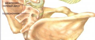
In total, there are 5 sections in the human spine, each of which has a certain number of vertebrae. There are 5 units in the lumbar region. The segments of this part of the spinal column are the largest when compared with others that are located in the rest of the spine. They consist of two elements - an arched rear part and a front part resembling a spool of thread. Between all the vertebrae there is a special body - the intervertebral disc, consisting of cartilage tissue and fluid. The presence of these elements allows the spine to remain flexible and flexible.
Lumbar vertebra
On a note! The vertebrae have a spongy structure. On the outside they are covered with dense bone tissue, and on the inside they have a lattice structure. Due to the presence of free space filled with blood, the vertebrae are quite light. If they consisted entirely of dense bone tissue, then a person would simply not be able to move freely.
The lumbar region is located between the chest and pelvis. It supports the entire upper half of the body, and this part of the spine experiences enormous load every day. This is why pain often occurs in this area.
The lumbar region of the spinal canal is considered the strongest, as it is exposed to heavy loads every day.
Cause 1. Spinal infections
Another name is spinal infections. These are viral lesions that affect the internal structures of the vertebrae or the interdisc space. Viruses can enter the body from the outside (wound infections - due to injuries, operations) or be complications of viral diseases (often various types of myelitis, coccal infections, etc.).
Symptoms of spinal column infection depend on the type of infection. This may be unexpressed aching pain in the back and chest area, or sudden intense pain. The focus is quite difficult to determine. The patient usually says “everything hurts.” The condition is accompanied by limited mobility, chills, fever, weakness, and body aches.
If you have these signs, we recommend calling an ambulance immediately. Spinal infections are fraught with paralysis and other irreversible (!) restrictions on mobility. But they are quite rare.
Reason 7. Spinal tumors
These are cystic formations and cancerous tumors.
A cyst is a bubble with blood. It appears as a result of various types of hemorrhages in the spine. The cyst is characterized by constant severe pain, which can only be relieved with the help of painkillers. Numbness and tingling in the extremities can also be symptoms of a cyst.
Cancer can be primary or secondary. Primary is cancer that has formed in the spinal column, secondary is metastases, that is, secondary tumors that form in late stages in all organs. With malignant tumors, pain may be accompanied by muscle weakness and loss of sensitivity in certain areas.
Neoplasms in the spine are studied by a vertebrologist, oncologist, and neurosurgeon.
