Lymphadenopathy is a condition that is characterized by an increase in the size of peripheral lymph nodes, a change in their consistency and mobility. It develops along with an increase in overall body temperature, an enlargement of the spleen and liver. Even a slight enlargement of the lymph nodes indicates that pathological processes are developing in the body that require adequate treatment. It can cover one or more structures and cause inflammatory processes.
The hematology department of CELT invites you to undergo diagnosis and treatment of lymphadenopathy in Moscow. You have the opportunity to consult with leading domestic hematologists, who have modern techniques and a powerful base that allows you to quickly and accurately establish a diagnosis. They provide treatment in accordance with international standards. You can find out the cost of services in the “Services and Prices” section. To avoid misunderstandings, do not forget to check the numbers during consultation or with our operators.
Lymphadenopathy: what is it?
Enlarged lymph nodes look like soft or dense rounded formations located under the lower jaw, in the cervical, axillary, groin or other areas. They can have a smooth or bumpy surface and very often develop after acute infections and inflammations. Sometimes their appearance is caused by traumatic injuries to the skin or the introduction of a vaccine.
Lymphadenopathy develops due to the accumulation of a certain type of cell in the lymphadenoid tissue. The reaction is often provoked by increased blood flow, an increase in the number of lymphocytes and macrophages in response to the appearance of foreign genes in the body. In just one week, the node can increase five to fifteen times. A pathological condition is spoken of if, along with an excess of size, there is a change in the density, mobility and surface of the structure. When palpated, it can be quite painful or painless.
Reasons for contacting our center for diagnosing lymphadenopathy in Moscow: independent identification of large nodes that do not hurt, a feeling of intense pain during palpation, other symptoms in the form of a rash, increased body temperature, weight loss, and rapid fatigue. Particularly dangerous can be nodes that do not go away for more than two months, located in different areas, with a diameter of more than two centimeters, the enlargement of which occurred for no apparent reason.
Lymphadenopathy: classification and causes
The classification of lymphadenopathy is based on a number of parameters, starting with the location of the affected structures and ending with their pain and size:
| Localization of the affected node/nodes | Etiological factors |
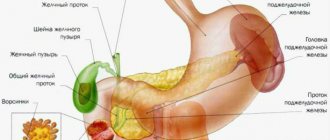
| Under the lower jaw | Diseases of the eyes, ear, throat, nose, damage to the skin in the head or neck area, dental problems. |
| Neck |
|
| Supraclavicular region | Malignant neoplasms:
|
| Armpits |
|
| Groin |
|
Classification
When determining the forms of lymphadenopathy, the location of enlarged lymph nodes is primarily taken into account. Lymphoid tissue is the main protective barrier against the spread of infectious pathogens and tumor cells. Therefore, the location of altered lymphatic formations facilitates the diagnosis of the disease that caused the lymphadenopathic reaction. Depending on the localization of the process, the following are distinguished:
- Enlargement of the submandibular lymph nodes
. Characteristic of pathological processes in the head and neck area - diseases of the eyes, ENT organs and paranasal sinuses, skin damage. Submandibular lymphadenopathy often signals dental problems and chronic tonsillitis. - Enlarged cervical lymph nodes
. Usually observed with respiratory infections, oral pathology, infectious mononucleosis, late stages of tuberculosis. Cervical nodes can be affected by lymphomas, lymphogranulomatosis, metastasis of thyroid cancer, lung cancer. - Enlarged supraclavicular lymph nodes
. Most often due to tumor causes. Detection of an enlarged node on the right is pathognomonic for cancer of the esophagus and lungs. The left lymph node is affected by malignant processes in the abdominal cavity, pelvis, and retroperitoneal space. - Enlarged axillary lymph nodes
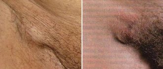
. Inflammatory lesions are possible in the presence of wound infections, cat scratch disease, and brucellosis. Damage to the nodes of the axillary group is typical for breast cancer, melanoma of the upper extremities, and the installation of silicone breast implants. - Enlarged inguinal lymph nodes
. As a rule, nodes in the groin react to the development of syphilis, gonorrhea, chancroid, and other genital infections. Inguinal lymphadenopathy is also a sign of malignant lesions of the pelvic organs, lymphoma, and bubonic plague.
Somewhat less frequently, lymph nodes of other groups are involved in the process - submental, cubital (in the area of the elbow), parotid, occipital, jugular. During a routine instrumental examination, an increase in internal lymph nodes can be determined - intrathoracic (mediastinal), bronchopulmonary, para-aortic, splenic, mesenteric, retroperitoneal.
In the diagnostic plan, it is important to take into account other criteria for the classification of lymphadenopathy - the characteristics of altered lymphoid formations, the extent of the lesion. This approach allows us to assume the type of pathological process occurring in the involved nodes and the body as a whole. Important criteria for the classification of enlarged lymph nodes are:
- Dimensions
. With I degree lymphadenopathy, the diameter of the affected formations is 0.5-1.4 cm, with II degree - 1.5-2.4 cm, and with III degree - 2.5 cm or more. A significant and prolonged increase in the size of lymph nodes is more typical for malignant processes. - Soreness
. Intense pain is often caused by inflammation of the lymph nodes, especially acute purulent lymphadenitis. Formations that have undergone malignant degeneration are often painless, except in cases of hemorrhage into the necrotic center. - Density
_ Enlarged, inflamed lymph nodes are usually soft; when they suppurate, fluctuation (fluctuation of fluid) is felt during palpation. The stony consistency of the formations is typical for the metastatic process, and tight elasticity is typical for lymphomas. - Communication with each other
. A pathological formation of lymph nodes that can be felt as a single unit and move together is called a conglomerate. Lymph nodes fused together are detected in tuberculosis, sarcoidosis, lymphogranuloma venereum, lymphomas and metastasis. - Quantity
. It can affect either one or two or several nodes in one zone. In the first case, they talk about single enlarged nodes, in the second - about local lymphadenopathy. The more active the process, the more formations are affected, however, with metastasis, one large node is often detected. - Prevalence
. With local lymphadenopathy, single nodes are identified in one area, with regional lymphadenopathy - several formations in 1-2 adjacent zones. A generalized (widespread) process is characterized by damage to lymphatic structures in three or more areas.
Taking into account the pathogenesis, enlargement of lymph nodes can be primary (systemic), secondary (reactive) and inflammatory. Primary polyadenopathies develop with systemic malignancy of lymphoid tissue (leukemia, lymphogranulomatosis, non-Hodgkin's lymphoma) and benign processes (sinus histiocytosis). Reactive lesions are a response to another pathology (infection, immune disease, proliferation of tumor cells, metabolic disorders). Inflammation (lymphadenitis) occurs when infectious agents multiply in the tissue of the node.
Lymphadenopathy: diagnosis
Before starting treatment of lymphadenopathy in adults, CELT hematologists conduct comprehensive studies to accurately diagnose and determine the cause of the development of the pathological condition. The following node parameters must be taken into account:
- Dimensions - determine the degree of the disease (first - 5 mm-14 mm, second - 15 mm-24 mm, third - 25 mm or more);
- Painful symptoms - most often it occurs during inflammatory processes, in particular purulent lymphadenitis;
- Quantity – one, two or several nodes located in one area can be affected. Multiple lesions are a sign of an active pathological process;
- Density - usually inflamed nodes are soft, their fluctuation is a sign of a purulent process, tight plasticity - lymphoma, hardness - metastases;
- Connectivity - several nodes combined into one whole can be a sign of tuberculosis, Besnier-Böck-Schaumann disease, or damage to lymphatic tissue.
In addition to the examination, the patient is prescribed diagnostic tests:
- Detailed blood test;
- Test for hepatitis and HIV;
- Ultrasound scanning of internal organs and affected nodes;
- Histology of biopsy samples from the affected node;
- X-ray examination;
- Magnetic resonance or computed tomography.
Causes
There are many reasons for increasing size, since any pathological process in the human body leads to a reaction of the lymphatic system.
- Infections. One of the most common causes of lymphadenopathy. This includes all infections that can be in the human body: bacterial, viral, parasitic and fungal.
- oncological diseases. The second most common reason. It may be a reaction to the primary cancer or metastases. Lymphadenopathy can develop both in a malignant and benign process.
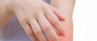
- immune diseases. This includes all systemic diseases: rheumatoid arthritis, dermatomyositis, systemic lupus erythematosus and others.
Regardless of the cause of the development of lymphadenopathy, consultation and examination by a doctor with a thorough diagnosis is necessary.
Lymphadenopathy: treatment
The development of treatment tactics is carried out by CELT specialists based on the diagnostic results obtained and the patient’s individual indicators. First of all, efforts are directed toward eliminating the pathological condition that has become the initiating factor for the development of lymphadenopathy. If purulent lesions of the lymphatic structures or bacterial infections are detected, the patient is prescribed antibacterial therapy. If indicated, surgical removal of the node is possible.
If the etiological factors involve damage by viral agents, the patient is prescribed antiviral and immunomodulatory pharmacological drugs. If he suffers from pain - painkillers. Most often, lymphadenopathy goes away within one to one and a half months after the cause that caused it is eliminated. Otherwise, the hematologist prescribes a biopsy and develops a new treatment strategy.
The hematology department of our clinic receives candidates, doctors and professors of medical sciences. Their practical and scientific work experience ranges from twenty-five years. You can make an appointment with them online or by contacting our information line operators. If necessary, you can make an appointment with specialists at any of our departments and undergo the necessary procedures. In particular, septoplasty is available in the otolaryngology department, which can improve the patient’s quality of life.
At CELT you can consult a hematologist.
- Initial consultation – 3,500
- Repeated consultation – 2,300
Make an appointment
By making an appointment with a hematologist, you can get a comprehensive consultation. The doctor is competent to treat various blood diseases, most of which can be identified in the early stages and prescribe timely treatment to cope with the disease quickly and easily.
Inflammation of the lymph nodes
Reactive lymphadenitis is the most common cause of palpable swelling of the neck or lymph nodes in children, adolescents, and adults of any age.
Lymph nodes are located throughout the body as part of the lymphatic system, which also includes lymph capillaries, vessels, and ducts. In healthy people, in the area of the lower jaw, neck, groin, etc., it is possible to palpate under the skin several small, less than 1 cm, seals that are visually invisible. Lymphadenopathy is a noticeable enlargement of a lymph node greater than 1 cm.
Patients of different ages come to see specialists at the Miracle Doctor clinic with symptoms of lymphadenitis, who either themselves discovered enlarged lymph nodes, or were referred by another specialist who discovered characteristic signs:
- painful subcutaneous thickening over 1 cm, located in the anatomical areas of the lymph nodes: under the jaw, on the neck, in the groin, under the armpit, etc.
- limited mobility and tight fit of the nodes to the surrounding tissues,
- the appearance of a lump is accompanied by headache, malaise, weakness, and changes in body temperature.
Causes of enlarged lymph nodes
Lymph nodes are “filtration stations” in the lymphatic system and are involved in activating the immune system when infectious pathogens enter the lymph flow.
Some components of blood plasma and foreign cells (for example, cancer cells, microorganisms) penetrate into the lymphatic vessels along with cellular material, antigens, etc.
Thanks to the filtration process, antigens also interact with lymphocytes contained in the lymph nodes. The immune response of these lymphocytes involves cell proliferation, which can cause enlarged nodes—reactive lymphadenopathy. Pathogenic microorganisms that enter the lymphatic fluid can directly infect nodes, causing lymphadenitis.
Thus, lymph node inflammation is primarily found in the context of inflammation and malignant diseases. If a patient has swollen lymph nodes, there are primarily two types of causes: benign causes, which are much more common, and malignant causes, which must be taken into account due to their serious consequences.
Depending on the duration of the course and manifestations, acute and chronic lymphadenitis are distinguished. Acute lymphadenitis can end in a purulent stage, with the formation of an abscess and the possible entry of purulent infected contents into adjacent tissues. Chronic develops after acute inflammation subsides and can be wavy in nature.
Diagnostics
The main goal is to establish the primary cause of the development of lymphadenopathy and differential diagnosis of other diseases with similar symptoms - phlegmon, purulent atheroma, etc.
Clinical examination and history taking are essential. Palpation is carried out to determine the number and degree of pain of the superficial inflamed lymph nodes, especially in the neck (including the occipital and supraclavicular regions), armpits and groin. The size of the nodes, sensitivity and degree of elasticity, mobility of the lymph nodes in the adjacent tissues are determined.
The duration of inflammation is established, the following are noted:
- possible skin injuries, especially cat scratches, rat bites;
- the presence of infections in the areas drained by the affected nodes: infectious and inflammatory diseases of the upper respiratory tract, lesions or discharge from the genitals, joint pain, swelling, local soft tissue infections, dental infection;
- information on recent travel to countries with endemic infections (eg, Middle East, Africa);
- history of taking medications that contain trigger components.
Doppler ultrasound allows you to determine the length, width, shape, echogenicity of blood vessels, and therefore the degree of damage to the lymphatic system and determine the causes of the pathology.
Histological examination after diagnostic biopsy of the lymph node is usually prescribed for the differential diagnosis of malignant tumors and destructive changes in tissues.
A complete blood count may reveal an abnormal number of white blood cells, which is a reason to prescribe a tuberculin skin test and serological tests for mononucleosis, toxoplasmosis, sexually transmitted diseases, and a test for antibodies to SLE.
As a rule, the treatment of lymphadenopathy is carried out by the attending physician, who can refer the patient for consultation with a specialist.
Depending on the true cause of lymphadenopathy, antiviral and antibacterial drugs and physical therapy sessions may be prescribed. For purulent formations, surgical sanitation, installation of drainage and drug treatment are performed.
Treatment
Since adenopathy itself is not curable, treatment is aimed at eliminating the cause that provokes the development of lymphadenopathy. Patients with generalized lymphadenopathy—numerous swollen lymph nodes—usually have a systemic disease causing the inflammation, while patients with localized adenopathy usually have a local disease.