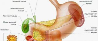
The cecum is the initial section of the large intestine. It is located in the right iliac region (lower right in the abdominal cavity) and, due to its structure, is compared to a vertical sac. In the lower part of the cecum there is a vermiform appendix.
The Yusupov Hospital has created all the conditions for the treatment of patients with cancer of the cecum:
- The wards are equipped with forced-air ventilation and air conditioning to provide patients with a comfortable temperature;
- Doctors examine patients using the latest equipment from leading European and American manufacturers;
- For operations on the cecum, surgeons use modern instruments and equipment;
- Oncologists masterfully perform the entire range of surgical interventions known today;
- Medical staff provides professional care before and after surgery;
- Doctors provide antitumor therapy with the latest chemotherapy drugs registered in the Russian Federation.
Patients, as part of scientific research conducted at the clinic, have the opportunity to receive the latest medicines. The growth of a malignant neoplasm can begin in any part of the gastrointestinal tract. Cecal cancer accounts for approximately one fifth of all colorectal cancers.
Causes
The immediate cause of cecal cancer is a cellular mutation that is not recognized by the immune system and not destroyed in time, as a result of which an endless cycle of division and growth of tumor cells is started. Malignant neoplasms of the cecum, according to experts from the World Health Organization, can develop under the influence of the following provoking factors:
- Excessive consumption of red meat that has undergone culinary processing;
- Age-related changes in tissues and weakening of intestinal motility;
- Hereditary predisposition;
- Chronic diseases and pathological conditions of the gastrointestinal tract (inflammatory, ulcerative, dyskinetic, benign neoplastic processes);
- Environmental factors;
- The presence and activity of oncogenic viruses in the body (human papillomavirus and some subtypes of the herpes virus);
- Smoking.
Scientists have not yet established a single reason why a tumor of the cecum develops.
Kinds
For cecal cancer, the TNM classification is used. In it, T denotes the size of the tumor, N – the presence of lymph node involvement, M – the presence of distant metastases. The disease occurs in 4 stages:
- Stage 0 is characterized by a very small tumor size, damage to only the upper layer of the wall of the cecum, and the absence of metastases in regional lymph nodes;
- In the first stage of cecal cancer, the pathological process spreads to the second and third layers of the colon, but does not grow to the outer side of the intestine, and there are no metastases in the lymph nodes;
- At stage II, the malignant tumor grows on the outer wall of the cecum, the lymph nodes are not affected and no metastases are observed;
- At the third stage of the disease, the tumor begins to grow into nearby organs and tissues, lymph nodes are affected, but doctors do not detect distant metastases;
- At the terminal, fourth, stage of cecal cancer, the malignant tumor begins to grow into adjacent tissues and organs, affecting the lymph nodes and revealing distant metastases.
The following histological types of cecal cancer are distinguished:
- Adenocarcinoma develops from epithelial cells of the intestinal mucosa;
- Signet ring cell carcinoma appears as blisters;
- Undifferentiated cancer is considered the most aggressive form of malignancy;
- Unclassified cancer is a malignant tumor that does not belong to any of the histological forms;
- Squamous cell carcinoma is a tumor of the cecum, which consists of squamous epithelial cells;
- Glandular squamous cell carcinoma is a neoplasm consisting of squamous and glandular epithelium.
Adenocarcinoma of the cecum can be an exophytic or endophytic tumor. It can grow into the lumen of the cecum or penetrate all layers of the intestinal wall. The tumor often grows into the bladder, uterus, appendages, prostate gland in men, small intestine, and abdominal wall. Adenocarcinoma metastasizes most often to the liver, rarely to the lungs, bone system, and brain. Adenocarcinoma can be highly differentiated and poorly differentiated. Cells of a highly differentiated tumor are close to healthy cells and are able to perform their functions. A poorly differentiated tumor consists of degenerated cells that are unable to perform their functions. Favorable prognosis in patients with well-differentiated tumors. Oncologists at the Yusupov Hospital make a diagnosis and prescribe treatment, taking into account the results of a histological examination of the biological material obtained during a biopsy.
Make an appointment
COLON TUMORS
12.11.12
Diagnosis of the disease
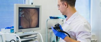
To diagnose colon tumors, X-ray examination (irrigoscopy), endoscopic examination (colonoscopy), digital and endoscopic examination of the rectum (sigmoidoscopy) are used.
Clinical manifestations of colon cancer
Clinical manifestations of colon cancer largely depend on the location of the malignant neoplasm, the degree of spread of the tumor process and the presence of complications aggravating the course of the underlying disease.
The most common symptoms: abdominal pain, impaired motor-evacuation function of the intestine, clinically manifested by alternating constipation and diarrhea, pathological discharge with feces, changes in the general condition of the patient and, finally, a tumor palpable through the anterior abdominal wall.
Abdominal pain is the most common symptom of colon cancer and is observed in almost 80% of patients. In clinical observations with right-sided tumor localization, pain, as one of the first symptoms of cancer, occurred 2-3 times more often than with cancer of the left half. This fact is explained by a violation of motor function: pendulum-like movement of intestinal contents from the small intestine to the cecum and back.
Spasmodic contractions of the intestine, pushing feces through the intestinal lumen partially blocked by the tumor, cause pain. Intratumoral and perifocal inflammation of the intestinal wall, often accompanying disintegrating infected tumors, aggravates pain.
Colon tumors can occur for a long time without pain, and only when the tumor spreads beyond the intestinal wall, when moving to the peritoneum and surrounding organs, pain appears, the intensity and frequency of which may vary. Depending on the location of the tumor, the pain syndrome can simulate chronic appendicitis, cholecystitis, gastric and duodenal ulcers, and chronic adnexitis.
Malignant neoplasms of the right half of the colon are characterized by a combination of pain, hyperthermic reaction (increase in temperature), leukocytosis and rigidity (tension) of the muscles of the anterior abdominal wall . The clinical manifestations of the disease resemble destructive appendicitis, and the correct diagnosis can only be established during an inspection of the abdominal organs during surgery. An analysis of the clinical course of cancer of the right half of the colon showed that in almost 60% of cases the presence of a tumor is accompanied by pain in the right abdomen, intestinal disorders, hyperthermia, symptoms of intoxication and anemia.
This combination of clinical symptoms is characteristic of the toxic-anemic form of colon cancer.
Violations of the motor-evacuation function of the colon lead to stagnation of intestinal contents and cause symptoms of discomfort such as a feeling of heaviness in the abdomen, loss of appetite, and nausea. An important role in the development of intestinal discomfort is played by reflex functional disorders of other organs of the digestive system. Absorption of decay products by the inflamed mucous membrane, a change in the normal composition of the intestinal microflora, accompanied by the appearance of pathogenic strains that secrete exo- and endotoxins, leads to the development of endogenous intoxication syndrome. Functional disorders of the gastrointestinal tract in patients with colon cancer are manifested by impaired passage of contents, constipation, bloating, and paroxysmal pain.
The accumulation of feces above the tumor is accompanied by increased processes of putrefaction and fermentation, leading to bloating with retention of stool and gases.
In cases where the course of the tumor process is complicated by the development of intestinal obstruction, the clinical picture of patients with colon cancer is dominated by symptoms such as bloating with difficulty passing feces and gases, nausea, belching, and vomiting. The pain is paroxysmal in nature. According to some authors, when a malignant tumor is localized in the left half of the colon, the stenotic nature of tumor growth leads to a narrowing of the intestinal lumen, as a result of which feces, accumulating above the tumor, can be palpated through the abdominal wall and are sometimes mistaken for a tumor.
One of the fairly common and relatively early clinical manifestations of colon cancer is pathological discharge from the rectum . These include mucus, blood, pus, tumor masses, etc. Most often, pathological impurities in stool are noted when the colon tumor is located on the left side, rather than when the tumor is located in the right half (62.4% and 18.5%, respectively). Discharges of pus and fragments of tumor masses, indicating the addition of an inflammatory process leading to tumor disintegration, infection and the formation of perifocal and intratumoral abscesses, are noted much less frequently. In any case, the presence of such discharge quite often indicates a widespread tumor process.
One of the symptoms indicating an advanced tumor process is a tumor palpable through the abdominal wall. The frequency of this symptom ranges from 40 to 60%.
Any of the symptoms listed above (pain, intestinal disorders, the presence of pathological impurities in the stool) can be present with any intestinal disease, not just tumors. Analysis of the clinical course of colon cancer indicates a significant percentage of diagnostic errors (up to 35%), leading to hospitalization in general therapeutic and infectious diseases clinics for the treatment of anemia of unknown etiology, dysentery, etc. The percentage of patients hospitalized in general surgical hospitals for emergency indications at altitude remains high obstructive intestinal obstruction.
The following clinical forms of colon cancer are distinguished:
- toxic-anemic, characterized by varying degrees of severity of anemia, general symptoms, intoxication;
- obstructive – characterized by the appearance of signs of intestinal obstruction and accompanied by paroxysmal abdominal pain, rumbling and increased peristalsis, stool retention and poor passage of gases;
- enterocolitic form, accompanied by bloating, alternating diarrhea with constipation, the presence of pathological impurities in the stool, dull, aching pain in the abdomen;
- pseudo-inflammatory form, characterized by low severity of intestinal disorders against the background of signs of an inflammatory process in the abdominal cavity;
- tumor (atypical) form, which is not characterized by general symptoms, intestinal obstruction, with a palpable tumor in the abdominal cavity;
- dyspeptic form, the characteristic features of which are symptoms of gastric discomfort (nausea, belching, feeling of heaviness in the epigastric region), accompanied by pain localized mainly in the upper floor of the abdominal cavity.
It must be emphasized that the identification of clinical forms is, to a certain extent, conditional and mainly characterizes the leading symptom complex. However, knowledge of the manifestations of colon cancer allows us to suspect the presence of a tumor even in cases where the disease occurs with mild intestinal disorders.
Complicated forms of colon cancer
Complications that quite often accompany colon cancer and have a direct impact on the course of the disease and the prognosis of the tumor process include intestinal obstruction of varying severity, perifocal inflammatory process, tumor perforation, intestinal bleeding, as well as tumor spread to surrounding organs and tissues.
According to the literature, the incidence of intestinal obstruction in patients with colon cancer ranges from 10 to 60%. Such pronounced differences in the frequency of this complication are largely due to the fact that the vast majority of patients with a complicated course of the tumor process end up in emergency surgical hospitals, and not in specialized medical institutions.
The clinical course of the disease largely depends on the severity of intestinal obstruction. In case of a decompensated form of intestinal obstruction (severe bloating with retention of stool and gases, vomiting, cramping pain throughout the abdomen against the background of severe metabolic disorders), emergency surgical intervention is indicated, the volume and nature of which depends not only on the location of the tumor, but also on the severity of the developed complications. In cases of compensated form of obstructive intestinal obstruction, conservative measures are often effective in preparing the patient for planned surgery.
The passage of liquid intestinal contents persists when the intestinal lumen narrows to 0.8-1 cm; with cancer of the right half of the colon, ileus (intestinal obstruction) usually occurs with large tumor sizes. As stenosis progresses, an expansion of the intestine above the tumor is formed, leading to the accumulation of feces and the appearance of aching pain in the abdomen, at times cramping and spastic in nature.
When the tumor is localized in the left parts of the colon, the development of intestinal obstruction is often preceded by constipation, alternating with copious foul-smelling loose stools. In cases of decompensated intestinal obstruction, the dysfunction of the gastrointestinal tract organs is quickly joined by metabolic disorders, leading to disruption of the vital functions of organs and systems.
Intratumoral and perifocal inflammatory processes pose a great danger in colon cancer. The frequency of such complications is quite high: from 12 to 35%.
Inflammatory changes in the tumor, caused by the presence of a large number of virulent microorganisms in the intestinal contents, the qualitative and quantitative composition of which changes with the disintegration of tumor tissue, lead to infection and the formation of inflammatory infiltrates and ulcers.
In most clinical observations, histological examination of removed specimens in patients with perifocal inflammatory process revealed ulceration of the tumor and signs of acute purulent inflammation with the formation of abscesses, necrosis and fistulas in the thickness of the adipose tissue, tumor stroma or lymph nodes.
Perforation of the intestinal wall and bleeding from a disintegrating tumor are the most dangerous complications of this disease. Long-term stasis of intestinal contents against the background of chronic intestinal obstruction in combination with trophic disorders of the intestinal wall lead to the formation of bedsores and perforation.
The most unfavorable prognosis is tumor perforation into the free abdominal cavity, leading to diffuse fecal peritonitis. When a segment of intestine devoid of peritoneal cover is perforated, an acute purulent focus forms in the retroperitoneal space. In a number of patients, the pinpoint perforation is covered by the omentum or a nearby organ, leading to the formation of a perifocal inflammatory process that spreads to nearby organs and tissues. Perifocal and intratumoral inflammation, complicating the course of the underlying disease on the one hand, and perforation of a colon tumor on the other, are parts of the same pathological process, which is based on infection of the affected part of the colon with conditionally pathogenic strains of microorganisms penetrating through the pathologically altered intestinal wall .
Diagnostics
Improving the methods of clinical examination of a patient using modern X-ray and endoscopic techniques, and the use of a wide arsenal of screening diagnostic methods, until recently, have not significantly improved the early detection of colon cancer. More than 70% of patients with colon cancer at the time of hospitalization had stages III and IV of the disease. Only 15% of them consulted a specialist within 2 months from the onset of the first symptoms of the disease. In less than half of the examined patients, the diagnosis was established within 2 months from the onset of the disease, and in every fourth case it took more than six months to determine the nature of the disease. Quite frequently occurring diagnostic errors led to unnecessary surgical interventions and physiotherapeutic procedures leading to dissemination of the tumor process.
The diagnosis of colon cancer is made on the basis of x-ray and endoscopic examinations. An equally important method of physical examination of the patient is palpation of the abdomen, which allows not only to identify a tumor in the abdominal cavity, but also to evaluate its consistency, size, and mobility.
According to a number of authors, in 60-70% of cases, a colon tumor is palpable.
Types of studies
- X-ray examination, along with colonoscopy, is leading in the diagnosis of colon cancer.
- Irrigoscopy allows you to obtain information about the localization of the tumor, determine the extent of the lesion, determine the form of tumor growth, assess its mobility, and sometimes judge the relationship with other organs. When performing irrigoscopy, it is also possible to identify synchronous tumors of the colon. The last circumstance is also important because with the stenosing nature of the growth of the neoplasm, endoscopic examination does not allow assessing the condition of the overlying parts of the colon before surgery.
- Endoscopic examination, along with visualization of a malignant tumor, allows one to obtain material for histological examination, which is a necessary attribute of the preoperative diagnosis of a malignant neoplasm.
- The simplest and most widespread method of endoscopic examination of the colon is sigmoidoscopy, in which it is possible to assess the condition of the lower part of the intestinal tube. When performing sigmoidoscopy, the researcher assesses the condition of the colon mucosa, vascular pattern, the presence of pathological impurities in the intestinal lumen, elasticity and mobility of the intestinal wall. When a colon tumor is detected, its size, appearance, consistency, mobility during instrumental palpation are studied, and a biopsy is performed.
Determining the degree of spread of the tumor process
The program for examining the patient before surgery, in addition to the traditional methods already listed, includes special x-ray and radioisotope studies.
Hematogenous metastasis is based on the process of embolization by cancer cells of the venous outflow pathways from the organ affected by the tumor process. Penetration of tumor cells into venous vessels occurs as a result of invasion and destruction of the vessel wall by the tumor. The bulk of venous blood in patients with colorectal cancer enters the portal vein through the system of the inferior and superior mesenteric veins, which explains the fact that distant metastases are mainly localized in the liver.
Ultrasound examination is widely used to assess the extent of tumor spread. It is based on the principle of recording a reflected ultrasonic wave from the interfaces of tissues that differ in density and structure. Having high resolution and information content, ultrasound is a practically harmless diagnostic method that allows you to visualize tumor nodes measuring 0.5-2.0 cm.
The anatomical and topographic structure of the liver and the good propagation of ultrasound in it determine the high information content of the study. It is important that ultrasound helps to determine not only the nature of pathological changes in the liver, but also to establish the localization and depth of focal changes. When performing ultrasound tomography, a layer-by-layer image of the internal structure of the liver is obtained and pathological space-occupying formations or diffuse changes are identified. Ultrasound of the liver can be repeated quite often without harm to the patient’s body, which makes it possible to evaluate the results of the treatment.
The use of X-ray computed tomography (CT) in medicine has contributed to significant improvements in the diagnosis of various pathological conditions.
Computed tomography has the following important advantages over other examination methods:
- presents an image of anatomical structures in the form of a cross section, excluding the combination of their images;
- provides a clear image of structures that differ slightly in density from each other, which is extremely important for diagnosis;
- provides an opportunity to quantitatively determine tissue density in each image area of the organ under study for differential diagnosis of pathological changes;
- It has a non-invasive diagnostic method, safety and low radiation exposure to the patient’s body.
According to the researchers, when analyzing the CT images of metastatic tumors of colorectal cancer, in 48% of cases the tumor nodes contained calcifications, and sometimes total calcification of metastatic tumors was revealed.
Radionuclide (isotope) methods for diagnosing and assessing the extent of colorectal cancer spread are used quite rarely in the daily practical work of medical institutions. One of these methods is positive scintigraphy, based on the use of specific drugs such as gallium in the form of a citrate complex, as well as bleomycin labeled with an indium isotope.
TREATMENT OF COLON CANCER
Choosing the type of surgical intervention and justifying its scope
The history of surgical treatment of colon cancer goes back more than 150 years. Reybard in 1833 performed the first resection of the colon for a malignant tumor with the formation of an interintestinal anastomosis. In Russia in 1886 E.V. Pavlov performed the first resection of the cecum for a malignant tumor with an anastomosis between the ascending colon and ileum . In contrast to manipulations on the small intestine, resection of the colon, according to V. Schmiden (1910), is one of the most important surgical interventions associated with the existence of such features as the presence of pathogenic microflora in the contents of a hollow organ, the absence of a mesentery in fixed areas of the colon intestines, a thinner layer of muscular tissue. These features of the colon predetermine increased demands on the reliability of the formation of interintestinal anastomoses, taking into account the anatomical features of various parts of the colon and the adequacy of the blood supply to the anastomosed segments.
The main disadvantage of these surgical interventions is the presence of a temporary colostomy - the removal of the intestine to the anterior abdominal wall. Therefore, in specialized oncoproctology clinics, the indications for performing two-stage surgical interventions are being rethought, considering them justified only in weakened patients with symptoms of decompensated intestinal obstruction.
The volume and nature of surgery for colon cancer depends on a number of factors, among which the most important are the location, extent of tumor spread, the presence of complications of the underlying disease, as well as the general condition of the patient.
Choosing the type of surgical intervention for complicated colon cancer
Most patients with colorectal cancer are admitted to specialized medical institutions in stages III and IV of the tumor process. Many of them experience various complications (obstructive form of intestinal obstruction, tumor perforation, bleeding and perifocal inflammatory process), often requiring emergency surgical intervention.
The results of surgical interventions in patients with complicated colorectal cancer depend to a certain extent on the qualifications of the operating surgeon, his ability to assess the degree and severity of the pathological process complicating the course of the underlying disease, and taking into account the general condition of the patient.
When choosing the type of surgical intervention, they strive not only to save the patient from an acute surgical complication, but also, if possible, to perform a radical operation.
One of the most dangerous complications of colon cancer is perifocal and intratumoral inflammation, often spreading to surrounding tissues. The frequency of this complication is quite high and ranges from 6% to 18%. This complication is manifested by the clinical picture of acute inflammation and intoxication, and the spread of the process to neighboring organs and surrounding tissues contributes to the formation of infiltrates, abscesses, and phlegmons. Often, a pronounced inflammatory process in the tumor and surrounding organs is interpreted as tumor infiltration, which is the reason for the inadequate scope of surgical intervention.
The presence of perifocal and intratumoral inflammation in colon cancer has a significant impact on the choice of the volume and nature of surgical intervention only in cases where the inflammatory process spreads to surrounding organs and tissues, and forces the use of combined surgical interventions.
Combined operations for colon cancer
Expanding the scope of surgical intervention due to the spread of a malignant tumor to nearby organs and tissues increases the duration of the operation, trauma and blood loss. Extension of the tumor beyond the intestinal wall indicates an advanced neoplastic process, but the absence of distant metastases makes it possible to perform a combined operation, which, while improving the quality of life of patients, eliminates severe complications of the tumor process and creates real preconditions for the use of specific methods of antitumor treatment.
Palliative surgical interventions in patients with colon cancer
Almost 70% of patients with colon cancer at the time of surgical intervention are diagnosed with stages III and IV of the disease, and in every third patient among those operated on, distant metastases are diagnosed, mainly in the liver and lungs. The development of intestinal obstruction forces one to resort to symptomatic surgical interventions - colostomy, formation of bypass anastomosis in patients with stage IV of the disease. However, more and more surgeons for advanced colorectal cancer prefer palliative resection or hemicolectomy .
Palliative resection of the colon or hemicolectomy significantly improves the quality of life, relieving the patient of such complications of the tumor process as purulent-septic complications, bleeding, tumor disintegration with the formation of a fecal fistula.
A comparative analysis of the immediate and long-term results of treatment of patients with colon cancer who underwent resection or hemicolectomy, regardless of whether the operation was radical or palliative, showed that the frequency and nature of postoperative complications were approximately the same.
Palliative surgical interventions in the form of resection or hemicolectomy are finding more and more supporters and are increasingly the operation of choice for metastatic colon cancer. This was facilitated by a decrease in the incidence of postoperative complications and mortality, and an expansion of indications for resection of organs affected by metastases (liver, lungs). When determining indications for palliative surgical interventions such as colon resection or hemicolectomy, both the general condition of the patient and the degree of tumor dissemination are taken into account.
One of the important factors influencing the prognosis of the disease in patients undergoing liver resection for metastases is the time interval between treatment for the primary tumor and detection of liver metastases. It has been established that the longer the duration of the relapse-free course of the tumor process, the more favorable the prognosis for surgical treatment of liver metastases.
When determining the extent of surgical intervention for metastatic colorectal cancer, studying the functional state of the liver plays an important role. Liver failure itself is one of the main causes of postoperative mortality in major liver resections. The liver is an organ with great compensatory capabilities. 10-15% of its healthy parenchyma is enough for the full functioning of the organ.
An important issue for determining surgical tactics is the number of metastatic nodes in the liver. Multiple nodes significantly worsen the prognosis and are one of the main reasons for refusing active surgical tactics. However, the presence of multiple nodes localized in one anatomical half of the liver is not a contraindication to surgical treatment, although, of course, the prognosis in such patients is much worse than with a single and single (2-3 nodes) metastases.
Combination treatment of colon cancer
The reasons for failure of surgical treatment of patients with colon adenocarcinoma are local relapses and distant metastases. Unlike rectal cancer, with this disease local relapses are relatively rare, and liver metastases predominate. In patients with stage III colon cancer, local relapses occur in 7% of cases, and distant metastases in 20%. The occurrence of these unfavorable secondary tumor formations is due to the dissemination of tumor cells during surgery. Preoperative radiation therapy, which has recently begun to be introduced into the practical activities of oncoproctology clinics, can increase the ablasticity of surgical interventions.
Depending on the sequence of application of ionizing radiation and surgical intervention, pre-, post- and intraoperative radiation therapy is distinguished.
Preoperative radiotherapy
Depending on the purposes for which preoperative radiation therapy is prescribed, two main forms can be distinguished:
- irradiation of operable forms of colon cancer;
- irradiation of inoperable (locally advanced) or doubtfully operable forms of tumors.
The death of tumor cells as a result of radiation exposure leads to a decrease in tumor size and separation from surrounding normal tissues due to the proliferation of connective tissue elements (in cases of prolonged preoperative irradiation and delayed operations). The realization of the positive effect of preoperative radiation therapy is determined by the magnitude of the radiation dose.
Clinical studies have shown that a dose of 40-45 Gy leads to the death of 90-95% of subclinical growth lesions. A focal dose of no more than 40 Gray, administered at 2 Gray daily for 4 weeks, does not cause difficulties in performing subsequent surgery and does not have a noticeable effect on the healing of the postoperative wound.
Postoperative radiotherapy
Certain advantages of postoperative radiotherapy are:
- planning the volume and technique of irradiation is carried out on the basis of data obtained during surgery and after a thorough morphological study of the removed tissues;
- there are no factors that have a negative impact on the healing of postoperative wounds;
- surgical intervention is performed as quickly as possible from the moment of clarifying diagnosis of the disease.
To achieve a therapeutic effect during postoperative radiation therapy, high doses are required - at least 50-60 Gray.
The presence of inflammatory phenomena in the surgical area, disruption of blood and lymph supply leads to a delay in the supply of oxygen to tumor cells and their complexes, which makes them radioresistant. At the same time, normal tissues in a state of regeneration become more radiosensitive, namely, they must be included in a larger volume in the target for postoperative irradiation, because it is necessary to influence the tumor bed, the entire postoperative scar and areas of regional metastasis.
Symptoms and signs
Malignant tumors localized in the cecum are characterized by a long asymptomatic course. Rapidly progressing subjective discomfort is observed in the later stages of the disease and does not have pathognomonic specificity that would indicate the localization of the tumor in the cecum. Patients present the following complaints:
- Nausea, poor appetite, belching;
- Flatulence, apparently inexplicable alternation of diarrhea and constipation;
- Pain in the right side of the abdomen.
As the tumor increases in size and is damaged by stool, bleeding progresses. It can remain hidden for a certain period and manifest itself with increasing symptoms of anemia and asthenia:
- Apathy;
- Unsteadiness;
- Weakness, fatigue;
- Pale earthy skin color;
- Emaciation.
Frequent or regular discharge of red blood during bowel movements is one of the typical symptoms of colorectal cancer of any location. In the terminal stage of the disease, the following signs of cecal cancer are usually associated:
- Intense pain caused by tumor growth into surrounding structures and pressure on neighboring organs;
- Phenomena of partial intestinal obstruction or complete occlusion of the intestinal lumen;
- Jaundice and liver failure, since the structure of the intestinal system of lymph circulation and blood supply determines the metastasis of the tumor process to the liver.
Diagnostics
The preliminary diagnosis of “cecal cancer” is established by specialists at the oncology clinic during the collection of complaints and anamnesis, and clinical examination. A large tumor can be detected during palpation of the abdomen. For the purpose of differential diagnosis, to clarify the location, shape and size of the tumor, and to identify metastases, additional research methods are carried out:
- Colonoscopy;
- Irrigoscopy;
- Computed tomography;
- Ultrasound screening;
- Diagnostic laparoscopy.
During an endoscopic or laparoscopic examination, doctors necessarily select material for histological analysis, which allows them to come to unambiguous diagnostic conclusions. The most informative method for diagnosing sigmoid colon cancer is rectoscopy. With sigmoidoscopy, up to 25 cm of the distal colon is examined.
The use of a flexible sigmoidoscope and colonoscope allows for more accurate preoperative diagnosis of cecal cancer. The X-ray method using a double contrast enema has great sensitivity. It allows you to detect small tumors. A malignant neoplasm manifests itself in the form of a characteristic narrowing or compaction, which is located in the contrast zone. In doubtful cases, doctors at the Yusupov Hospital repeat the examination or perform a colonoscopy.
Scanning computed tomography with air contrast is becoming increasingly widespread. This method is used when making a final decision about the need for surgical intervention. At the Yusupov Hospital, spiral computed tomography with a small slice thickness, the so-called “virtual colonoscopy,” is widely used to detect cancer of the cecum.
Colon carcinoma cells produce carcinoembryonic antigen (CEA), a tumor marker for cancer. However, it is not specific enough to serve as a reliable indicator of the existence of a tumor. Carcinoembryonic antigen is also found in pancreatitis, inflammatory bowel processes, in smokers and in people who abuse alcohol. The CEA test is used in patients with initially high levels of this tumor marker after surgery. Its level decreases after successful surgery, and an increase in CEA concentration in the postoperative period may be the first sign of tumor relapse.
Doctors at the Yusupov Hospital carry out differential diagnosis of cecal cancer with the following diseases:
- Diverticulosis of the colon;
- Ulcerative and ischemic colitis;
- Irritable bowel syndrome.
Other diseases manifested by rectal bleeding (hemorrhoids, polyposis) make diagnosis difficult. Pain in the right half of the abdomen may indicate the development of acute appendicitis. If the patient has positive symptoms of an “acute abdomen,” he undergoes urgent surgery, during which the true cause of the pain syndrome is determined.
Tubular adenoma of the cecum is a benign neoplasm. It may present with symptoms that resemble those of a cancerous tumor.
Make an appointment
Symptoms and diagnosis of cecal tumors
It is necessary to identify the problem as early as possible. Chronic constipation, feelings of bloating, heaviness in the stomach and incomplete bowel movements, mucus, pus or blood in the stool, nausea and vomiting - with these signs you need to consult a doctor to establish a diagnosis. As colorectal cancer develops, symptoms include weakness and fatigue, sudden loss of appetite and weight, a slight increase in temperature, severe pain, intestinal obstruction and disturbances in the functioning of other organs.
For diagnosis, tapping, palpation, digital examination, urine, blood, stool tests, CT and MRI, colonoscopy, irrigoscopy, sigmoidoscopy, cytoscopy, and ultrasound are used. This allows you to clarify the location and size of the tumor, its features, identify damage to other organs and the presence of metastases.
Our expert in this field:
Ivanov Anton Alexandrovich
Medical director, oncologist-surgeon, candidate of medical sciences
Call the doctor
Call the doctor
Treatment
One of the key features of cecal cancer is that it is completely and relapse-free in almost 93-97% of cases. This applies only to the initial stages of the disease. Early diagnosis of a malignant tumor is a vital issue. For this reason, doctors at the Yusupov Hospital, if there are symptoms of intestinal diseases, first of all exclude cancer.
Treatment of a tumor of the cecum is always intensive and combined. Oncologists at the Yusupov Hospital use all three main areas of modern oncology - surgery, radiation treatment and chemotherapy.
The priority treatment for this type of cancer is surgical removal of the tumor. Laparoscopy is used to perform surgeries in the initial stages of tumors. During endoscopic operations, surrounding tissues are minimally damaged.
For growing tumors, a wide resection of the affected cecum and mesentery is performed, and the lymphatic apparatus is removed. A right hemicolectomy is performed: the surgeon removes up to 20 cm of the terminal ileum, the right half of the colon, including the ascending colon, the cecum, the right third of the transverse colon and the hepatic curvature.
In case of multiple damage to organs and tissues by metastases, surgery to remove tumors is not performed; severe symptoms - intestinal obstruction - are eliminated; antibiotic therapy, chemotherapy and palliative treatment are prescribed. Good prognosis for patients with no metastases in regional lymph nodes.
Conservative therapy for cecal cancer at the Yusupov Hospital is carried out if surgical intervention is not possible. Doctors at the Oncology Clinic use the latest equipment from leading global manufacturers for radiation therapy and the latest generation of antitumor drugs. They are effective and have a minimal range of side effects.
Exposure to cytostatic drugs can reduce the size of the tumor, which increases the chances of its successful removal. Systemic chemotherapy destroys the smallest cancerous foci and prevents tumor recurrence after surgery. Depending on the histological nature of the tumor, the patient’s response to chemotherapy drugs, doctors at the Yusupov Hospital use monochemotherapy (prescribe one cytostatic drug that effectively affects a given type of cancer cell) or polychemotherapy - a combination of several different types of drugs that have a destructive effect on the tumor.
Radiation techniques are used in preparation for surgery as a way to reduce tumor mass. At the Yusupov Hospital, radiotherapy is performed using modern equipment. Linear accelerators generate radiation that, with extreme precision, only affects mutated tissue without affecting healthy cells. The power of the equipment allows you to reduce the time of the session, thereby the effect on the entire body is more gentle.
Treatment of cecal cancer includes radiosurgical methods, in particular, the use of a Cyber Knife. The installation allows you to focus 150-300 thin rays at one point. They hit the desired target from different angles, without having a detrimental effect on healthy tissue. Where the rays intersect (in the tumor of the cecum) a high level of radiation is created. Thanks to high-dose precision irradiation, the number of sessions can be reduced to 2-5.
In some cases, doctors use specialized radiation therapy methods: intraoperative radiation therapy or brachytherapy. These types of radiation treatments help get rid of small tumors that cannot be removed with surgery. Intraoperative radiation therapy is performed during surgery. In this case, a high single dose of radiation therapy is used to destroy a hard-to-reach tumor of the cecum.
Brachytherapy is carried out using tiny SIR spheres containing a radioactive substance called yttrium-90. It is used for secondary liver cancer arising from a malignant tumor of the cecum, when surgery is not an option. The method allows you to slow down the growth of cancer cells.
If cecal cancer is at the first stage of development, there is no tumor growth into neighboring organs or metastases, laparoscopic tumor removal is performed at the Yusupov Hospital. This is a minimally invasive method that is most gentle for the patient. The surgeon does not need to make extensive incisions. Surgical intervention is carried out through several punctures of the anterior abdominal wall.
For more extensive lesions, oncologists excise the damaged area of the intestine with adjacent tissues and perform an anastomosis - restoring the integrity of the intestine, creating a bypass for food, suturing the two sections of the intestine. In some cases of cecal cancer, in order to avoid tumor spread to neighboring organs, surgeons perform resection of regional lymphatic vessels and nodes along with the tumor, en bloc. If it is not possible to save the organ, surgeons remove not only the cecum, but also a section of the small or large intestine. Next, a colostomy is formed - an opening for the removal of intestinal contents. Through it, feces enter the colostomy bag, which the patient wears.
In some cases, a colostomy is performed for a short period of time to optimize the bowel healing process. It can also be permanent. With modern surgical methods used by oncologists at the Yusupov Hospital, and the use of radiation therapy, treatment with modern chemotherapy drugs before surgery, most patients with cecal cancer do not require a permanent colostomy. The competent use of antiblastic techniques by surgeons at the oncology clinic, cleaning the tumor removal site from the slightest remaining atypical cells, significantly reduces the risk of relapse in patients at the Yusupov Hospital.
4.Treatment
One of the key features of cecal cancer is that it is completely and relapse-free in almost all cases (various sources give estimates of 93-97%). However, this applies only to the initial stages - which makes early diagnosis a matter of vital importance - and does not apply to any non-medical practices, the only proven result of which is financial profit for some people and a suicidal waste of time for others.
Treatment is always intensive and combined and involves the use of all three main areas of modern oncology - surgery, radiation therapy and chemotherapy.
Prognosis for cecal cancer
The prognosis for malignant neoplasms of the cecum depends on the depth of germination of the primary tumor, the presence of regional and distant metastases. The five-year survival rate directly depends on the stage of cecal cancer. This figure has increased over the past few decades. This is due to careful diagnosis and modern treatment methods used by oncologists at the Yusupov Hospital.
The patient's prognosis after tumor excision depends not simply on the presence or absence of metastases to regional lymph nodes, but on the number of affected lymph nodes. Unfavorable factors that worsen the prognosis for cancer of the cecum include tumor growth into fatty tissue, perforation of the colon, low degree of differentiation of cancer cells, spread of cancer to adjacent tissues and organs, and spread of the tumor into the lumen of large veins.
The five-year survival rate for stage II cecal cancer is 85%. If a diagnosis of stage 3 cecal cancer is established, the prognosis for five-year survival is 74%, and with the fourth stage of the tumor process, 6% of patients survive up to five years. For this reason, oncologists at the Yusupov Hospital do not recommend postponing a visit to the doctor if signs of intestinal discomfort appear. Call at any time of the day. The contact center of the Yusupov Hospital is open seven days a week and without a lunch break.
Make an appointment
1.General information
There are several main sections in the human large intestine: colon, the longest intestine; the sigmoid colon as the lower continuation of the colon and the transition to the rectum; the rectum ending in the anus. The cecum refers to the initial section of the large intestine. It is located on the right and, due to its structure, is most often compared to a vertical bag. In the lower part of the cecum there is a vermiform appendix (literally “appendage”), which for a long time - and, as later studies showed, erroneously - was considered a functionally useless rudiment.
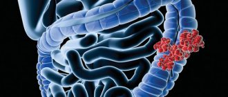
Colon cancer today is an acute, globally significant problem, occupying a leading position in the overall volume of oncopathology and in the lists of causes of mortality; Moreover, mortality in a given localization of the tumor process tends to increase. Even the most developed countries of the world, forced to expend enormous resources and efforts on implementing national screening programs for early diagnosis (which is critically important in this case), are with great difficulty and so far only slightly restraining this alarming trend, due to a number of factors of modern civilization.
It is known that the growth of a malignant neoplasm can begin in any part of the gastrointestinal tract (as, in fact, in any zone of the body in general), however, due to anatomical and functional features, this probability, etiopathogenesis and risk factors for different localizations are not the same. Cecal cancer accounts for approximately one fifth of all colon cancers.
A must read! Help with treatment and hospitalization!