Pityriasis versicolor or pityriasis versicolor is a superficial skin lesion caused by a yeast-like fungus of the genus Malassezia.
These microorganisms are representatives of typical skin microflora, are found in most people and do not cause the development of the disease. But under the influence of external or internal factors, the pathogen can transform from a non-pathogenic form into a pathogenic one: the fungus penetrates the upper layers of the skin and damages melanocytes - the cellular structures responsible for the pigmentation of the skin.
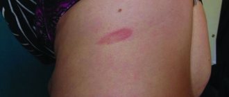
As a result, the patient develops small spots of different colors: from pale pink to red-brown, with noticeable peeling in the center. When touched, the scabs flake off easily and resemble flakes, which is what gives pityriasis versicolor its name.
What is pityriasis versicolor
Pityriasis versicolor, or pityriasis versicolor, is a chronic skin disease of a fungal nature. It appears in the form of specifically pigmented spots of various sizes and shades, without signs of inflammation. Most often, the areas affected by pityriasis versicolor are located on the body (back, abdomen, neck, shoulders) and scalp. Representatives of both sexes are most susceptible to the disease at a young age, and children under 7 years of age are the least susceptible.
External manifestations of the disease usually do not cause concern, so they are often perceived as harmless cosmetic defects. For this reason, the pathology becomes chronic: spots periodically appear, then disappear, and after some time, under the influence of provoking factors, the disease worsens again.
annotation
The genus consists of 14 Malassezia
of yeast-like fungi, 13 of which are lipophilic and 1 is non-lipophilic.
They are commensals and in predisposed individuals they usually cause a range of chronic, recurrent infections. In rare cases, they cause such serious diseases as infections that occur during vascular catheterization, during hemodialysis, and can cause peritonitis, etc. Although these mushrooms have been known to mankind for more than 150 years, due to the whimsical nature of mushrooms, their bulky cultures, and the ability to speciate, they have not been sufficiently studied. Since the last taxonomic revision, 7 new species have been added to this genus. Their ability to evade host immunity and increase in virulence, the range of diseases they cause has increased. Recently, Malassezia
have been implicated as a causative agent in the common disease atopic dermatitis. Although research on fungal cultures is complex, new molecular analysis techniques and advances in instrumentation have prompted an increase in research in this area. Therefore, we can pay more attention to this type of fungi to study in detail their characteristics and their growing impact on the disease clinic.
What was known?
Malassezia
causes superficial mycoses, but systemic infections are rare. They do not have descriptions of drug resistance.
Introduction
Malassezia
(formerly known as Pityrosporum) forms a cutaneous synanthropic flora that is associated with a variety of clinical manifestations, ranging from benign skin diseases such as pityriasis versicolor to hematogenous fungal infection in the immunocompromised individual.[1]
Currently, 14 species have been described, namely Malassezia furfur
,
Malassezia pachydermatis
,
Malassezia sympodialis
,
Malassezia globosa
,
Malassezia
o
btusa
,
Malassezia restricta
,
Malassezia slooffiae
,
Malassezia equina
Malassezia dermatis
, Malassezia japonica
, Malassezia nana
,
Malassezia capre
,
Malassezia yamatoensis
, recently
Malassezia cuniculi
was discovered .[2] Due to their lipophilic nature, these fungi are able to colonize seborrheic areas of the skin and survive on the fatty acids normally present in sebum.
They cause the following skin diseases: hyperkeratosis, invasion of hair follicles and inflammation. Although, by definition, superficial mycoses do not extend beyond the horny epithelium, these fungi are detected in the mouth, central and deep layer of the hair follicle. Malassezia
infections can be superficial and localized, and can also be systemic in immunocompromised people.[3]
Spread on skin
Malassezia skin colonization density
depends on age, body area and comorbid skin conditions, as well as on the person’s place of residence.
Being lipophilic, the highest prevalence of Malassezia
is found in sebaceous areas of the skin such as the scalp, face and upper torso.
Malassezia
most often found in younger people, who tend to have oilier skin.[3]
Factors affecting the reproduction of Malassezia
Geographical factor: depending on the geographical area, the density of different species of the genus Malassezia
on the skin.
The most favorable zones for the growth of Malassezia
are subtropical and tropical climates.
Research shows that the growth of Malassezia globosa
increases during the summer when temperatures are high and sweating is increased.[4]
Age factor: pityriasis versicolor affects people from 20 to 40 years old. However, in India, it is seen in people in the age group of 10 to 30 years. Pityriasis versicolor is rare in children and the elderly. [5,6,7,8]
Hormonal factor: corticosteroid therapy, malnutrition, increased serum cortisol concentrations contribute to the occurrence of pityriasis versicolor in patients.[9]
Pathogenesis: Human sebum, which is a complex mixture of lipids, is the source of nutrition for this type of yeast. Sebum consists of triglycerides of fatty acids, waxes, esters, sterol esters, cholesterol, cholesteryl esters and squalene. Sebum is utilized by breaking down triglycerides and esters into diglycerides, monoglycerides and free fatty acids. Thus, changes in the secretion of sebum and its breakdown causes the development of dandruff. Malassezia infection
, is also associated with increased sweating.[10
a
,
b
,11,12,13]
Clinical scenarios
Yeast of the genus Malassezia
cause the development of chronic recurrent dermatoses such as pityriasis versicolor, seborrheic dermatitis and folliculitis.
Malassezia
has recently been shown to play a role in the pathogenesis of atopic dermatitis and psoriasis, especially in cases involving the scalp.
In addition, studies mention Malassezia
, which is the causative agent of confluent reticular papillomatosis and onychomycosis.
This yeast is also associated with catheter-associated infections among neonates receiving total parenteral nutrition and in frail older adults receiving parenteral lipid supplements. Also, such patients may experience hematogenous fungal infection and sepsis caused by Malassezia furfur
and
Malassezia pachydermatis
.[1,14]
Pityriasis versicolor
Malassezia infection
.
Pityriasis versicolor, also known as pityriasis versicolor, is a common superficial mycosis that is a chronically recurring infection of the stratum corneum of the skin. It is characterized by small white scales, hypo- or hyperpigmented macules, which are unevenly distributed throughout the body and are most often found on oily areas of the skin on both the trunk and extremities. Some patients may experience itching, but most patients are asymptomatic.[1] The Latin name for pityriasis versicolor (or multicolored) is pityriasis versicolor
.
It comes from the Greek. “ P ityron
” - “flakes, scales.”[3,15]
Back in 1871 Neumann
et al.[16]
revealed that it is the mycelial forms of Malassezia
that are responsible for the appearance of pityriasis versicolor.
Successful cultivation of Malassezia
is described in the works
of Neumann
et al.
and Gordon
et al.[17]. It was also found that yeast forms can be seen both on areas of skin affected by pityriasis versicolor and on healthy areas of skin. It was found that in order for the clinical picture of pityriasis versicolor to manifest itself, the necessary conditions are a large proliferation of fungal filaments, and they must secrete several serovar A antigens.[1]
Differential diagnosis
Skin lesions caused by pityriasis alba, pityriasis rosea, vitiligo, hypopigmented mycosis fungoides, erythrasma and seborrheic dermatitis look similar.
Diagnosis
Pityriasis versicolor is diagnosed clinically; only in rare cases is a biopsy or Malassezia
. Under a Wood's lamp, pityriasis versicolor fluoresces yellow. Examination of mushrooms with 10% potassium hydroxide solution shows single-celled, short, curved, straight mycelium filaments and spore clusters. All of the above should resemble “meatballs and spaghetti.”
Histopathological examination
Although pityriasis versicolor is rarely biopsied, it has spores (yeast form) and short hyphae (mycelium) in the stratum corneum of the skin, which are detected using diazonium blue, periodic acid-Schiff, or silver nitrate-methenamine complex. Moderate hyperkeratosis and acanthosis are rare.[3]
Malassezia species that do not cause pityriasis versicolor
Pityriasis versicolor is caused by several species of Malassezia
[18,19,20,21],
M. globosa
is the most common causative agent of lichen with a probability of 58.2%, 55%, 55% and 53.3%, respectively.
A study conducted in north central India found 54% M. globosa
, and 30%
M. furfur
.
Several studies have shown that M. furfur
predominantly causes pityriasis versicolor.
Makimura
et al.
identified M. furfur
and
M. sympodialis
in temperate climates;
however, in tropical countries, they isolated M. globosa
.[22,23,24]
Pathogenicity of M. globosa
due to its esterase and lipolytic activity. Unlike other mycoses, here, in the center of the lesions there are more viable cultures.[25]
Seborrheic dermatitis
Seborrheic dermatitis is the second most common infection associated with Malassezia
, which was originally described by
Unna PG
in 1887. It is a superficial eczematous dermatitis, can be subacute or chronic, and is characterized by the presence of an erythematous plaque with dry or greasy scales. It is found in areas of the skin rich in sebaceous glands, such as the scalp, face, ears, chest and armpits. Weak corticosteroid drugs have proven effective in treatment.
However, the relapsing nature of this disease makes it difficult to treat. Antifungal therapy should be the mainstay in the treatment of this disease, since the use of corticosteroids does not reduce the number of Malassezia
.[3,26]
Seborrheic dermatitis is common during periods of a person's life when their glands are most active:[15]
- First 3 months of life (infantile seborrheic dermatitis)
- During puberty
- When sebum production declines after age 50.[15]
This disease affects 3-5% of the population, with more men than women. In immunocompetent individuals, seborrheic dermatitis usually begins during periods of high sebum secretion, i.e. during puberty and become chronic and recurrent forms. Stress is known to aggravate this type of dermatitis. In patients with AIDS, resistance to antifungal therapy for seborrheic dermatitis is observed from 30 to 80%, in contrast to immunocompetent individuals. [27]
In HIV-infected patients, the appearance of seborrheic dermatitis is a surrogate marker of CD
4+ T lymphocytes.
M.
restricta
and
M. globosa
are observed primarily on the scalp, both during health and illness.[28]
Pathogenesis
The immunopathogenesis of seborrheic dermatitis is complex and multifactorial.
It has been proven that the increased density of colonies is higher in damaged skin than in undamaged skin. However, techniques must be standardized to prove this. Recently, “quorum sensing”—the ability of some organisms to communicate and coordinate their behavior through the secretion of molecular signals—has been actively discussed in science. Malassezia
also operates according to the “quorum” principle.
Also Malassezia
has a complex antigenic structure, represented by both protein and carbohydrate antigens, which are associated with the stages of their life cycle;
protein antigens appear in the early stages of growth and are associated with the development of cell walls and cytoplasmic organelles, while carbohydrate antigens are derived from mannan and mannoproteins. Research by B ergbrant
et al.
showed an increase in NK
cells, impaired T cell function in addition to an increase in total serum
IgA
and IgG among
patients
with seborrheic dermatitis.
However, the titer of Malassezia
-specific antibodies was not elevated, so it was suggested that hypergammaglobulinemia may occur as a reaction to toxins contained in yeast and to lipases.
People suffering from dandruff have loosely connected corneocytes or a small number of dysmosomes. The level of secreted lipids is reduced, which in turn affects the epidermal water barrier. Itching associated with histamine also increases, causing damage and reducing the effectiveness of the skin's defense mechanism. All this makes it easier for Malassezia
into the skin.
Malassezia
produces lipases that break down sebum into arachidonic and oleic acids, which are irritants and have a desquamative effect on keratinocytes. Thus, this contributes to the onset of the inflammatory process and creates vicious circles.
In some cases Malasseziae
have the ability to exist as pathogenic microorganisms that cause seborrheic dermatitis, and in other cases exist as normal flora. They are capable of causing an immune response. They activate the classical and alternative pathways of the complement cascade. Complement component C3 has been found in damaged skin and has been postulated as one of the causes of inflammatory reactions. Although yeast should ideally be phagocytosed and eliminated by our immune system, it is capable of suppressing the secretion of cytokines and interleukins.
Malassezia
alters and evades the local immune response by producing secondary metabolites that contribute to seborrheic dermatitis.[28] Environmental factors, stress, UV radiation, climate change, hormonal changes and trauma also likely contribute to the maintenance of seborrheic dermatitis.[28]
Milky crusts: Infantile or neonatal seborrheic dermatitis is also called pityriasis capitis, which is a nonpruritic dermatosis ( Cradle
cap directory
,
WebMD
.
Retrieved
August
26 , 2012).
Infantile seborrheic dermatitis usually occurs from the 2nd week of life to 6 months. The rashes can be eczematous and psoriasis-like in nature, rare localizations of seborrheic dermatitis are the face, torso and sternum area. The lesions can be single or multiple, and can merge, especially on the face in the fold area.[15]
Neonatal cephalic pustulosis (neonatal acne)
A child has skin colonization with Malassezia
begins on the first day of life.
Malassezia
is transmitted to the baby from the mother and from health care workers, and colonization increases during the first weeks of life.
Environmental factors and maternal hormonal levels influence the colonization process. The high prevalence of M. sympodialis
in newborns is observed from birth and is the cause of neonatal acne.[29]
Folliculitis caused by Malassezia
Synonyms: pityrosporum folliculitis.
This intensely pruritic, follicular papulopustular rash is seen usually on the upper torso in the young age group and is rare in older people.
This disease was first described in 1969 by Weary
etal., based on the histological ultrastructure
of Potter
etal. in 1973 it was discovered that it was a separate type of disease. This is a chronic infection in which patients typically present with erythema, papules and pustules on the chest or back, and a follicular pattern. The distinctive symptom is itching. Rare localizations are the face, neck, shoulders and arms.[6,15,30,31]
Risk factors for folliculitis:[32,33,34,35,36,37]
- Immunosuppression
- Diabetes
- Broad-spectrum antibiotics
- Steroids
- Puberty,
- Pregnancy
- Cosmetics, lotions, sunscreens, emollients, olive oil, which are the cause of skin clogging.
Diagnosis: The diagnosis of folliculitis is based on the clinical picture and positive response to antifungal therapy.
Some differences in the distribution of Malassezia
according to epidemiological data:
Low number of Malassezia
, compared to normal skin, causes other diseases.
There are pathogenic strains that are not isolated in psoriasis.
Histopathological findings: According to microscopy, the discharge from the pustules represents budding yeast forms and spores, and not hyphae, as in the study of pityriasis versicolor. to diagnose pityrosporum
folliculitis
, but histology shows dilated hair follicle openings with large amounts of cellular material and keratin plugs.
The inflammatory response is caused by lymphocytes, histiocytes and neutrophils. The mouth of the sebaceous gland ducts is surrounded by monocytes and mucin deposits. Yeasts of the genus Malassezia
can be detected in hair follicles; however, they are more often found in the orifices of the sebaceous gland ducts. Periodic acid-Schiff and Grocott-Gomori staining can be used to identify unipolar budding yeast cells; mycelial forms were not detected by these methods. [38],[39],[40]
Treatment
Treatment for pityrosporum
follicilitis
uses oral antifungal medications, since topical medications do not penetrate the hair follicles. Since there is a high relapse rate, there is a need for maintenance therapy.
Atopic eczema/dermatitis syndrome
Atopic dermatitis is a chronic, inflammatory skin disease. Patients with atopic dermatitis often have bronchial asthma. The pathogenesis is not entirely clear. Patients suffer from itching, which may be the body's response to irritants or microorganisms, due to the loss of surface barrier mechanisms in dry skin [Fig. 1]. [3],[41],[42]
Rice. 1: Malassezia
in the pathogenesis of atopic eczema/dermatitis syndrome
After the successful therapeutic use of ketocanazole in the treatment of atopic dermatitis, a number of studies have proven the role of Malassezia
as one of the etiological factors in the occurrence of exacerbation of atopic dermatitis. [43]
Their multilayer membranes and cell wall mannans contain antigens. of Malassezia among patients
-specific
IgE
, and they have positive skin prick tests.
Malassezia
may act as a persistent allergic irritant.
The combined cellular and humoral immune response leads to increased sensitization. The cellular response leads to the activation of T h
2 lymphocytes, this leads to an increase in the levels of IL 4 and IL5 compared to the control group of healthy people.
Due to allergen-specific IgE
and cytokines release histamine from mast cells.
Symptoms of itching due to histamine release lead to further sensitization of Malassezia
.
Antifungal drugs lead to a decrease in the yeast population on the skin, thereby reducing the antigenic load, which prevents the inflammatory process.
IgE
specific are determined to
M. sympodialis
,
M. furfur
and, more recently, to
M. globosa
.
Isolation and speciation
Several studies have been conducted on M. globosa
,
M. restricta
,
M. sympodialis
and
M. furfur
.
M. globosa
the allergen is responsible for the production of heat shock protein 70.
In 2002 Sugita
et al.
described new species
of
M. dermatis
isolated from patients with atopic dermatitis in Japan.
However, the researchers suggest that more studies should be conducted in other countries as well to confirm the fact
that
M. dermatis
can be detected in patients with atopic dermatitis not only in Japan.
The pathogenesis of the disease is not very clear. Genus Malassezia
promotes the production of
IgE
-specific antibodies, and this plays an important role in pathogenesis.
More research is needed to understand the role of Malassezia
in the mechanisms of disease, chronicity and exacerbation. [3],[15],[41],[42],[43],[44],[45],[46],[47],[48],[49],[50],[51 ],[52]
Psoriasis
It has been documented that chronic plaque psoriasis and psoriasis vulgaris are caused and aggravated by Malassezia
.
Rivolta
et al.
in 1873 he first proved this fact. Later, in 2007, Narang
et al. [53] treatment with fluconozole is effective for psoriasis.
Among patients with psoriasis, especially those whose affected area involves the scalp, eyebrows, ears, and trunk Malassezia
plays a big role.
Elewski
et al.
in 1990 [54] reported a case of guttate psoriasis in areas affected by Malassezia folliculitis
.
Some differences have been noted in the epidemiological data and distribution of the genus Malassezia
.
The pathogenesis is not fully understood. Triggers of T cell activation and cytokine release are multifactorial - stress, infections, trauma, drug use, etc.
Autoantigens: 13 keratin has homology to K17, which is an identified candidate autoantigen that is not normally found in adults but appears in response to injury.
This explains the Koebner phenomenon, where new lesions appear at sites of injury. Neutrophils are found in psoriasis in the skin infiltrate and increase chemotoxic reactions.
This phenomenon does not occur when the skin is damaged by staphylococci.
Superantigens are those antigens that cause a cytokine storm. Superantigens are staphylococci and streptococci, which aggravate the course of atopic dermatitis, guttate psoriasis and chronic psoriatic plaques.
It can be said that the superantigen promotes inflammation by activating complement, producing inflammatory cytokines and recruiting neutrophils, but this fact is not fully proven.
Malassezia colonies
on skin affected by psoriasis may be due to the fact that keratinocytes produce antimicrobial peptides.
These peptides destroy the normal bacterial microbiota in the affected area and allow only selective overgrowth of Malassezia
.
This proliferation itself promotes an increase in the number of keratinocytes and an increase in the level of LL
37 (human cationic antimicrobial protein).
This can cause the development of psoriatic rashes. However, the number of colonies is always lower compared to the healthy control group. The reason for this is not very clear, but may be due to the inhibitory effect of LL
37 noted in patients with psoriasis.[3,15,55]
Indirubin, an indole produced by Malassezia
, have been successfully used in treatment, further strengthening the association
of Malassezia
with this disease.[3,15,55,56]
Onychomycosis
In Malassezia
there are no enzymes with keratolytic activity.
They are lipophilic and so nails are not a good source of lipids, there are no lipids under the nail plate, Malassezia
does not colonize them.
Malassezia
is not considered to be the primary causative agent of onychomycosis.[2,56]
Table 1. Diseases rarely caused by Malassezia
.
Superficial infections | Deep infections |
| Confluent reticular papillomatosis | Mastitis[61] |
| Dacrocystitis[57] | Sinusitis[62] |
| Vulgar acne[58, 59] | Septic arthritis[63] |
| Seborrheic blepharitis[59, 60] | Malignant otitis externa[64] |
| Nodular hair infection |
The role of Malassezia in the occurrence of systemic diseases
There have been reports of a role for Malassezia
as an infectious agent during peritoneal dialysis in a patient with chronic renal failure.
Several cases have been reported implicating Malassezia
in the development of peritonitis.
Hassal
et al.
notes pulmonary embolism and pulmonary infection due to Malassezia
in a patient who was on urokinase therapy.
Role of Malassezia in catheter-associated blood infections
Catheter-associated sepsis due to Malassezia
, was found in low birth weight infants and in intensive care unit patients who were on total parenteral nutrition and in those with predisposing conditions.
two
species, namely M. furfur
and
M. pachydermatis
are proven causative agents of systemic disease.[65,66,67,68,69,70,71,72,73]
Involvement of Malassezia pachydermatis in the development of human infections
This species was isolated in 1925 by Weidman
from a captured Indian rhinoceros.
He named it pityrosporum pachydermatis
. [74]
Dodge
et al.
in 1935 assigned it to the genus Malassezia
. This species exists as normal flora in domestic animals such as dogs, cats, and also in wild animals. Skin colonization is common among pet owners
For the first time M. pachydermatis
was described in 1983 as a causative agent of peritonitis during peritoneal dialysis in a patient with insulin-dependent diabetes.
M. pachydermatis
was the cause of a hematogenous fungal infection in the neonatal intensive care unit in 8 low birth weight preterm infants receiving total parenteral nutrition with lipid supplements.
M. pachydermatis
was isolated from the blood of 6 of 8 newborns, from the tracheal aspirate of 2 of 8, from the eyes of 1 of 8, from nasal secretions in 1 of 8, and from urine in 1 of 8 newborns.[75]
M. pachydermatis
may lead to outbreaks as it can persist on the surface of the incubator for up to 3 months, despite standard disinfection.
Thus, the following were implemented: careful personal hygiene of medical and neonatal care personnel, as well as modifications to cleaning procedures.[72] Colonization of pet owners by Malassezia
was demonstrated by high rates of amplified DNA compared to those who did not own pets.
Thus, M. pachydermatis
is a potential pathogen.[15]
Malassezia
pachydermatis
was isolated from a facial granuloma in a healthy woman and her dog from skin scrapings and earwax. The skin lesions healed after oral fluconazole and cryotherapy.
conclusions
Genus Malassezia
known to mankind for more than 150 years as a synanthropic group of opportunistic fungi.
Although they are generally not life-threatening, causing only chronically recurring superficial mycoses in most cases, this fact is taken into account when searching for the causes of a wide range of diseases, including systemic infections. It is a potential pathogen among predisposed patients and among patients receiving intensive care. The taxonomy of Malassezia
has been supplemented with 7 new species. More research is needed to understand their behavior in clinical settings. The minimum inhibitory concentration of antifungal drugs used against newly identified species should also be studied.
What's new?
New relationship between pathogens and diseases such as atopic dermatitis and psoriasis, etc.
Documentation of high minimum inhibitory concentrations obliges us to understand the spectrum of diseases caused by the genus Malassezia
, including knowledge of rare diseases caused by these pathogens.
Symptoms of the disease
When infected with pityriasis versicolor, the pathogens multiply in the superficial layers of the skin, disrupting the normal functioning of cells. First of all, melanocytes, which are responsible for the production of the pigment melanin, are affected, thanks to which the skin has one color or another. This means that under the influence of pathogenic microorganisms, the affected areas acquire an uncharacteristic, painful shade. Hence the second name for pityriasis versicolor - versicolor.
The main symptoms of pityriasis versicolor include:
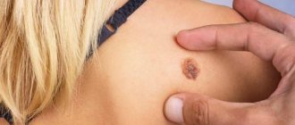
- Spots of yellow, coffee, scarlet or dark brown shades, located mainly on the back, head and neck, shoulders and stomach. In children, they also appear on the limbs, armpits and scalp. The affected areas can be of different sizes, but usually their size does not exceed 1 cm. Subsequently, the spots merge with each other, forming significant areas of the affected skin.
- No signs of inflammation in the form of redness, swelling, hyperemia and pain to the touch.
- Severe peeling of the skin in infected areas, aggravated by touch.
- No tanning on the affected areas.
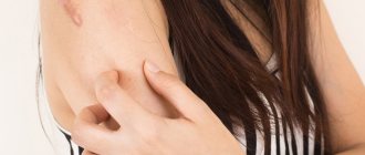
Pityriasis versicolor is not characterized by itching and discomfort, so patients often mistake the manifestations of the disease for minor cosmetic defects, postponing a visit to the doctor and full treatment.
Such spots begin to itch and scratch only if a secondary infection occurs.
Prices for services
Initial appointment with a dermatologist (assessment of patient complaints, history taking, examination, preliminary diagnosis, consultation)
Primary appointment – visiting a doctor of a specific specialty for the first time. Make an appointment
1500 ₽
Repeated appointment with a dermatologist
Make an appointment
800 ₽
Appointment with a dermatologist during removal of tumors (evaluation of complaints, medical history, dermatoscopy if indicated)
Make an appointment
500 ₽
Reasons for development
Versicolor versicolor is a type of fungal skin infection that affects the stratum corneum of the epidermis and hair follicles. Its causative agents are two types of fungi, and infection is possible only through prolonged and close contact with the patient. And in this case, provoking factors play a big role. These include:
- weakened immunity;
- hyperhidrosis;
- disruption of the sebaceous glands;
- diseases of the endocrine system (obesity, diabetes, Itsenko-Cushing syndrome, etc.);
- hormonal imbalance due to pregnancy, menopause or taking hormone-containing medications;
- vegetative-vascular dystonia;
- abuse of antibacterial personal hygiene products;
- excessive exposure to ultraviolet rays (intense tanning, frequent visits to the solarium) and regular overheating of the body.
It is noteworthy that patients with pityriasis versicolor over 60 years of age are extremely rare. This is due to natural age-related changes in the skin, which make it less susceptible to pathogens.
In children under 10 years of age, the main causes of pityriasis versicolor infection are neglect of personal hygiene rules or improper skin care. At this age, with the protective functions of the skin intact, the body independently copes with pathogenic microorganisms attacking it, so the development of the disease does not occur. But closer to adolescence, when hormonal changes begin, the body’s susceptibility to bacteria, viruses and fungi increases, so children over 10 years old become infected with pityriasis versicolor just like adults.
How does infection occur?
The contagiousness of pityriasis versicolor is believed to be very low. But not all dermatologists-mycologists support this opinion. Pityriasis versicolor can be contracted from a loved one through personal contact or through household contacts through objects used by the infected person, for example, bedding, towels, clothing, washcloths. You can also pick up a fungus in the fitting room of a store. The incubation period for pityriasis versicolor ranges from two weeks to several months.
Make an appointment
Treatment of pityriasis versicolor
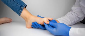
The diagnosis of pityriasis versicolor is made to the patient after examination by a dermatologist and dermatoscopy. Additionally, an iodine test and laboratory testing of scrapings can be used.
Treatment of pityriasis versicolor is carried out on an outpatient basis until the symptoms of the disease completely disappear. If measures were taken on time, the patient is prescribed local therapy using antifungal ointments and special agents for exfoliating dead cells.
Additionally, immunomodulators, vitamin complexes, antifungal shampoos and antihistamines are used if the patient is bothered by itching. In the most advanced cases, antimycotic agents are prescribed for oral administration. In addition, in order to avoid relapse of the disease and infection of others, the patient’s clothing and bedding are treated with disinfectant compounds.
Disease prevention also plays an important role. The following will help prevent the development of the disease:
- timely solution to the problem of hyperhidrosis (use of medicinal deodorants, creams, powders, compliance with personal hygiene rules, frequent changes of underwear, etc.);
- use of high-quality soaps and skin care products;
- regular water procedures;
- wearing clothes made from natural, hypoallergenic materials;
- avoiding stress;
- balanced diet rich in vitamins and microelements.
Experts also recommend avoiding overheating of the body and promptly seeking advice from cosmetologists and dermatologists in order to identify the problem and begin proper treatment.
Diagnostics
Diagnosis of this pathology is carried out by a dermatovenerologist and is based on objective examination data and additional research methods. Most often, the Balser iodine test is used to clarify the diagnosis. The essence is to stain the spots with a 2% iodine solution, under the influence of which the foci of pityriasis versicolor become colored. The method with an ultraviolet Wood lamp is also used.
Additional examination for the presence of fungi in the layers of the skin is possible.
What is erythrasma
Erythrasma is a chronic bacterial disease affecting the epidermis layer in the deep folds of the skin. It is characterized by a long course - in some cases, symptoms develop for at least 10 years, without causing significant discomfort to the patient. The clinical picture of erythrasma is similar to a fungal infection of the skin, but modern dermatology classifies it as a group of pseudomycoses.
The following main stages are distinguished in the development of the disease:
- Progression. The first characteristic spots appear on the skin, their size slowly increases, and additional symptoms develop. In some cases, secondary infections occur. The spots gradually merge with each other, forming large areas of damage.
- Stabilization. New spots do not appear, and existing ones stop growing. Peeling of the skin begins. This stage is usually associated with a change in external conditions, for example, cold weather, during which the intensity of sweating decreases and the skin condition stabilizes.
- Exacerbation or relapse. Usually associated with the beginning of the warm season. But in the case of prolonged erythrasma, the disease constantly develops in waves, and after a slight decline its symptoms again actively appear.
- Remission. Occurs with a favorable microclimate, compliance with preventive measures and proper skin care. The color of the affected areas gradually returns to normal, itching and flaking disappear, and the skin is restored.
Without timely, well-chosen treatment, erythrasma can lead to the development of serious complications.
For example, it can provoke eczema and secondary infection in patients with diabetes or obesity. Also, the course of the disease is aggravated by increased humidity and contamination of the affected areas. As a result, its typical symptoms are complicated by burning, itching and pain.
Signs of the disease
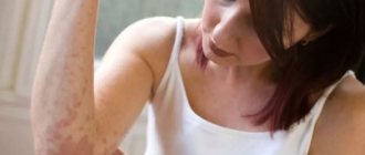
Externally, erythrasma manifests itself in the form of light brown, brick-red, brown or yellow-brown spots on the skin, most often round in shape and without signs of inflammation. The diameter of the lesions can reach several centimeters, and they tend to merge, forming large affected areas. First of all, erythrasma spots appear in the folds of the skin, where there is a favorable environment for the proliferation of bacteria.
In addition to spots, erythrasma is characterized by:
- Peeling of the skin on the affected areas, aggravated by touch. Usually this is where the development of the disease begins.
- Mild, irregular itching. It intensifies and begins to cause significant discomfort only if a secondary infection is added to the primary disease.
- Absence of fever, wounds, ulcers and ulcers with copious discharge. This distinguishes erythrasma from most bacterial skin pathologies.
- Getting wet. An optional symptom, the manifestation of which depends on the amount of sweating and the quality of skin care.
It is noteworthy that in children, symptoms of erythrasma appear extremely rarely. The risk group includes adults, primarily men, who are predominantly overweight and prone to excessive sweating. In this case, in men, the skin in the groin, navel and inner thighs is usually affected, and in women, the entire abdomen, armpits and areas under the breasts are affected.
Causes of pathology
Corynebacteria, which are the causative agents of the disease, are normally always present on human skin. Moreover, they provoke the development of pathology only under certain, favorable conditions. Corynebacteria do not penetrate deeper than the epidermis, and also do not affect nails and hair. Since the appearance of erythrasma is directly related to increased sweating, the disease most often manifests itself in the hot summer season.
Among the main reasons for the development of erythrasma are:
- hyperhidrosis;
- deviation of the normal pH of the skin to the alkaline side;
- diaper rash, constant friction and mechanical damage to the skin;
- dermatitis and other skin diseases;
- neglect of personal hygiene rules;
- wearing synthetic, overly warm clothing;
- the use of low-quality care products or the abuse of soap with an antibacterial effect, which destroys the natural protection of the skin.
Erythrasma is transmitted by contact, most often after the use of clothing, bedding and personal hygiene products of the patient. You can also become infected during sexual intercourse, when visiting a pool or bathhouse, and when walking barefoot on the ground or beach. At the same time, it is not always possible to accurately determine the source of infection, because the carrier may not have obvious external manifestations of the disease in the form of characteristic spots and peeling.
Are there predisposing factors?
The fungus, which is the causative agent of pityriasis versicolor, can live on the skin for quite a long time without appearing outwardly. In order for its signs to be revealed, a push is needed. Such an impetus for the active development of fungus can be endocrine pathologies, increased sweating, weakened immunity, stressful situations for the skin that disrupt the protective function of the skin - solarium, excessive exposure to the sun, frequent use of antibacterial soaps and shower gels.
IMPORTANT!
The thought of self-medicating pityriasis versicolor is tempting. It seems so simple - I bought antifungal ointment, did it as indicated in the instructions for use, and it’s done. Alas! Without an individual, qualified course of therapy, the fungus will simply adapt to the drugs used, and further treatment will be complicated. Unsystematic or symptomatic self-treatment is highly undesirable.
Treatment of erythrasma
To diagnose a patient with erythrasma, a dermatologist first uses a visual examination. This is especially true for rashes in the groin area, which have characteristic distinctive features in the form of pronounced protrusions and bubbles along the edges. Also, the affected areas of the skin are illuminated with a Wood's lamp and a microscopic examination of the scraping is performed to exclude other diagnoses: pityriasis versicolor or pityriasis rosea, candidiasis, dermatitis or eczema.
Treatment of erythrasma is primarily based on the use of antibacterial ointments that are used to treat the affected areas of the skin.
Under their influence, corynebacteria die, and the spots gradually lighten, decrease in diameter and disappear. On average, such therapy takes at least 7-10 days. Used in parallel:
- Antiseptics. Treatment with them is carried out before applying antibacterial ointment, as well as after it, to maintain dryness of the affected areas and prevent re-infection.
- Antifungal drugs. They are prescribed together with antibacterial drugs, since corynebacteria are similar in structure to fungal micelles.
- Exfoliating ointments. They help cleanse the skin of a layer of dead cells, activating its regeneration.
- Ultraviolet irradiation. Promotes skin disinfection and restoration. Patients benefit from both natural sunbathing and physiotherapeutic UV irradiation.
If the disease has not reached an advanced stage, the use of external medications is sufficient to solve the problem. But in some cases, with multiple skin lesions, to obtain the desired result, patients are prescribed systemic antibacterial therapy.