Where is increased fat content possible?
The content of the article
Problem areas:
- upper back;
- facial part, especially the chin, forehead and cheeks;
- cervical and thoracic parts of the body;
- genitals in both sexes;
- peripapillary zone.
These are the most common and most common, but there may be others. The only places on the body where acne cannot appear are the palms and feet. Due to the too thick and rough layer of the epidermis, incapable of this kind of inflammation.
Additional care
- It is better to leave ordinary creams, cream or milk in the past, as they are little compatible with the original data. It is optimal to select special gels, micellar water, tonics.
- It is recommended to use soft exfoliants if there are no inflammatory processes on the surface. It is advisable to choose options that have the specification “non-comedogenic” in the instructions.
- It is advisable to select all basic activities with a focus on regulating the functioning of the sebaceous glands. Moreover, preference should be given not to those procedures that are vaguely “intended for this type of skin,” but specifically those that narrow pores.
Causes
- Dry skin;
- Hormonal changes in the whole body;
- Infection;
- Too much sebum production.
In addition to these reasons, there is another factor that was recently established by scientists - heredity.
Let's look at each reason in more detail.
Dry skin
One of the most common reasons. Due to dryness, the skin exfoliates and its particles clog the pores, which causes inflammation. It is very important to consider several points that cause excessive dry skin:
- Age. Over the years, the skin gradually loses many of its important properties, which include moisture retention.
- Environment. Dry and cold air, low humidity, and so on.
- Shower with hot water. Leads to the destruction of the protective layer of the skin.
- Allergic diseases.
- Hygiene products. Using regular soap on sensitive areas of the skin: facial skin, intimate areas, can cause dryness and cause acne.
Hormonal background
The reason lies in the incorrect proportion of sex hormones in the blood. Androgens (male hormones) play a very important role here. If their level is exceeded, then excess secretion begins from the sebaceous and sweat glands, which is the reason for the appearance of acne. You can also isolate progesterone, a hormone secreted in pregnant women. Leads to clogging of pores and, as a result, sebaceous inflammation appears.
Lack of estrogen can also cause excessive oily skin.
What do people with oily skin suffer from?
If you have oily skin, it means that the sebaceous glands on your face produce too much sebum. In addition, they can expand and become clogged with comedones. Such skin appears to be covered with mini-craters and looks unaesthetic. At the same time, the oiliness of the skin surface itself does not indicate that you will definitely have acne.
In some cases, after washing, a feeling of tightness may occur, this indicates a violation of the lipid barrier of the epidermis - a lack of ceramides in it, which are responsible for creating a protective film on the skin.
The lipid barrier could be damaged as a result of improper care, for example, frequent use of aggressive lotions containing alcohol.
Sebum production
There are 3 factors that can affect the amount of sebum production. Androgen, or rather its excess in the blood, is one of the reasons. Rarely, but still there is a special disease called “seborrhea”. And of course, a very common cause in the 21st century is poor nutrition.
Here is a list of products that promote the active secretion of sebum:
- carbonated drinks;
- alcoholic drinks;
- products in the production of which dyes were used;
- caffeinated drinks (black tea, coffee;
- sweet and flour;
- spicy, smoked food.
What are sebaceous glands?
Each hair is equipped with a sebaceous gland, and sometimes more than one. Their excretory ducts open in the hair follicle and release about 20 g of sebum per day. The composition of sebum includes29:
- fatty acid;
- glycerol;
- alcohols;
- wax esters;
- cholesterol;
- metabolites of steroid hormones, etc.
Sebaceous glands are located almost throughout the body, but their largest number is on the face, back, and scalp. These zones are called seborrheic. At the same time, their smallest number is on the border of the lips, the back of the hands, but on the soles and palms there are none at all. 29
In seborrheic areas, the density of sebaceous glands can reach 900 per 1 cm2. The sebaceous glands begin to manifest their functions as actively as possible in adolescence; this period can last up to 25 years. 29
When secretory function is disrupted, skin problems begin. If there is not enough sebum secreted, it flakes off, and signs of aging appear faster. And when too much content of the sebaceous glands is secreted, rashes may appear. 29
Sebocytes (cells of the sebaceous glands) are sensitive to sex steroid hormones. The cytoplasm of their cells contains enzymes that convert androgens into dihydrotestosterone, which promotes the secretion of more sebum. 29
Treatment and prevention
It would be best and safest to contact a competent doctor - an endocrinologist, so that further treatment takes place under his strict supervision. You will have to start with a healthy diet that limits the consumption of foods that are harmful to good skin condition. And it won’t hurt to start special cosmetic skin care.
ONLINE REGISTRATION at the DIANA clinic
You can sign up by calling the toll-free phone number 8-800-707-15-60 or filling out the contact form. In this case, we will contact you ourselves.
How to fight?
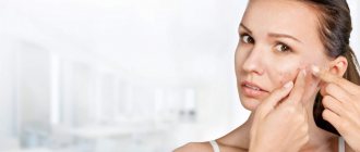
One of the most common methods of dealing with acne is squeezing it out. Beauty salons even provide deep facial cleansing services, which supposedly helps get rid of acne forever. However, such procedures do not affect any of the factors in the formation of acne. The improvement is only temporary
and does not affect the further development of the disease.
Some cosmetic products allow you to gently remove the keratin plug from the sebaceous gland duct, improving the outflow of secretions and preventing the proliferation of bacteria. There are also products with an antibacterial effect that continue to act even when inflammation has already begun in the sebaceous gland. A qualified dermatologist will select products
suitable for your specific skin type and severity of symptoms. But if these measures are not enough, then the treatment of acne requires serious medical intervention and the use of special medications.
Let's sum it up
There can be many reasons for the occurrence of increased oiliness in the dermis, but the solutions to all problems are basically the same. Therefore, it is very important to figure out how to reduce or remove, get rid of excess oily skin on the face, what to do when the chosen gentle products no longer help, and what you can count on. Modern cosmetology offers many different options for influencing the dermis, but not all of them are equally safe and effective, and many problems can be completely solved with high-quality home care.
Epidermis
Rice.
2. Epidermis The epidermis is a constantly exfoliating epithelial layer of the skin, which consists mainly of 2 types of cells - keratinocytes and dendritic cells. Melanocytes, Langerhans cells, Merkel cells, and intraepidermal T lymphocytes are present in small quantities in the epidermis. Structurally, the epidermis is divided into 5 layers: basal, spinous, granular, shiny and horny, differing in the position and degree of differentiation of keratinocytes, the main cell population of the epidermis (Fig. 2).
Keratinization. As keratinocytes differentiate and move from the basal layer to the stratum corneum, their keratinization (keratinization) occurs - a process that begins with the phase of keratin synthesis by keratinocytes and ends with their cellular degradation. Keratin serves as a building block for intermediate filaments. Bundles of these filaments, reaching the cytoplasmic membrane, form desmosomes necessary for the formation of strong contacts between neighboring cells. Further, as epithelial differentiation progresses, epidermal cells enter the degradation phase. The nuclei and cytoplasmic organelles are destroyed and disappear, metabolism stops, and cell apoptosis occurs when it is completely keratinized (turns into horny scales).
The basal layer of the epidermis consists of a single row of mitotically active keratinocytes, which divide on average every 24 hours and give rise to new cells in the overlying epidermal layers. They are activated only in special cases, such as when a wound occurs. Next, a new cell, a keratinocyte, is pushed into the spinous layer, where it spends up to 2 weeks, gradually approaching the granular layer. The movement of the cell to the stratum corneum takes another 14 days. Thus, the lifespan of a keratinocyte is about 28 days.
It should be noted that not all cells of the basal layer divide at the same rate as keratinocytes. Epidermal stem cells under normal conditions form a long-lived population with a slow proliferation cycle.
The spinous layer of the epidermis consists of 5-10 layers of keratinocytes, differing in shape, structure and intracellular content, which is determined by the position of the cell. Thus, closer to the basal layer, the cells have a polyhedral shape and a round nucleus, but as the cells approach the granular layer, they become larger, acquire a flatter shape, and lamellar granules appear in them, containing various hydrolytic enzymes in abundance. Cells intensively synthesize keratin filaments, which, gathering into intermediate filaments, remain unconnected on the nuclear side, but participate in the formation of multiple desmosomes on the membrane side, forming connections with neighboring cells. The presence of a large number of desmosomes gives this layer a spiny appearance, for which it received the name “spinous.”
The granular layer of the epidermis is composed of still living keratinocytes, distinguished by their flattened shape and a large number of keratohyalin granules. The latter are responsible for the synthesis and modification of proteins involved in keratinization. The granular layer is the most keratogenic layer of the epidermis. In addition to keratohyalin granules, keratinocytes of this layer contain large quantities of lysosomal granules. Their enzymes break down cellular organelles during the transition of the keratinocyte to the phase of terminal differentiation and subsequent apoptosis. The thickness of the granular layer can vary; its value, proportional to the thickness of the overlying stratum corneum, is maximum in the skin of the palms and soles of the feet.
The shiny layer of the epidermis (so named for its special shine when viewing skin preparations on a light microscope) is thin, consists of flat keratinocytes, in which the nuclei and organelles are completely destroyed. The cells are filled with eleidin, an intermediate form of keratin. It is well developed only in some parts of the body - on the palms and soles.
The stratum corneum of the epidermis is composed of corneocytes (dead, terminally differentiated keratinocytes) with a high protein content. The cells are surrounded by a waterproof lipid matrix, the components of which contain compounds necessary for exfoliation of the stratum corneum (Fig. 3). The physical and biochemical properties of cells in the stratum corneum vary depending on the position of the cell within the layer, directing the exfoliation process outward. For example, cells in the middle layers of the stratum corneum have stronger water-binding properties due to the high concentration of free amino acids in their cytoplasm.
Rice. 3. Schematic representation of the stratum corneum with the underlying granular layer of the epidermis.
Regulation of proliferation and differentiation of epidermal keratinocytes. Being a continuously renewing tissue, the epidermis contains a relatively constant number of cells and regulates all interactions and contacts between them: adhesion between keratinocytes, interaction between keratinocytes and migrating cells, adhesion with the basement membrane and underlying dermis, the process of terminal differentiation into corneocytes. The main mechanism for regulating homeostasis in the epidermis is supported by a number of signaling molecules - hormones, growth factors and cytokines. In addition, epidermal morphogenesis and differentiation are partially regulated by the underlying dermis, which plays a critical role in maintaining postnatal skin structure and function.
