- Acute maxillitis
Sinusitis (maxillary sinusitis, maxillitis) is inflammation of the mucous membrane of the maxillary (maxillary) sinuses. The sinuses are connected by common bony walls to the nasal cavity, mouth and orbit (eye socket) and are normally filled with air.
Sinusitis is an inflammation of the mucous membrane of the maxillary sinuses
The main functions of the maxillary sinuses, along with the frontal, sphenoid and ethmoid sinuses, are:
- formation of individual voice sound;
- warming and purifying inhaled air;
- equalization of pressure in the cavity formations of the skull in relation to external atmospheric pressure.
All sinuses communicate with each other through small holes, but if for some reason these holes are closed, their cleansing and ventilation stops. This promotes the accumulation of microbes and the development of inflammation.
The development of maxillitis is accompanied by an increase in body temperature, swelling of the cheek and eyelid on the affected side, intense pain in the bridge of the nose and at the wings of the nose, mucopurulent discharge from the nasal passages and difficulty in nasal breathing. With timely initiation of therapy prescribed by an otolaryngologist (ENT), serious complications can be avoided - osteomyelitis, orbital phlegmon, brain abscess, meningitis, otitis media, as well as kidney and myocardial damage.
Inflammation of the mucous membrane of the maxillary sinuses occurs in people of all ages, but in children under the age of 5 years, pathology develops extremely rarely, since their paranasal sinuses are underdeveloped.
Types of sinusitis
Sinusitis code according to ICD-10 (the international classification of diseases, 10th revision, developed by the World Health Organization):
- acute sinusitis : J01 (class - respiratory diseases, heading - acute respiratory infections of the upper respiratory tract);
- chronic sinusitis : J32 (class - respiratory diseases, heading - other diseases of the upper respiratory tract).
Maxillitis can be exudative or catarrhal. These forms of the disease are accompanied by a large amount of mucus or purulent discharge. Depending on the nature of the discharge, purulent, mucous and serous sinusitis are distinguished.
According to the prevalence of the process, maxillitis is unilateral, which, depending on the affected side, is divided into right-sided and left-sided, as well as bilateral.
Sinusitis can be unilateral or bilateral
Classification according to the course of the disease:
- acute : symptoms are similar to a runny nose, acute respiratory viral infection and other colds. Typically, the duration of inflammation varies from 14 to 21 days;
- chronic : can develop in the absence of adequate treatment for acute sinusitis. The duration of this form of the disease is usually 2 months or more. Symptoms may disappear almost completely, and then reappear with renewed vigor;
- recurrent : characterized by symptoms occurring two, three, or more times per year.
Classification by etiological factor:
- viral;
- traumatic;
- bacterial, divided into bacterial aerobic and bacterial anaerobic;
- fungal;
- endogenous, divided into vasomotor, otogenic, odontogenic;
- mixed;
- allergic;
- perforated;
- Iatrogenic.
Often chronic sinusitis is accompanied by a night cough that does not respond to conventional therapy. The reason for its appearance is pus flowing down the back wall of the pharynx from the maxillary sinus.
Classification according to the route of infection:
- hematogenous : the infectious agent enters through the blood. Most often, this form of sinusitis develops in children;
- rhinogenic : infection enters through the nasal cavity. Usually occurs in adults;
- odontogenic : microbes enter the maxillary sinus from the molars of the upper jaw;
- traumatic.
Chronic sinusitis, according to the nature of morphological changes, is divided into the following types:
- productive (parietal-hyperplastic, atrophic, necrotic, polypous, purulent-polypous, etc.). Against this background, changes in the mucous membrane of the maxillary sinus are observed (hyperplasia, atrophy, polyps and others);
- exudative (purulent and catarrhal), in which pus is formed.
In the chronic course of the disease, small pseudocysts and true cysts of the maxillary sinus often form due to blockage of the mucous glands. The most common forms of chronic inflammation are polyposis and polyposis-purulent. Catarrhal allergic and parietal hyperplastic forms occur in rare cases, and necrotic, oseotic, cholesteatoma and caseous forms occur in very rare cases.
Treatment of acute rhinosinusitis
Symptomatic therapy is indicated for patients with ARRS.
- Analgesics and antipyretics - ibuprofen or paracetamol can be used to relieve pain and reduce fever as needed.
- Saline irrigation therapy - Irrigation of the nasal cavity with saline or hypertonic saline can reduce the need for pain medications and improve overall health. It is important that nasal irrigation solutions are sterile, as there is evidence of amoebic encephalitis due to the use of tap water for nasal irrigation.
- Intranasal glucocorticoids (inGCS) - studies have shown small symptomatic benefits and minimal side effects with short-term use of inGCS in patients with both viral and bacterial ARS. InGCS are more effective in patients with concomitant allergic rhinitis. A meta-analysis of three studies in patients with ARS found that intranasal steroids increased the rate of symptom resolution compared with placebo. However, there is also evidence that only 1 out of 15 patients will have a significant reduction in clinical symptoms during treatment with inhaled corticosteroids.
- Ipratropium bromide is an anticholinergic spray that may help reduce rhinorrhea in patients with underlying cold symptoms.
- Oral decongestants may be useful for eustachian tube dysfunction.
- Intranasal vasoconstrictors are often used by patients as symptomatic therapy. Sprays containing oxymetazoline or xylometazoline may provide temporary relief of nasal congestion, but there is no evidence that they are significantly effective for ARS.
In cases of mild, uncomplicated ABRS, there are no indications for prescribing systemic antibiotics; treatment is carried out symptomatically, as in cases of viral infection.
For ABRS, a wait-and-see approach is recommended for 7 days. Systematic reviews and meta-analyses have shown that about 80% of patients with clinically diagnosed ABRS recover without antibiotic therapy within two weeks.
If health does not improve within 7 days or worsens after symptomatic treatment, systemic antibacterial therapy should be added.
The drugs of choice for ABRS are amoxicillin or amoxicillin-clavulanate. The use of an antibiotic with clavulanic acid in its composition expands the spectrum of action of the drug, including against ampicillin-resistant bacteria Haemophilus influenzae or Moraxella catarrhalis. This is not necessary in every case. There is evidence that the use of amoxicillin-clavulanate is preferable in children than in adults. It should also be understood that resistance rates vary depending on the region, which must also be taken into account when selecting therapy.
Preference should be given to amoxicillin-clavulanate in the following cases.
- The patient lives in a region with high rates of penicillin-resistant S. pneumoniae strains (more than 10%).
- Age ≥ 65 years.
- Hospitalization in the last five days.
- Use of antibiotics in the previous month.
- Immunocompromised patients.
- Patients with underlying medical conditions such as diabetes, chronic heart, liver or kidney disease.
- Severe or complicated course of the disease.
If you are allergic to penicillin antibiotics, options for prescribing doxycycline, cephalosporin antibiotics, and clindamycin are considered. Another alternative for patients allergic to penicillins is respiratory fluoroquinolones. However, these drugs should only be used when there is no alternative due to serious side effects.
The duration of the course of antibacterial therapy in adults is usually 5-7 days, in children - 10-14 days. There are studies showing that short courses of antibiotic therapy are not inferior in effectiveness to long courses.
If there is no effect from the first course of antibiotics, the next drug prescribed should have a broader spectrum of activity and/or belong to a different class of drugs.
How is acute rhinosinusitis treated at the Rassvet Clinic?
- We carefully collect anamnesis and conduct an examination.
- We do not prescribe antibiotics unless there is clear evidence that a bacterial infection is developing.
- We do not routinely order x-rays, bacteriological examinations or blood tests. It is not necessary.
- We adhere to the principles of evidence-based medicine, which means that if there are indications for antibiotic therapy, we always give preference to systemic antibiotics, usually oral. Administration of the drug intramuscularly has no proven advantages over tablet forms.
- Not a single local antibiotic has been shown to be effective for ARS, so we do not perform “cuckoo” or multiple punctures of the sinuses and rinse them with “complex solutions.”
- At the Rassvet clinic, you do not need to come to the doctor for control every 2-3 days in cases of the standard course of the disease.
Causes and risk factors for the development of inflammation
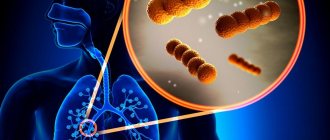
The causative agents of sinusitis can be viruses, chlamydia, fungi, staphylococci, streptococci, Haemophilus influenzae and mycoplasma. In adults, maxillitis is most often caused by viruses, pneumococci and Haemophilus influenzae, in children - by mycoplasma and chlamydia. In case of impaired immunity and in weakened patients, inflammation can occur due to saprophytic and fungal microflora.
Possible causative agents of the disease are staphylococci, streptococci, viruses, chlamydia, fungi, mycoplasma and hemophilus influenzae
Risk factors for the development of maxillitis are pathologies and conditions that complicate the ventilation of the maxillary sinus and contribute to the penetration of infection into its cavity. These include:
- congenital narrowness of the nasal passages;
- acute respiratory viral infection, acute and chronic rhinitis of any origin;
- chronic tonsillitis, pharyngitis;
- adenoids (in children);
- deviated nasal septum;
- surgical interventions performed on the alveolar process of the upper jaw or teeth;
- carious lesions of the upper molars.
The risk of developing the disease increases in the autumn-winter period, which is due to the natural seasonal decrease in immunity.
Symptoms
Symptoms of acute sinusitis
The inflammation begins acutely. The patient experiences an increase in body temperature to febrile (38–39 °C), severe signs of general intoxication and, possibly, chills. In some cases, body temperature may remain normal or subfebrile (37.1–38 °C). The patient's main complaints are pain in the affected maxillary sinus, forehead, root of the nose and zygomatic bone. On palpation, the pain intensifies; it can radiate to the corresponding half of the eyelid and temple. It is also possible to experience a diffuse headache of varying intensity.
On the side of inflammation, nasal breathing is impaired, and in cases of bilateral sinusitis, nasal congestion forces the patient to breathe through the mouth. Due to blockage of the tear duct, the development of lacrimation is sometimes observed. Nasal discharge from serous and liquid gradually becomes greenish, cloudy and viscous.
Symptoms of chronic sinusitis
Typically, chronic sinusitis develops as a result of an acute process. During the period of remission, the general condition, as a rule, does not worsen. During an exacerbation, symptoms of general intoxication occur in the form of headache, weakness and fatigue, and the body temperature can rise to febrile or subfebrile.
With exudative forms of maxillitis, the amount of discharge increases during the period of exacerbation, and when the patient’s condition improves, it decreases. Catarrhal sinusitis is characterized by a liquid and serous discharge, with an unpleasant odor; in the purulent form, it is a thick, yellowish-green, abundant, viscous mucus, which dries out and turns into crusts.
As a rule, headache develops only during the period of exacerbation of the chronic form of maxillitis or against the background of a violation of the outflow of discharge from the maxillary sinus. The patient may experience a pressing or bursting headache, which is localized behind the eyes and intensifies with pressure on the infraorbital areas and when the eyelids are lifted. When lying down or during sleep, the severity of the pain syndrome decreases, since in a horizontal position the outflow of pus resumes.
To avoid the development of side effects and to obtain a high concentration of the drug at the site of inflammation, topical antibiotics are used.
Often chronic sinusitis is accompanied by a night cough that does not respond to conventional therapy. The reason for its appearance is pus flowing down the back wall of the pharynx from the maxillary sinus.
With chronic maxillitis, skin lesions (weeping, maceration, swelling or cracks) in the vestibule of the nasal cavity are often detected. Many patients experience concomitant keratitis and conjunctivitis.
Publications in the media
Sinusitis is an inflammatory disease of the paranasal (paranasal) sinuses associated with infection or allergic reactions. Frequency : 10% of the population. Most often, the cells of the ethmoid bone are affected, then the maxillary, frontal and, finally, the sphenoid sinuses.
Classification of acute sinusitis • Acute sinusitis • Acute ethmoiditis • Acute frontal sinusitis • Acute sphenoiditis.
Classification of chronic sinusitis • Exudative sinusitis •• Purulent form •• Catarrhal form •• Serous form • Productive sinusitis •• Parietal-hyperplastic form •• Polypous form •• Cystic form • Cholesteatoma sinusitis • Necrotic sinusitis • Atrophic sinusitis • Mixed forms.
Etiology • Infection of the sinuses by various microflora •• Acute sinusitis is characterized by a monoculture: bacterial infection (pneumococci, streptococci, staphylococci; only in 13% of patients), viral infection (influenza virus, parainfluenza, adenoviruses) •• Chronic sinusitis is characterized by a mixed microflora: more often staphylococcus, Pseudomonas aeruginosa, Proteus, Escherichia coli, fungal infection (fungi of the genera Aspergillus, Penicillium, Candida) • Previous ARVI • Nasal tamponade with nosebleeds.
Risk factors • Complicated allergy history • Immunodeficiency conditions • Diseases of the dental system • Swimming in contaminated water.
Ways of infection to enter the nasal sinuses • Rhinogenic (through the natural anastomosis of the sinuses) • Hematogenous • Odontogenic • With sinus injuries.
Clinical picture
Acute sinusitis • General symptoms of acute sinusitis •• Nasal congestion •• Headache •• Fever •• Nasal discharge •• Cold symptoms • Acute sinusitis •• Nasal congestion •• Feeling of heaviness, tension in the cheek area, especially when bending the torso forward • • Feeling of pressure on the eyes •• Pain in the teeth on the affected side •• Headache of uncertain localization •• Nasal discharge of a mucopurulent or purulent nature •• Deterioration of smell •• Lacrimation (due to obstruction of the nasolacrimal duct) • Acute ethmoiditis. The symptoms differ little from acute sinusitis. Additionally, pain is noted in the root of the nose and orbit • Acute frontal sinusitis - headache in the forehead, especially intense in the morning (due to difficulty in outflow from the sinus when the patient is horizontal) • Acute sphenoiditis •• Headache in the back of the head, deep in the eye •• Drainage of purulent discharge from the nasopharynx along the back wall of the pharynx •• Unpleasant odor.
Chronic sinusitis • The clinical picture of chronic sinusitis without exacerbation is less pronounced than in acute sinusitis • Fungal sinusitis is characterized by: •• pronounced unilateral or bilateral nasal congestion; •• pain in the area of the affected sinus; •• pronounced feeling of pressure in the sinus; •• toothache (with sinusitis) • The nature of the discharge depends on the pathogen: •• with mold mycoses - viscous, grayish-white or dark, jelly-like; •• with aspergillosis - gray with blackish dots (reminiscent of cholesteatoma); •• with candidiasis - yellow or yellow-white color (resembles cheesy masses) • More often than with other forms, swelling of the soft tissues of the face, and sometimes fistulas, are observed. Usually they occur as monosinusitis, most often the maxillary sinus is affected.
Research methods.
• Rhinoscopy •• Acute sinusitis ••• Hyperemia of the nasal mucosa, most pronounced in the middle meatus. Purulent discharge drains from the middle turbinate ••• Palpation of the anterior wall of the maxillary sinus is painful •• Acute ethmoiditis. Purulent discharge is usually found in the middle and upper nasal passages (since all groups of cells of the ethmoid bone are affected). Painful palpation of the nasal slope area at the inner corner of the eye •• Acute frontal sinusitis - characterized by pronounced changes in the area of the anterior section of the middle turbinate. The mucous membrane in this area is hyperemic and edematous. Localization of accumulations of pus in the anterior sections of the middle nasal meatus. Painful palpation of the anterior and especially the lower walls of the sinus •• Acute sphenoiditis - with anterior rhinoscopy after anemization of the mucous membrane, a strip of pus is visible in the most posterior parts of the upper nasal passage. The posterior sections of the nasal cavity are hyperemic and edematous. Posterior rhinoscopy reveals accumulation of pus in the nasopharynx.
• X-ray of the sinuses - accumulation of fluid, fluid level, thickening of the mucous membrane in the affected sinuses.
• Diagnostic puncture - determining the presence of the nature of the discharge.
• CT scan in some unclear cases of chronic sinusitis.
Differential diagnosis • Viral rhinitis • Allergic rhinitis • Tumors • Foreign bodies • Wegener's granulomatosis.
TREATMENT
Acute sinusitis • For uncomplicated sinusitis, treatment is usually conservative •• Antibiotic therapy (for example, benzylpenicillin 500 thousand units 4-6 times / day) for 7-10 days •• Sulfonamide drugs (for example, sulfadimethoxine on the first day 2 g, then 1 g/day, co-trimoxazole 1 tablet 3 times a day after meals) •• Non-narcotic analgesics •• Vasoconstrictor nasal drops, for example 0.05–0.1% solutions of naphazoline or xylometazoline; instillation is carried out with the patient lying on his side. The vasoconstrictor effect gradually decreases, so after 5–7 days of use a break for several days is recommended. The drugs are contraindicated in arterial hypertension, tachycardia and severe atherosclerosis •• Physiotherapy (with good outflow from the sinus), for example, microwave therapy (LUCH-2 device), UHF currents, Sollux lamp •• On an outpatient basis for acute sinusitis, it is advisable to perform a sinus puncture with subsequent washing with nitrofural solution (1:5,000), iodinol, 0.9% sodium chloride solution and the introduction of antibacterial agents into it, for example benzylpenicillin (2 million units), 1% hydroxymethylquinoxylin dioxide solution (prescribed only to adults , before starting use, a tolerance test is carried out, contraindicated in pregnancy), 20% sulfacetamide solution •• In case of severe swelling, 1-2 ml of hydrocortisone suspension, 1% diphenhydramine solution are simultaneously injected into the sinus •• In acute frontal sinusitis, ethmoiditis or sphenoiditis and the absence of effect from conservative therapy, hospitalization is indicated for puncture or probing of these sinuses • For complicated acute sinusitis - surgical treatment •• Radical sinus surgery •• Endoscopic sinus surgery.
Chronic sinusitis
• In case of exacerbation - a combination of general and local treatment. Features •• For staphylococcal lesions, antibiotic therapy is not always effective. Anti-staphylococcal plasma is used (250 ml 2 times a week), staphylococcal g-globulin (1 ampoule every other day, 5 injections in total) •• For fungal sinusitis and without exacerbation - sulfonamide drugs, antifungal drugs, for example nystatin 3-4 million units/ day or levorin 2 million units/day for 4 weeks •• For allergic sinusitis - see Allergic rhinitis.
• Drainage of the maxillary sinus is performed using puncture - either a Kulikovsky needle is first inserted into a polyethylene tube, or after puncture a smaller tube is passed through the needle into the sinuses. Drainage is inserted into any sinus in a similar manner. To drain the frontal and sphenoid sinuses through natural openings, it is advisable to use a probe-conductor on which a tube is placed. After probing, the tube is left in place and the probe is removed. The outer end of the tube is attached to the skin with an adhesive plaster. Antibacterial agents are introduced into the sinuses through drainage, taking into account the sensitivity of the microflora to them •• To liquefy the pus, enzymes can be simultaneously introduced into the sinus (chymotrypsin 25 mg or chymopsin 25 mg) •• For allergic sinusitis, a suspension of hydrocortisone (2–3 ml) or antihistamines •• For fungal sinusitis, levorin sodium salt or nystatin is injected into the sinus at the rate of 10 thousand units per 1 ml of 0.9% sodium chloride solution, quinozole solution 1:1,000 or amphotericin B.
• Physiotherapy: microwaves, mud therapy (contraindicated in case of exacerbation of sinusitis). Physiotherapy is contraindicated for hyperplastic, polyposis and cystic sinusitis.
• Surgical treatment - for polypous, mixed forms, as well as in case of ineffectiveness of conservative treatment of exudative forms •• Radical operations on the sinuses for the purpose of their sanitization by applying an artificial anastomosis with the nasal passage (for sinusitis - methods according to Caldwell-Luc, Dliker-Ivanov, with frontitis - according to Killian) •• Osteoplasty with a closed method (Mishenkin N.V., 1997) •• Ultrasound surgery.
Complications • Orbital (orbital) •• Cellulitis •• Optic neuritis (rare) •• Orbital periostitis •• Edema, abscess of retrobulbar tissue • Panophthalmos (inflammation of all tissues and membranes of the eye) - very rare • Intracranial •• Meningitis •• Arachnoiditis • • Extra- and subdural abscesses •• Brain abscess •• Thrombophlebitis of the cavernous sinus •• Thrombophlebitis of the superior longitudinal sinus •• Septic cavernous thrombosis.
Concomitant pathology • Rhinitis • Barosinusitis • Pansinusitis.
Prognosis: for acute sinusitis, favorable with timely treatment and prevention of complications; for chronic sinusitis, it can be favorable if the allergen is eliminated and good drainage is ensured.
Age characteristics • Children and adolescents •• The incidence of acute and chronic sinusitis increases in late childhood •• An increase in incidence is observed among children with tonsillitis and adenoids •• The presence of chronic sinusitis indicates the need to determine the cause underlying the disease (nasal deformity, infection, adenoids) • Elderly •• Incidence increases by age 75, then decreases •• Sinusitis is more difficult to cure in this age group.
ICD-10 • J01 Acute sinusitis • J32 Chronic sinusitis
Diagnostics
To diagnose sinusitis, it is necessary to collect the patient’s complaints, externally examine him, including determining the reflex dilatation of the skin vessels in the infraorbital region, and examine the mucous membrane of the nasal cavity in order to identify swelling, inflammation and purulent discharge from the sinus opening.
X-rays may be prescribed to diagnose the disease.
An x-ray reveals darkening of the maxillary sinus. If the information content of these research methods to determine whether the patient is contagious or not is not enough, a puncture of the maxillary sinus is performed.
Acute sinusitis - symptoms and treatment
May include:
- anterior rhinoscopy;
- radiography of the sinuses;
- ultrasonography;
- diagnostic puncture;
- laboratory research;
- microbiological examination;
- diaphanoscopy.
Anterior rhinoscopy
The main method of objective diagnosis of acute sinusitis [1][6][7]. It is carried out by an otorhinolaryngologist using a nasal speculum. Before its administration, the nasal mucosa is lubricated with a solution of a vasoconstrictor drug (anemization). This is done in order to reduce swelling of the mucous membrane.
The doctor assesses the condition of the mucous membrane of the nasal turbinates and passages, the absence or presence of discharge. Signs of sinusitis are the presence of purulent or mucopurulent discharge in the area of the outlet openings of the affected sinuses. It is accompanied by redness and swelling of the nasal mucosa.
A pathological secretion can be visible on the posterior wall of the oropharynx and in the nasopharynx when examined with a spatula and a nasopharyngeal speculum (posterior rhinoscopy) and examination of the pharynx (pharyngoscopy).
Endoscopic examination of the nasal cavity
Allows you to examine in detail the nasal passages and turbinates to the nasopharynx, identifying the smallest anatomical changes. Often, video endoscopy is performed and the result is recorded, which later helps to assess how the disease progresses.
X-ray of the sinuses
Use only for prolonged runny nose and headache for about 7–10 days [1][6][7]. During the procedure, the head is fixed in a certain position - in the nasomental, nasofrontal and lateral projections. The position of the head is determined by the radiologist. With sinusitis, the mucous membrane thickens, a horizontal fluid level is revealed and the pneumatization of the sinus is significantly reduced.
Computed tomography (CT) is used for chronic pathology of the paranasal sinuses, orbital and intracranial complications. It is not advisable to use it to diagnose acute sinusitis. Neither X-ray nor CT scan can differentiate between viral and bacterial sinusitis.
Ultrasound examination (ultrasound)
Non-invasive screening method. Allows you to identify effusion in the lumen of the frontal and maxillary sinuses and evaluate the effectiveness of the therapy [2]. It is actively used in examining children and pregnant women.
The action of ultrasound is based on the reflection of an ultrasonic signal at the boundary of two substances with different acoustic properties (bone - air, air - exudate, etc.) in a linear and two-dimensional mode. In the first case, ultrasound scanners are used for the paranasal sinuses (“Sinuscope”, “Sinuscan”), in the second case, standard ultrasound machines are used.
Diagnostic puncture
Sinus puncture (from Latin “puncture”) is not a routine diagnostic method due to the high risk of complications. It is used if there are contraindications to radiography, for example during pregnancy.
Laboratory diagnostics
Includes a complete blood count and determination of C-reactive protein (CRP).
Allows:
- distinguish viral sinusitis from bacterial sinusitis, and therefore determine whether you need to take antibiotics;
- assess the severity and dynamics of the disease.
Microbiological examination
It is carried out in cases of prolonged forms of sinusitis and the ineffectiveness of antibiotic therapy. For the study, you will need nasal cavity discharge or material from the affected sinus, extracted by puncture.
The reliability of the method is relative, and the information content is small. Firstly, the microflora of the nasal cavity and sinuses are initially different. Taking a smear, even if all conditions are met, does not guarantee that the bacterium cultured on the media is the cause of inflammation in the sinus, and not an accidental “fellow traveler” when removing a cotton probe from the nose.
In children, the information content of microbiological research is even lower. This is due to the child’s negative reaction to the manipulation and the inability to carry out the procedure correctly [11].
Secondly, the reliability may be affected by non-compliance with the storage and transportation conditions of the biomaterial [12]. Therefore, assessing the results of microbiological research is complex and ambiguous.
Diaphanoscopy
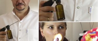
An outdated non-invasive method for diagnosing sinusitis of the maxillary sinus. This is done using a special lamp. If the cavity is airy, then the sinus will be highlighted in red. [5]. If the sinus contains pus, the glow becomes black.
Conservative treatment of sinusitis
Acute maxillitis
In order to reduce swelling of the mucous membrane of the maxillary sinuses and to restore normal ventilation, local vasoconstrictors (for example, xylometazoline hydrochloride, naphazoline) are used for a course of up to 5 days.
If the patient has significant hyperthermia, antipyretics are prescribed; in cases of severe intoxication, drugs with antibacterial action are prescribed.
To avoid the development of side effects and to obtain a high concentration of the drug at the site of inflammation, topical antibiotics are used.
After normalizing body temperature, physical therapy is recommended, for example, UHF therapy (ultra high frequency), Sollux infrared lamp.
Chronic maxillitis
In the chronic course of the disease, a sustainable therapeutic effect can be achieved only by eliminating the cause that caused inflammation in the maxillary sinus (sore teeth, deviated nasal septum, chronic pathologies of the ENT organs, adenoids, and others). In case of exacerbation of the disease, in order to avoid atrophy of the mucous membrane of the maxillary sinuses, local vasoconstrictor drugs are used in short courses.
Patients are prescribed drainage of the maxillary sinus. Sinus lavage is carried out using sinus evacuation or cuckoo (vacuum transfer method). For procedures, disinfectant solutions are used (for example, Potassium permanganate, Furacilin). Solutions of antibacterial agents and proteolytic enzymes are injected into the cavity. Of the physiotherapeutic procedures, ultraphonophoresis with hydrocortisone, diathermy, inhalations, and UHF therapy are most often prescribed. Speleotherapy is also effective.
Patients with necrotic, cholesteatoma, caseous, polypous and purulent-polypous forms of chronic maxillitis are advised to undergo surgery - maxillary sinusotomy.
Treatment of sinusitis at home
Traditional medicine can be used as an auxiliary therapy for maxillary sinusitis at home.
Decoctions and infusions of medicinal herbs are often used at home as an auxiliary therapy.
An herbal infusion can be used orally. To prepare it, add 2 tbsp to an enamel or glass container with a lid. spoons of St. John's wort, eucalyptus, lavender, chamomile and sage, 1 tbsp. spoon of yarrow and string, mix thoroughly. From the resulting mixture take 3 tbsp. spoons, pour 2 liters of boiling water over them, close the container with a lid and leave for half an hour at room temperature, then filter. The finished infusion is taken orally, 100 g every 3 hours.
Also, in the treatment of the chronic form of the disease, horseradish root is often used in the form of grated gruel in combination with lemon juice (1/3 cup of gruel and the juice of three lemons). The prepared mixture is taken orally every morning, 1/2 teaspoon, 20 minutes before meals. Treatment is carried out in courses, repeating them in the fall and spring until complete recovery.
If the patient has significant hyperthermia, antipyretics are prescribed; in cases of severe intoxication, drugs with antibacterial action are prescribed.
When therapy at home, topical agents are often used (the sinuses are washed with a solution of table salt or sodium chloride before the procedure):
- clay compresses : 50 g of clay is diluted in hot water to the consistency of plasticine. Moisten gauze in warm vegetable oil and place it on both sides of the nose (on the area of the maxillary sinuses). Place warm clay cakes on top of the gauze and keep them for 1 hour;
- honey ointment : 1 tbsp. grate a spoonful of fragrance-free baby soap. Mix 1 tbsp. spoon of honey, milk and vegetable oil and add them to the grated soap. The resulting mixture is heated in a water bath until the soap melts. Add 1 tbsp to the resulting product. a spoonful of alcohol, pour the whole mixture into a glass jar and let it cool. Using a cotton swab, the ointment is introduced into the nasal passages and left for 15 minutes. The duration of treatment is 21 days. The ointment should be stored in a closed container in the refrigerator;
- inhalation with sea buckthorn oil : 10 drops of sea buckthorn oil are added to a pan of boiling water. The released steam is inhaled for approximately 15 minutes;
- Shilajit drops : 10 crushed Shilajit tablets (0.2 g each) are thoroughly mixed with 1 teaspoon of glycerin and 4 teaspoons of water. The resulting product is instilled into the nose 3 times a day. The duration of therapy is 21 days. The course of treatment with breaks of 5 days is repeated several times until complete recovery.
Traditional medicine is recommended to be used with caution, especially if the components they contain can cause allergic reactions. If there is no therapeutic effect within several days, or the patient’s condition worsens, you should consult an otolaryngologist for advice.
