General information
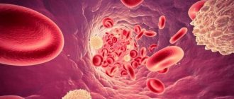
A condition called thrombocytopenia is characterized by a decrease in the number of platelets in the blood to less than 150 x 109/L. As a result, the patient experiences increased bleeding and difficulty stopping the bleeding . Thrombocytopenia (ICD-10 code - D69.6 Thrombocytopenia, unspecified) may accompany hematological diseases. However, an independent disease is also diagnosed - idiopathic thrombocytopenic purpura (ICD-10 code - D69.3), as well as an autoimmune form of the disease.
Speaking about thrombocytopenia - what kind of disease it is, it should be noted that it develops as a result of a disruption in the production of platelets or too active destruction of them. The autoimmune form of the disease is most often diagnosed. This condition can also develop against the background of a number of diseases: herpes , hepatitis , HIV, infectious mononucleosis , influenza . It can also be provoked by some exogenous factors. What thrombocytopenia is, the causes and treatment of this disease are described in detail in this article.
Pathogenesis
Platelets are blood cells that are produced in the bone marrow. They live in the body for a little more than a week, after which they disintegrate in the spleen and liver. The main functions of these cells in the body are to prevent the development of bleeding. The cells stick together and a blood clot forms, blocking the wounded vessel. In addition, they determine the synthesis of growth factors that affect tissue repair processes. Platelet levels may fluctuate depending on certain factors. However, such fluctuations are physiological and temporary. Normally, the level of these cells is 150 thousand per microliter (150 x 109/l). If the platelet count decreases, we are talking about thrombocytopenia.
Currently, the pathogenesis of this disease has not been sufficiently studied.
The mechanism of development of the disease may be associated with the following factors:
- impaired platelet production by the bone marrow;
- disruption of their distribution in the vascular bed;
- platelet deficiency and increased destruction.
Idiopathic thrombocytopenic purpura develops due to an autoimmune reaction that is directed against one’s own platelets. This immune reaction is a complex cyclic process in which B lymphocytes, T lymphocytes, macrophages , NK cells, and cytokines participate. Antibodies to platelets are not always detected in the blood.
In children, the development of ITP often occurs after acute respiratory viral infections. When the platelet count falls below 10 x 109/L, the bleeding period prolongs.
How does thrombocytopenia manifest?
Clinical cases of thrombocytopenia
- Home> Useful articles>Clinical cases of thrombocytopenia
S.V. Kuleshova, A.A. Altaeva, E.A. Kuznetsova
Clinical cases of false and true thrombocytopenia in outpatient practice.
Federal State Budgetary Institution "Polyclinic No. 2" of the Administration of the President of the Russian Federation, 119146, Moscow, Russia
The article is devoted to the laboratory nuances of diagnosing thrombocytopenia in outpatient practice. Using the example of two different patients, the importance of knowledge of basic research in the differential diagnosis of false and true thrombocytopenia is shown.
Key words: thrombocytopenia, EDTA-dependent thrombocytopenia, platelet count according to Fonio.
In the activities of an outpatient clinical diagnostic laboratory, a general blood test accounts for up to 30% of all tests performed.
Typically, a general clinical blood test consists of data from a hematological analyzer, and if there is a deviation in the leukocyte formula, a differentiated count of leukocytes in a blood smear using light microscopy.
The current level of hematology analyzers allows you to provide up to 70% of test results without smear microscopy.
However, the doctor should not blindly trust laboratory analyzers, but should be the final authority, after whose review and approval the results will be validated.
There are no small details in the work of a clinical laboratory diagnostics doctor. In the flow of work, choosing from the data of a hematology analyzer those that require additional research is not easy. An example of the need for a wide range of knowledge and skills of a CDL doctor is thrombocytopenia - a decrease in the number of platelets in the absence of other abnormalities in the count of formed elements and in the blood smear. Thrombocytopenia includes conditions in which the number of peripheral blood platelets is less than 150 x 109/L. A platelet count of 50x109/l still allows abdominal surgery while maintaining hemostasis. Platelet count ≤ 20 x109/l. refers to critical values from the point of view of clinical laboratory diagnostics [1]. For such patients, according to the national standard, transfusion of platelet concentrate is indicated [2]. To calculate the number of platelets, the following are recognized as unified:
- blood count using a hematology analyzer;
- counting in smears using the Fonio method.
A decrease in platelet count in a blood test may be true and will then require immediate action based on the severity of the thrombocytopenia. A decrease in platelet count in a blood test may not be true. Then it will reflect the individual characteristics of the patient or will be the result of an error at the preanalytical stage. In addition to true thrombocytopenia, a decrease in the number of platelets is possible, which usually occurs due to untimely passage of blood through the analyzer, or due to spontaneous aggregation of platelets. False decreases in platelet counts can occur in a wide range of disorders, such as autoimmune diseases, viral and bacterial infections, and chronic inflammatory diseases. Spontaneous platelet aggregation can be a manifestation of factors such as immunological (antiplatelet antibodies), chemical (anticoagulants) and physical (temperature). Venous blood is preferable for performing a complete blood count. Today, potassium salts of ethylene-diamine-tetra-acetic acid (EDTA) are used as an anticoagulant. EDTA-dependent thrombocytopenia is a consequence of the interaction of antiplatelet antibodies with platelet antigens in the presence of EDTA and when exposed to low temperatures. According to foreign authors, EDTA-dependent pseudothrombocytopenia accounts for 0.07-0.11% of all blood tests [3]. EDTA-dependent pseudothrombocytopenia is manifested by a decrease in the number of platelets, and these phenomena progress as the time elapses after blood collection increases. To avoid aggregation, it is recommended to pass blood through the analyzer in the interval of 0-5 minutes. or 1 hour or more after blood collection [4]. In the interval of 5 min. — 1 hour, temporary platelet aggregation occurs, which can lead to a false decrease in platelet levels in the blood sample. Immediately after blood collection, the possibility of spontaneous platelet aggregation is excluded.
We want to demonstrate the importance of such a routine technique as platelet counting using the Fonio method. And also two clinical cases emphasize the need for a clinical laboratory diagnostics doctor to be not only an analyst, but also to work with a microscope.
The first clinical case is common in outpatient practice.
Patient M., born in 1962. I applied for a certificate from the pool as part of the annual medical examination. At the time of examination, he is not actively complaining. General condition is satisfactory. Body temperature 36.6°C. Consciousness: clear. Skin: pink. Visible mucous membranes are pink. The food is ok. Lymph nodes: not enlarged. The thyroid gland is not enlarged. There is no swelling. The osteoarticular system is without pathology. The tones are sonorous, the rhythm is correct. Pathological noises are not heard. Heart rate 68/min. SBP 120/80 Hg. DBP 120/80 mm Hg Abdomen: not enlarged, involved in breathing, soft and painless on palpation. The liver is not enlarged. Physiological functions are normal. Urinary system: Urination is not impaired. The effleurage symptom is negative on both sides.
When performing a general blood test, a sharp decrease in the number of platelets was revealed - up to 15x109/l.
| GENERAL BLOOD ANALYSIS No._________ Date of referral February 5, 2014 12:59 | |
| F.,I.,O.M, born in 1962 Institution of the Federal State Budgetary Institution "Polyclinic No. 2" Diagnosis upon referral: K29.30 | ist. bol. No. 287 On doctor's orders |
| Parameter name | Result | Norm | Unit |
| ESR according to Panchenkov | 5 | 2 — 10 | mm/h |
| Hemoglobin (HGB) | 145.00 | 130.0 — 160.0 | g/l |
| Red blood cells (RBC) | 4.75 | 4.00 — 5.00 | 1012/l |
| Hematocrit (HCT) | 40.6 | 40 — 48 | % |
| (MCV) Average volume of erythr. | 85.5 | 80 — 103 | fl |
| (MCH) Average heme content in one erythrocyte | 30.5 | 26 — 34 | pg |
| (MSHC) Average heme concentration in one erythrocyte | 35.7 | 30 — 38 | g/dl |
| Platelets (PLT) | 15 | 150 — 400 | 109/l |
| Lymphocytes (LYM) | 35.7 | 5 — 55 | % |
| Bas., eos., monocytes (MXD) | 5.1 | 1 — 20 | % |
| Neutrophils (NEUT) | 59.2 | 5 — 95 | % |
| LYM# - absolute number of lymphocytes | 2.1 | 0.8 — 2.7 | |
| MXD# - absolute number of basof., eosin., monocytes | 0.3 | 0.1 — 1.5 | |
| NEUT# - absolute number of neutrophils | 3.5 | 1.2 — 5.3 | |
| RDW-SD- distributor erythrocyte size | 40.7 | 33.4 — 49.2 | |
| Relative volume of large thrombi. (P-LCR) | 47.7 | 13 — 43 | % |
| Mean platelet volume (MPV) | 13.4 | 9 — 13 | fl |
| RDW-CV-red blood cell distribution by particle size | 18.5 | 10.8 — 14.9 | |
| PDW(platelet distribution weight) | 24 | 9.8 — 18.0 | % |
| White blood cells (WBC) | 5.90 | 4.00 — 9.00 | 109/l |
| Rod-nuclear (neutrophils) | 3 | 1.00 — 6.00 | % |
| Segmented nuclear (neutrophils) | 59 | 47.00 — 72.00 | % |
| Eosinophils | 1 | 0.50 — 5.00 | % |
| Lymphocytes | 34 | 19.00 — 37.00 | % |
| Monocytes | 3 | 3.00 — 11.00 | % |
The analysis was performed on a Sysmex KX-21N hematology analyzer manufactured by Roche Diagnostics (Switzerland). The microscopy specimen was prepared and stained using a HemaTek automated blood smear stainer, manufactured by Bayer Diagnostics. When calculating the leukemia formula, frequent and large accumulations of platelets were detected, which suggested pseudothrombocytopenia. The patient was called for a repeat blood test. Blood was drawn and slides were prepared for platelet counting according to Fonio. The unified method of counting in blood smears (according to Fonio) is based on calculating the number of platelets in stained blood smears per 1000 red blood cells, followed by calculation per 1 μl (or 1 l) of blood, based on the known content of red blood cells in this volume [5]. Prepared, fixed and stained preparations according to Romanovsky-Giemsa were microscoped with an immersion lens, counting the number of platelets in thin areas of the preparation where red blood cells are located in isolation. In each field of view, the number of red blood cells and platelets was counted by moving the smear until 1000 red blood cells were counted. Then we recalculated the number of red blood cells obtained from the analyzer.
The blood was passed through the analyzer at 4 minutes:
| FULL NAME. M, born 1962 Institution of the Federal State Budgetary Institution "Polyclinic No. 2" Diagnosis upon referral: K29.30 | ist. bol. No. 287 On doctor's orders |
| Parameter name | Result | Norm | Unit |
| ESR according to Panchenkov | 5 | 2 — 10 | mm/h |
| Hemoglobin (HGB) | 145.00 | 130.0 — 160.0 | g/l |
| Red blood cells (RBC) | 4.7 | 4.00 — 5.00 | 1012/l |
| Hematocrit (HCT) | 40.6 | 40 — 48 | % |
| (MCV) Average volume of erythr. | 85.5 | 80 — 103 | fl |
| (MCH) Average heme content in one erythrocyte | 30.5 | 26 — 34 | pg |
| (MSHC) Average heme concentration in one erythrocyte | 35.7 | 30 — 38 | g/dl |
| Platelets (PLT) | 225 | 150 — 400 | 109/l |
| Lymphocytes (LYM) | 35.7 | 5 — 55 | % |
| Bas., eos., monocytes (MXD) | 5.1 | 1 — 20 | % |
| Neutrophils (NEUT) | 59.2 | 5 — 95 | % |
| LYM# - absolute number of lymphocytes | 2.1 | 0.8 — 2.7 | |
| MXD# - absolute number of basof., eosin., monocytes | 0.3 | 0.1 — 1.5 | |
| NEUT# - absolute number of neutrophils | 3.5 | 1.2 — 5.3 | |
| RDW-SD- distributor erythrocyte size | 40.7 | 33.4 — 49.2 | |
| Relative volume of large thrombi. (P-LCR) | 21.7 | 13 — 43 | % |
| Mean platelet volume (MPV) | 9.4 | 9 — 13 | fl |
| RDW-CV-red blood cell distribution by particle size | 12.5 | 10.8 — 14.9 | |
| PDW(platelet distribution weight) | 11.8 | 9.8 — 18.0 | % |
| White blood cells (WBC) | 5.90 | 4.00 — 9.00 | 109/l |
| Rod-nuclear (neutrophils) | 1,5 | 1.00 — 6.00 | % |
| Segmented nuclear (neutrophils) | 58 | 47.00 — 72.00 | % |
| Eosinophils | 1 | 0.50 — 5.00 | % |
| Lymphocytes | 35 | 19.00 — 37.00 | % |
| Monocytes | 4,5 | 3.00 — 11.00 | % |
The normal platelet count was confirmed by the Fonio count – 235x109/l.
This pseudothrombocytopenia did not pose a hemorrhagic or thrombotic risk to the patient.
The following example is an illustration of true thrombocytopenia, which, according to the standard of patient management of the American Society of Hematology, is subject to hospitalization for intravenous immunoglobulin and glucocorticosteroids [6].
Patient L, born in 1991. I went to the clinic to see a dermatovenerologist about hemorrhagic rashes and bloody discharge from the nose. I feel satisfactory. From the anamnesis it became known that after a long stay in the cold, for two hours at a temperature of -24 0C, he independently began to take Paracetamol + Phenylephrine + Pheniramine + Ascorbic acid prophylactically. Two days before treatment, the first rashes appeared on the skin of the legs, which over the next days spread throughout the body. Denies chronic diseases. Denies bad habits. Allergy history is not burdened.
Objective condition of the patient: The condition is relatively satisfactory. Body temperature is 36.1 C. The skin is of normal color, hemorrhagic rashes of the type of multiple petechiae measuring 1-3 mm. on the skin of the trunk, limbs, oral mucosa. There is no swelling. Peripheral lymph nodes are not enlarged. The pharynx is moderately hyperemic, there are no plaques. Respiratory system: There is a pulmonary sound over the lungs percussion, vesicular breathing on auscultation, no wheezing. Chd 16 in 1 min. Cardiovascular system: Heart sounds are sonorous and rhythmic. The pulse is equal to heart rate - 82 per 1 min. Blood pressure 110/70 mmHg. Digestive system: The tongue is moist, covered with a white coating. The abdomen is soft, painful in the right and left hypochondrium. Liver + 2 cm, painful on palpation. The spleen is enlarged. The stool is normal. Urinary system: Urination is not impaired. The effleurage symptom is negative on both sides.
Basic laboratory data:
A general blood test performed on a Sysmex KX-21N analyzer (Sysmex KX-21N) manufactured by Roche Diagnostics (Switzerland) revealed leukopenia, relative lymphocytosis and a complete absence of platelets.
| FULL NAME. L, 05.11.1991 Institution of the Federal State Budgetary Institution "Polyclinic No. 2" Diagnosis upon referral: L95.80 | ist. bol. No. 8 On doctor's orders |
| Parameter name | Result | Norm | Unit |
| ESR according to Westergren | 10 | 2 — 15 | mm/h |
| Hemoglobin (HGB) | 138.00 | 130.0 — 160.0 | g/l |
| Red blood cells (RBC) | 4.61 | 4.00 — 5.00 | 1012/l |
| Hematocrit (HCT) | *37.4 | 40 — 48 | % |
| (MCV) Average volume of erythr. | 81.1 | 80 — 103 | fl |
| (MCH) Average heme content in one erythrocyte | 29.9 | 26 — 34 | pg |
| (MSHC) Average heme concentration in one erythrocyte | 36.9 | 30 — 38 | g/dl |
| Platelets (PLT) | *0 | 150 — 400 | 109/l |
| Lymphocytes (LYM) | *73.1 | 5 — 55 | % |
| Bas., eos., monocytes (MXD) | 2.4 | 1 — 20 | % |
| Neutrophils (NEUT) | 24.5 | 5 — 95 | % |
| LYM# - absolute number of lymphocytes | 2.2 | 0.8 — 2.7 | |
| MXD# - absolute number of basof., eosin., monocytes | 0.1 | 0.1 — 1.5 | |
| NEUT# - absolute number of neutrophils | *0.7 | 1.2 — 5.3 | |
| RDW-SD- distributor erythrocyte size | 33.9 | 33.4 — 49.2 | |
| RDW-CV-red blood cell distribution by particle size | 12.2 | 10.8 — 14.9 | |
| White blood cells (WBC) | *3.00 | 4.00 — 9.00 | 109/l |
| Rod-nuclear (neutrophils) | 3 | 1.00 — 6.00 | % |
| Segmented nuclear (neutrophils) | *18 | 47.00 — 72.00 | % |
| Eosinophils | 5 | 0.50 — 5.00 | % |
| Lymphocytes | *69 | 19.00 — 37.00 | % |
| Monocytes | 5 | 3.00 — 11.00 | % |
When counting platelets using the Fonio method, their number was 4x10⁹ l.
Based on severe thrombocytopenia, clinical picture and anamnestic data, the patient was urgently hospitalized at the Central Clinical Hospital of the UD PRF. Blood and bone marrow studies revealed pronounced disturbances in megakaryocytopoiesis and the autoimmune nature of thrombocytopenia. A diagnosis of autoimmune thrombocytopenic purpura was made.
We believe that the above examples were useful for repeating the algorithm for performing a general blood test and interpreting thrombocytopenia.
If the doctor’s internal attitude that all thrombocytopenias are false and do not need additional research wins, then the life of the next patient will be in danger. The platelet critical values must be among the laboratory's other approved critical laboratory values. And responsibility for them must be borne personally by the clinical laboratory diagnostics physician.
The introduction of new technologies into laboratory practice is inevitable, centralization of laboratory services is a global trend, but basic knowledge and skills should not fade into the background in the daily work of a clinical laboratory diagnostics doctor.
- National Standard of the Russian Federation Clinical laboratory technologies. Ensuring the quality of clinical laboratory tests GOST R 53079.3-2008 4.7.3
National'nyj Standart Rossijskoj Federacii Tehnologi laboratornye klinicheskie. Obespechenie kachestva klinicheskih laboratornyh issledovanij GOST R 53079.3-2008 4.7.3
- National Standard of the Russian Federation Donor blood and its components. GOST R 53470-2009.
National'nyj Standart Rossijskoj Federacii Krov' donorskaja i ee komponenty. GOST R 53470-2009
- Alan D. Michelson Platelets. Academic press.2012;989-1011.
- Methodological recommendations Hematological analyzers. Interpretation of blood test. 2007.
Methodology rekomendacii Gematologicheskie analizatory. Interpretacija analisa krovi. 2007.
- Directory. Laboratory research methods in the clinic. Edited by V.V. Menshikov. Medicine. 1987.
Reference. Laboratory metody issledovanija v clinic. Pod red.VVMen'shikova. Medicina. 1987.
- George JN, Woolf SH, Raskob GE, Wasser JS, Aledort LM, Ballem PJ, et al. Idiopathic thrombocytopenic purpura: a practice guideline developed by explicit methods for the American Society of Hematology. Blood 1996;88:3-40.
- Kuleshova Svetlana Vyacheslavovna, head of the clinical laboratory of the Federal State Budgetary Institution "Polyclinic No. 2" of the Administration of the Russian Federation, Moscow, 119146, st. 2-ya Frunzenskaya, 4, (assistant of the Department of Clinical Laboratory Diagnostics, Faculty of Internal Affairs, Russian National Research Medical University named after N.I. Pirogov)
- Altaeva Alexandra Andreevna, Candidate of Medical Sciences, Head of the Dermatology and Venereology Room of the Federal State Budgetary Institution "Polyclinic No. 2" of the Administration of the Russian Federation
- Kuznetsova Elena Alekseevna, doctor of the clinical care department of the Federal State Budgetary Institution "Polyclinic No. 2" of the Department of Public Administration of the Russian Federation
Classification
The disease is divided according to several characteristics. Taking into account the origin, it is determined:
- Primary thrombocytopenia - development is associated with a violation of hematopoietic processes, the production of antibodies in the body that destroy platelets.
- Secondary thrombocytopenia develops against the background of other diseases. The secondary form of the disease can also be the result of serious poisoning, radiation, or alcoholism.
The types of thrombocytopenia are determined by:
- Immune thrombocytopenia - this condition develops if the immune system begins to perceive platelets as foreign agents. As a result, immune thrombocytopenia leads to the destruction of platelets by the body's immune system.
- Essential thrombocytopenia - this form of the disease most often affects older people, after 50 years. The essential form often develops after surgical operations, as well as against the background of chronic diseases of internal organs and iron deficiency.
- Idiopathic thrombocytopenic purpura is most often diagnosed in children. The disease is associated with blood clotting disorders.
- Thrombocytopenia in newborns can accompany congenital pathological conditions, and also develop as a secondary disease during asphyxia during childbirth, infection of the child, etc.
Taking into account the severity of the disease, it is determined:
- Easy . The platelet level is the lower limit of normal (30-50 x 109/l), symptoms of pathology do not appear. Sometimes nosebleeds occur. As a rule, this condition is determined by chance, during preventive tests. In this case, no treatment is required; the doctor monitors the patient’s condition.
- Average. The platelet level is reduced (20-50 x 109/l). There are hemorrhages under the skin, even with minor blows noticeable hematomas occur, and frequent nosebleeds are a concern. Small rashes may appear on the body. Treatment of the condition is carried out if there is a threat of internal bleeding.
- Heavy. The platelet level is very low, sometimes critical (less than 20 x 109/l). Extensive internal bleeding and multiple skin rashes develop. Treatment is mandatory and the patient should be hospitalized.
Based on the nature of the disorders, the following forms of thrombocytopenia are distinguished:
- isoimmune;
- transimmune;
- heteroimmune;
- autoimmune.
The nature of the flow determines:
- acute (up to six months);
- chronic:
- with rare relapses;
- continuously relapsing.
How dangerous is the disease and who is at risk?
The causes of the disease depend on the pathological processes occurring in the body:
- Platelet deficiency in the bone marrow. It is observed in aplastic and megablastic anemia, metastasis of malignant tumors into the body of the red brain, acute and chronic leukemia, and viral invasion. It can be a consequence of taking medications and a course of radiation therapy.
- Massive bleeding or increased platelet destruction.
- Impaired distribution of platelets or their excessive deposition.
- Excessive rate of destruction of blood plaques and attenuation of compensatory mechanisms.
- Anomalies in the development of the skeletal system.
The mechanics of the development of the disease are identical for all cases - the concentration of platelets decreases, which provokes disturbances in the nutrition of the capillary walls, and this leads to their excessive fragility.
The result is spontaneous or traumatic bleeding due to rupture of small vessels. Since the platelet count is low, a platelet plug does not appear inside the damaged vessel, and this causes a massive release of blood outside the vessel into the surrounding tissue.
The greatest danger for the patient is the fact that a decrease in platelets to 100x10 in 9 g/l is asymptomatic (at a normal level of 180-320x10 in 9 g/l). A person feels quite comfortable, but at the same time life-threatening conditions, for example, anemia, are already developing in his body.
The prognosis for the outcome of the disease is determined by several factors:
- severity of clinical symptoms and duration of the disease;
- presence of complications;
- timeliness and correctness of treatment;
- the severity of the underlying disease.
All patients undergoing treatment for thrombocytopenia should undergo a complete blood count every 6 months for preventive purposes.
Thrombocytopenia is insidious with sudden relapses. Against the background of complete well-being, a sharp drop in platelets is possible. This is mainly characteristic of thrombocytopenia in blood diseases. Such patients, in addition to laboratory monitoring, require self-monitoring. Even slight bleeding of the gums should alert you. In this case, you do not need to undergo a scheduled examination; you must immediately make an appointment.
Causes of thrombocytopenia
The reasons for the decrease in platelet levels in children and adults are due to the fact that they are being destroyed too actively, or an insufficient number of them is formed in the body.
The following conditions can lead to platelet destruction:
- idiopathic thrombocytopenic purpura;
- other autoimmune diseases - lupus erythematosus , rheumatoid arthritis ;
- previous surgical operations ( heart bypass surgery );
- side effects of certain medications;
- preeclampsia in pregnant women.
The causes of thrombocytopenia in adults, leading to decreased platelet production, may be the following:
- viral diseases - hepatitis C , chickenpox , HIV , mononucleosis , etc.;
- oncological diseases – leukemia , lymphoma ;
- bone marrow irradiation;
- chemotherapy;
- alcoholism;
- deficiency of folic acid , vitamin B12 .
Symptoms
Thrombocytopenia in adults is manifested by a number of symptoms that depend on the severity of the disease. Signs may be as follows:
- the appearance of spontaneous hemorrhages in the mucous membranes and under the skin;
- nausea, dizziness ;
- prolonged bleeding after tooth extraction;
- increasing the period of bleeding cessation;
- spontaneous appearance of hematomas in different parts of the body;
- severe drowsiness , loss of strength;
- increased nosebleeds;
- very heavy periods.
Speaking about what thrombocytopenia is in adults, it should be noted that the results of a blood test for thrombocytopenia show from 0 to 50 per 10 in 9 platelets / l.
Tests and diagnostics
The main test to determine thrombocytopenia is a clinical blood test that determines platelet levels.
If, based on the test results, the patient is diagnosed with congenital thrombocytopenia , a bone marrow examination is performed. megakaryocytes is determined . If they are absent, this indicates a disruption in the process of platelet formation, if they were present - we are talking about the deposition of platelets in the spleen or their peripheral destruction.
To determine the causes of the development of pathology, the doctor may prescribe the following studies:
- antibody tests;
- genetic tests;
- Ultrasound;
- electrocardiogram;
- endoscopic examination;
- radiography.
Publications in the media
Thrombocytopenia is a low platelet count in the peripheral blood, the most common cause of bleeding. When the platelet count decreases to less than 100´109/l, the bleeding time lengthens. In most cases, petechiae or purpura appear when the platelet count drops to 20–50´109/l. Serious spontaneous bleeding (eg, gastrointestinal) or hemorrhagic stroke occurs when thrombocytopenia is less than 10´109/L.
Etiology and pathogenesis
• Thrombocytopenia can occur as a manifestation of drug allergies (allergic thrombocytopenia), caused by the production of antiplatelet antibodies (autoimmune thrombocytopenia), caused by infections, intoxications, thyrotoxicosis (symptomatic).
• In newborns, thrombocytopenia can be caused by the penetration of autoantibodies of a sick mother through the placenta (transimmune thrombocytopenia).
• Pathology of thrombocytopoiesis •• Maturation of megakaryocytes is selectively suppressed by thiazide diuretics and other drugs, especially those used in chemotherapy, ethanol •• A special cause of thrombocytopenia is ineffective thrombopoiesis associated with the megaloblastic type of hematopoiesis (occurs with deficiency of vitamin B12 and folic acid, as well as with myelodysplastic and preleukemic syndromes). Morphologically and functionally abnormal (megaloblastic or dysplastic) megakaryocytes are identified in the bone marrow, giving rise to a pool of defective platelets that are destroyed in the bone marrow •• Amegakaryocytic thrombocytopenia is a rare cause of thrombocytopenia caused by a congenital deficiency of megakaryocyte colony-forming units.
• Anomalies in the formation of the platelet pool occur when platelets are eliminated from the bloodstream, the most common cause is deposition in the spleen •• Under normal conditions, the spleen contains a third of the platelet pool •• The development of splenomegaly is accompanied by the deposition of a larger number of cells with their exclusion from the hemostasis system. With a very large size of the spleen, it is possible to deposit 90% of the total platelet pool •• The remaining 10% in the peripheral bloodstream has a normal circulation duration.
• Increased platelet destruction in the periphery is the most common form of thrombocytopenia; Such conditions are characterized by a shortened platelet lifespan and an increased number of bone marrow megakaryocytes. These disorders are referred to as immune or nonimmune thrombocytopenic purpura • • Immune thrombocytopenic purpura • • • Idiopathic thrombocytopenic purpura (ITP) is the prototype of thrombocytopenia due to immune mechanisms (no obvious external causes of platelet destruction). See Idiopathic thrombocytopenic purpura ••• Other autoimmune thrombocytopenias caused by the synthesis of antiplatelet antibodies: post-transfusion thrombocytopenia (associated with exposure to isoantibodies), drug-induced thrombocytopenia (for example, caused by quinidine), thrombocytopenia caused by sepsis (the incidence can reach 70%), thrombocytopenia in combination with SLE and other autoimmune diseases. Treatment is aimed at correcting the underlying pathology. It is necessary to stop taking all potentially dangerous drugs. GK therapy is not always effective. Transfused platelets undergo the same accelerated destruction • • Nonimmune thrombocytopenic purpura ••• Infections (eg, viral or malaria) ••• Massive transfusion of preserved blood with a low platelet count ••• DIC ••• Prosthetic heart valves ••• Thrombotic thrombocytopenic purpura .
Genetic aspects
• Thrombocytopenia (*188000, Â). Clinical manifestations: macrothrombocytopenia, hemorrhagic syndrome, rib aplasia, hydronephrosis, recurrent hematuria. Laboratory tests: autoantibodies to platelets, shortened platelet life, increased clotting time, normal tourniquet test, defects in the plasma component of hemostasis.
• May–Hegglin anomaly (Hegglin syndrome, 155100, Â). Macrothrombocytopenia, basophilic inclusions in neutrophils and eosinophils (Döhle bodies).
• Epstein syndrome (153650, Â). Macrothrombocytopenia in combination with Allport syndrome.
• Fechtner family syndrome (153640, Â). Macrothrombocytopenia, inclusions in leukocytes, nephritis, deafness.
• Congenital thrombocytopenia (600588, deletion 11q23.3–qter, Â). Clinical manifestations: congenital dysmegakaryocyte thrombocytopenia, mild hemorrhagic syndrome. Laboratory studies: 11q23.3-qter deletion, increased number of megakaryocytes, giant granules in peripheral blood platelets.
• Cyclic thrombocytopenia (188020, Â). Hemorrhagic syndrome, cyclic neutropenia.
• Thrombocytopenia Paris–Trousseau (188025, deletion 11q23, defect of the TCPT gene, Â). Clinical manifestations: hemorrhagic syndrome, thrombocytopenia, hypertelorism, ear abnormalities, mental retardation, coarctation of the aorta, developmental delay in the embryonic period, hepatomegaly, syndactyly. Laboratory studies: giant granules in platelets, megakaryocytosis, micromegakaryocytes.
• TAR syndrome (from: t hrombocytopenia – a bsent r adius - thrombocytopenia and absence of the radius, *270400, r). Congenital absence of the radius in combination with thrombocytopenia (pronounced in children, later smoothed out); thrombocytopenic purpura; in the red bone marrow there are defective megakaryocytes; Anomalies of kidney development and congenital heart disease are sometimes noted.
The clinical picture is determined by the underlying disease that caused thrombocytopenia.
Diagnostics • Thrombocytopenia is an indication for examining the bone marrow for the presence of megakaryocytes; their absence indicates a violation of thrombocytopoiesis, and their presence indicates either peripheral destruction of platelets, or (in the presence of splenomegaly) platelet deposition in the spleen • Pathology of thrombocytopoiesis. The diagnosis is confirmed by identifying megakaryocytic dysplasia in a bone marrow smear • Abnormalities in the formation of the platelet pool. The diagnosis of hypersplenism is made when there is moderate thrombocytopenia, a normal number of megakaryocytes is detected in the bone marrow smear, and a significant enlargement of the spleen • Diagnosis of idiopathic thrombocytopenic purpura requires the exclusion of diseases associated with thrombocytopenia (for example, SLE) and thrombocytopenia caused by taking medications (for example, quinidine). Available but nonspecific methods for detecting antiplatelet antibodies are known.
TREATMENT
• Pathology of thrombocytopoiesis. Treatment is based on eliminating the offending agent, if possible, or treating the underlying disease; The half-life of platelets is usually normal, allowing platelet transfusions in the presence of thrombocytopenia and signs of bleeding. Thrombocytopenia caused by deficiency of vitamin B12 or folic acid disappears when their normal levels are restored.
• Amegakaryocytic thrombocytopenia responds well to treatment; antithymocyte immunoglobulin and cyclosporine are usually prescribed.
• Anomalies in the formation of the platelet pool. There is usually no treatment, although splenectomy may resolve the problem. During transfusions, some platelets are deposited, making transfusions less effective than in states of reduced bone marrow activity.
• Treatment of idiopathic thrombocytopenic purpura - see Idiopathic thrombocytopenic purpura.
Complications and associated conditions • Decreased platelet production is associated with aplastic anemia, myelophthisis (replacement of bone marrow by tumor cells or fibrous tissue) and some rare congenital syndromes • Evans syndrome (Fisher-Evans syndrome) - a combination of autoimmune hemolytic anemia and autoimmune thrombocytopenia.
ICD-10 • D69 Purpura and other hemorrhagic conditions
Treatment with folk remedies
If the treatment of thrombocytopenia is carried out according to the regimen prescribed by the doctor, then after consultation with a specialist, you can try to improve the general condition of the body using folk remedies. However, it is important to understand that treatment of thrombocytopenia with folk remedies should under no circumstances replace therapy prescribed by a doctor.
You can use the following traditional methods:
- Verbena infusion. Pour 5 g of verbena into a glass of boiling water. Leave for half an hour. Drink a glass every day for a month.
- Sesame oil. Every day you should consume 10 g of oil, adding it to a salad or consuming it separately from food.
- Stinging nettle. Pour 10 g of dry raw material into a glass of boiling water and boil for 10 minutes. Drink 20 ml 3 times a day.
- Burnet decoction. An effective remedy for stopping bleeding. 2 tbsp. l. pour the product into a glass of water and boil for 15 minutes. After 3 hours, strain, drink 20 minutes before meals, 3 tbsp. l.
- Herbal collection. Mix 30 g of yarrow flowers and shepherd's purse. 1 tbsp. l. Pour 0.5 liters of boiling water over this mixture and leave for several hours. Drink half a glass three times a day before meals.
- Rose hip decoction. Pour 50 g of rose hips into 0.5 liters of water and cook for 5 minutes. Pour into a thermos and leave overnight. Drink instead of tea, adding honey.
- Viburnum bark. 2 tbsp. l. dry raw materials, pour 300 ml of water, cook for half an hour. Strain and add water to get the original volume. Drink 4 times a day, 2 tbsp. l.
- Dried fruits. It is necessary to chop 200 g of dried apricots, figs, raisins, and prunes. Add 200 g of honey and the juice of 2 lemons to the mixture. Place the mixture in small jars and store them in the refrigerator. In the morning, take 1 tbsp. l. facilities.
- Beetroot juice. Mix fresh beet juice with equal amounts of cucumber and carrot juice. Take a glass of this remedy in the morning, then have breakfast an hour later.
In children
When discussing the treatment and causes of thrombocytopenia in children, it should be noted that this condition can be congenital, accompanying pathologies in newborns. Thrombocytopenia may also be a secondary disease. The reasons for this are related to infection of the child, premature birth or asphyxia during childbirth.
The causes of thrombocytopenia in children depend on the form of the disease.
- Idiopathic thrombocytopenic purpura develops with a frequency of 3-5 cases per 100 thousand children. Most often diagnosed in school-age children. Most often it manifests itself in children who have recently suffered a viral disease - mumps , measles , chickenpox , rubella , etc.
- Secondary thrombocytopenia can develop against the background of certain diseases ( hepatitis , cytomegalovirus infection ), taking a number of medications ( Heparin , Abciximab , etc.).
In order to recognize the disease in a child in time, parents should pay attention to the characteristic manifestations of the disease. In this condition, bleeding develops - pinpoint hemorrhages appear on the skin, frequent nosebleeds, gum bleeding, and gastrointestinal bleeding are disturbing. It is important to understand that internal bleeding can threaten the baby's life.
If the diagnosis has been confirmed through research, it is important to properly treat thrombocytopenia in children. His regimen is prescribed by a doctor.
Treatment is prescribed if the platelet count is less than 20-30 thousand per microliter. But most often, the doctor is guided by the presence of pronounced clinical manifestations before prescribing a treatment regimen. The specialist may prescribe corticosteroids ( Prednisolone , Dexamethasone ). In severe cases, cytostatics ( Vincristine , Vinblastine ) and immunosuppressants ( Azathioprine , Cyclosporine ) are prescribed. In rare cases, the spleen is removed.
But most often, the platelet count in children becomes normal over time on its own, without treatment. According to statistics, in 60% of children the condition returns to normal after a month and a half, in 80% of children - after 6 months.
But in any case, over a certain period it is necessary to visit a doctor and conduct control blood tests.
In addition, parents must adhere to some important rules to protect their baby:
- It is important that the child does not engage in activities that could cause injury. He should not engage in traumatic sports.
- You should choose your toothbrush carefully - it should be soft.
- In some cases, it is advisable to take laxatives to avoid intestinal trauma.
- Do not use medications that increase bleeding ( Aspirin ).
- Provide hospitalization of the child in case of bleeding, in the presence of severe hemorrhages in the mucous membranes and skin.
Parents should be aware that the most serious complication of thrombocytopenia is cerebral hemorrhage. It develops if the platelet count is 10-20 thousand per microliter. Symptoms that should be alarming are a sharp bursting headache , convulsions , vomiting, and facial asymmetry. If such signs occur, you should immediately call an ambulance. However, such complications rarely develop.
Theoretical and clinical aspects of thrombocytopenia in newborns
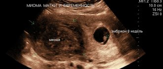
Thrombocytopenia includes conditions in which the number of peripheral blood platelets is less than 150 × 109/L. This is a fairly common hematological syndrome in the neonatal period. In 1–5% of children, thrombocytopenia is registered at birth, but in only 0.1–0.5% of newborns it is severe (platelet count less than 50.0 × 10⁹/L) [1, 2]. In approximately half of cases, thrombocytopenia manifests itself as hemorrhagic syndrome.
The causes of thrombocytopenia in newborns are highly variable [3]. These may be primary thrombocytopenias, which, as a rule, are based on immunopathological processes. Secondary (symptomatic) thrombocytopenia occurs against the background of various conditions, among which more often viral or bacterial infections, severe hypoxic manifestations, immunodeficiency states, intravascular coagulation syndrome, etc. All forms of thrombocytopenic purpura, according to the mechanism of occurrence, are acquired, even in cases of birth of a child with clinical picture of thrombocytopenic purpura.
The mechanisms of development of thrombocytopenia in the neonatal period are different. Among them are:
1. Increased destruction of platelets - as a result of immune processes:
a) isoimmune (alloimmune) thrombocytopenic purpura. The development of this disease is similar to hemolytic disease, however, the immunological conflict is caused by the incompatibility of the fetus and mother in platelet antigens (most often the PLA1 antigen, which is absent in the mother). Antiplatelet antibodies appear in the sensitized maternal body, which, penetrating the placenta, cause the destruction of platelets in the fetus. Isosensitization can occur both during the second and during the first pregnancy. Occurs with a frequency of 1 case per 1000 newborns [4]; b) transimmune thrombocytopenic purpura develops in children born to mothers suffering from autoimmune thrombocytopenia. This happens with idiopathic thrombocytopenic purpura, systemic lupus erythematosus, autoimmune thyroiditis, Evans syndrome, etc. Maternal autoantibodies penetrate the placenta and cause the destruction of platelets in the fetus. Since after the birth of a child the penetration of antibodies from mother to child stops, this form of the disease has a favorable prognosis.
Mechanical destruction of platelets as a result of their increased consumption in cavernous vascular formations occurs in Kasabach-Merritt syndrome.
2. Increased consumption of platelets in intravascular coagulation syndrome, artificial ventilation, hemolytic-uremic syndrome.
3. Insufficient production of platelets - a-, hypomegakaryocytosis. There are exclusively amegakaryocytic thrombocytopenias, which are not combined with any other types of pathology. Hypomegakaryocytoses are combined either with absent radius syndrome (TAR syndrome), or with other bone and organ defects and dysplasias. Pancytopenia - when a-, hypomegakaryocytosis is combined with aplasia of other hematopoietic germs, with various pigmentation disorders, congenital anomalies of the skeleton, eyes, ears, heart.
In addition to the above mechanisms and forms of the disease, heteroimmune thrombocytopenia is distinguished, in which antibodies are produced against a foreign antigen located on the surface of platelets. Examples of foreign antigens are drugs and viruses. It is also possible to develop thrombocytopenia as a result of changes in the antigenic structure of platelets, under the influence of viral exposure. After the medicine is removed from the body or the viral infection is recovered, the signs of the disease disappear and the patient recovers.
A number of toxic and dosage forms used by pregnant women also contribute to the development of thrombocytopenia in newborns. These include alcohol, thiazide drugs, hydralazine, sulfonamides, furazolidone, phenylbutazone, estrogens, heparin therapy, etc.
The mechanism of development of thrombocytopenia during intrauterine infections is quite complex and is associated with insufficient production of platelets by the bone marrow, inhibition of the megakaryocyte lineage by pathogen toxins, hypersplenism, and disseminated intravascular coagulation syndrome.
The diagnostic search algorithm in case of detection of thrombocytopenia in a newborn includes:
1) collection of anamnestic data: the mother has autoimmune thrombocytopenia (idiopathic thrombocytopenic purpura, systemic lupus erythematosus, autoimmune thyroiditis, Evans syndrome, etc.); abnormalities of the placenta (chorioangiomatosis, abruption, thrombosis), use of medications. The newborn has a premorbid background (hypoxic conditions, prematurity, intrauterine growth retardation (IUGR), postmaturity, etc.); the presence of an underlying disease (intrauterine infection, immunodeficiency conditions, giant hemangioma, etc.);
2) determination of the nature of thrombocytopenia - primary or secondary;
3) study of clinical data: hemorrhagic syndrome in the first days of a child’s life in the form of skin manifestations (petechiae, ecchymosis), bleeding from the microvasculature (nasal, gingival, etc.), bleeding from the umbilical cord, melena. Hemorrhagic syndrome with thrombocytopenia is characterized by spontaneity, asymmetry, polymorphism and polychrome. The localization of hemorrhages in the sclera and conjunctiva should be regarded as a prognostically unfavorable sign in terms of the occurrence of hemorrhages in the brain. In the transimmune form, the hemorrhagic syndrome gradually declines, since the supply of antiplatelet antibodies to the child from the mother stops and platelet destruction does not occur [5]. In the case of secondary thrombocytopenia, hemorrhages develop against the background of the underlying disease; a characteristic symptom is hepatoslenomegaly. The severity of hemorrhagic syndrome in newborns with TORCH infections depends not only on the pathogen, but also on the gestational age at which the fetus was exposed to the infectious agent. Kasabach–Merritt syndrome is characterized by the development of neonatal melena;
4) assessment of laboratory parameters. The presence of thrombocytopenia is indicated by:
- the number of platelets in peripheral blood is less than 150 × 109/l against the background of normal other indicators;
- increase in duration of bleeding according to Duque more than 4 minutes;
- reduction in blood clot retraction less than 60%;
- hyperplasia of the megacryocyte lineage (more than 54–114 in 1 μl) in the myelogram;
- absence of deviations in laboratory tests characterizing the coagulation link of hemostasis.
The detection of antiplatelet antibodies confirms the immune nature of the disease. Detection of a diagnostically significant increase in antibody titer to any pathogen (cytomegalovirus (CMV), Epstein-Barr virus, rubella virus, herpes simplex, etc.) confirms the secondary nature of thrombocytopenia (heteroimmune forms of the disease).
Only consistent and thorough conduct of all stages of the diagnostic search allows the doctor to obtain a clear understanding of the processes occurring in the child’s body and develop the correct treatment tactics.
The presence of hemorrhagic syndrome, regardless of the nature of thrombocytopenia (primary or secondary), makes the relief of hemorrhagic manifestations the basis of treatment measures.
For immune thrombocytopenia in the case of a mild disease accompanied only by skin syndrome. The drugs of choice are angioprotectors - etamsylate 12.5% solution 0.1 ml/kg IV or IM 2-4 times a day or 500 mg 4-6 times a day orally. The pharmacological effect of the drug is associated with increasing the stability of capillaries, reducing their permeability, improving microcirculation, as well as stimulating the formation of blood coagulation factor III and normalizing platelet adhesion [6]. Fibrinolysis inhibitors - epsilon-aminocaproic acid at a dose of 50 mg/kg intravenously once a day. Its action is based on inhibiting the activating effect of streptokinase, urokinase and tissue kinases on fibrinolysis, neutralizing the effects of kallikrein, trypsin and hyaluronidase, and reducing capillary permeability [6]. Suppression of the immunopathological process is achieved by using glucocorticoids - prednisolone at a dose of at least 2 mg/kg per day in two doses. Alternative therapy is possible - normal human immunoglobulins (Immunovenin, Pentaglobin, Octagam) at a dose of 0.4 g/kg per day IV for 5 days. The positive effect of immunoglobulins is due to the blockade of Fc receptors of macrophages, which reduces opsonization of platelets and prevents their destruction in the bloodstream. The use of immunoglobulins allows you to obtain a faster (after 24–48 hours), but less lasting effect than with the use of glucocorticoids. Therefore, they often resort to the combined use of these drugs. However, according to foreign authors, the use of prednisolone and immunoglobulins in the alloimmune form of the disease remains controversial [7, 8].
In case of life-threatening bleeding, the use of platelet concentrate is indicated. Its use requires a careful and balanced approach, since in immune forms, transfusion of platelet concentrate is contraindicated, since its use can further aggravate the process due to excessive formation of antibodies. The indication for platelet transfusion is a platelet count of less than 20/nl without signs of bleeding and less than 30/nl with signs of bleeding [7]. The estimated dose for transfusion of platelet concentrate is 10 ml/kg, however, the optimal dose can be obtained by performing an individual mathematical calculation in terms of one of the parameters (body weight, circulating blood volume, body area). After transfusion, platelet levels must be monitored. The clinical criterion for the effectiveness of transfusions of platelet concentrate is the relief of hemorrhagic syndrome, an increase in the patient’s platelet count 1 hour after transfusion by at least 50–60 × 109/l and retention of the result after 24 hours.
In case of secondary thrombocytopenia, it is necessary to treat the underlying disease, since the prognosis is determined precisely by its course.
Thus, the diagnosis of thrombocytopenia in newborns is a very complex process, the competent implementation of which largely determines the success of therapeutic measures, and therefore the prognosis and quality of life of the patient.
We present our own clinical observation of a newborn diagnosed with isoimmune thrombocytopenic purpura, which was diagnosed in a child with a concomitant disease - congenital heart disease.
Georgy V., was admitted to the department for newborn children of the Municipal Children's Hospital of Belgorod on the 8th day of life from the maternity hospital.
Anamnesis of life. From the first pregnancy, it occurred against the background of candidal colpitis. Examination for prenatal infections, IgG antibodies to herpes simplex virus type 1 were detected. Delivery at term, birth weight 3060 g, length 53 cm. Attached to the breast immediately, suckling actively. At birth, multiple elements from petechiae to ecchymoses, sometimes confluent, were noted on the skin. In the early neonatal period, physiological jaundice appeared on the third day of life, the level of total bilirubin was 170 µmol/l, glucose - 4.3 mmol/l, C-reactive protein (CRP) less than 5 ng/ml. The maximum loss of initial body weight was 8.5%; the umbilical remnant fell off on the fourth day of life. Mild neurological symptoms were noted in the form of decreased reflex activity and muscle hypotension. Hemogram on the first day of life: hemoglobin 150 g/l, erythrocytes - 4.6 × 10¹²/l, color index - 0.9, leukocytes - 9.4 × 10⁹/l, band neutrophils - 3%, segmented neutrophils - 65%, lymphocytes - 27%, monocytes - 4%, eosinophils - 1%. ESR 3 mm/hour. During the early neonatal period, cutaneous hemorrhagic syndrome persisted and tended to intensify in the form of the appearance of new elements. Treatment was carried out: etamsylate, menadione sodium bisulfite (Vikasol), phototherapy. Due to the lack of positive dynamics, the child was transferred to the second stage of treatment.
On admission. The condition is serious. The skin is pale pink, acrocyanosis, periorbital cyanosis at rest, hemorrhages throughout the body from small punctate to ecchymoses 3–4 cm in diameter, polychrome. The mucous membranes of the palate, pharynx, conjunctiva, and sclera are clean. On the part of the internal organs: breathing through the nose is free, upon auscultation, puerile breathing is performed over the entire surface of the lungs, there are no wheezes, percussion - a box-shaped percussion sound, respiratory rate is 45 per minute. The area of the heart is not visually changed; upon auscultation, the heart sounds are muffled, the rhythm is preserved, the heart rate is 123 per minute. The abdomen is soft on palpation, the liver is at the edge of the costal arch, the spleen is not palpable. The umbilical area is without features. Stool and diuresis are not disturbed. Over the next three days, the same condition remained: there were multiple hemorrhages on the torso, including “fresh” ones, and the appetite was reduced. From the side of the heart, upon auscultation in the second intercostal space on the left of the sternum, a systolic murmur of moderate intensity was heard. Liver +3.5 cm from under the edge of the costal arch, spleen +1.0 cm. Body weight 2850 g.
Examination. The dynamics of hemogram results are presented in the table.
The ESR did not exceed 6 mm/hour throughout the hospitalization. Urinalysis without deviations from the norm (five times). In a biochemical blood test, the level of CRP is less than 10 ng/ml, liver tests are normal, moderate hypoproteinemia. Enzyme-linked immunosorbent assay (ELISA) for specific antibodies to CMV antigens - negative, ELISA for specific antibodies to Ebstein-Barr virus antigens - negative, ELISA with herpes viruses type 1, 2 - IgM - negative, IgG - positive, avidity index 72 %. ELISA for specific antibodies to chlamydial antigen - negative, ELISA with mycoplasma antigen - negative, ELISA for toxoplasma antigen - negative. In the coagulogram: prothrombin test - international normalized ratio (INR) - 1.0, prothrombin index (PTI) - 100%, prothrombin time - 15.3 seconds; fibrinogen - 1.62 g/l, thrombin time - 16 seconds. Myelogram on the 14th day of life (5th day of hospitalization): blast cells 0.5%, neutrophil maturation index - 1.0, erythrokaryocyte maturation index - 0.8, leukoerythroblastic ratio - 1.3: 1.0 (normal 2.1:4.5). Conclusion: the type of hematopoiesis is normoblastic. The lacing of platelets is weakly expressed; there are many megakaryocytes of varying degrees of maturity.
A blood test of the mother did not reveal any abnormalities in the hemogram.
The electrocardiogram shows signs of hypertrophy of the right ventricle and interventricular septum. ECHO-CG study: additional trabecula of the left ventricle, atrial septal defect of 6 mm, and blood discharge from left to right were detected. The contractility of the ventricular myocardium is satisfactory, the ejection fraction is 68%. Ultrasound of the abdominal organs: liver 53 mm, homogeneous structure. The spleen measures 44 × 26 mm, the structure is homogeneous. Kidneys: on the left - pelvis 7 mm, size 46 × 21 mm, on the right - size 45 × 22 mm. Neurosonography: hypoxic changes and signs of immaturity of brain structures.
Treatment was carried out: etamsylate 12.5% solution 12.5 mg/kg IV 4 times a day for 14 days, normal human immunoglobulin 400 mg/kg/day IV for 6 days, prednisolone 2 mg/kg/day orally for 7 days , fresh frozen plasma 10 ml/kg IV twice, pentoxifylline 0.1 ml/kg IV, inosine 10 mg/kg IV, furosemide 1 mg/kg/day IV, levocarnitine 75 mg 3 times a day orally, recombinant human interferon alpha-2 150,000 2 times a day. The child was fed with human milk substitutes and breastfed from the sixth day of hospitalization. During treatment, by the fifth day of hospitalization, the platelet count increased sevenfold, and the cutaneous hemorrhagic syndrome did not recur. The child was discharged in satisfactory condition on the 26th day of life (18th day of hospitalization).
Thus, this clinical example demonstrates the difficulty of interpreting clinical and laboratory parameters in children of the neonatal period. Laboratory diagnosis of immune thrombocytopenia in many medical institutions in our country is not yet sufficiently developed. Only a clear diagnostic search algorithm will allow the doctor to quickly and clearly assess the variety of causes leading to the development of thrombocytopenia in newborns and promptly and correctly prescribe adequate therapy. Therefore, dissemination of information about the pathogenesis and existing methods for studying various forms of immune thrombocytopenia and the introduction of these methods into clinical practice is a necessary condition for improving diagnosis and increasing the effectiveness of treatment of these diseases.
Literature
- Maschan A. A., Rumyantsev A. G. Immune-mediated thrombocytopenia in newborns: differential diagnosis and principles of therapy // Issues of hematology/oncology and immunopathology in pediatrics. 2010, vol. 9, no. 3, p. 13–18.
- Pshenichnaya K.I. Congenital thrombocytopathies in children: features of diagnosis, symptoms and treatment: Abstract of thesis. diss. Dr. med. Sci. St. Petersburg, 2002. 28 p.
- Shabalov N.P. Neonatology: Textbook. allowance. T. 2. M.: MEDpress-inform. 2004. 640 p.
- Durand-Zaleski I., Schlegel N., Blum-Boisgard C. et al. Screening primiparous women and newborns for fetal/neonatal alloimmune thrombocytopenia: a prospective comparison of effectiveness and costs. Immune Thrombocytopenia Working Group // Am J Perinatol. 1996; 13 (7): 423–431.
- Golovko O.K., Linchevsky G.L., Vorobyova O.V. Clinical aspects of immune thrombocytopenia in neonatology // Child’s Health. 2006. No. 2. pp. 115–122.
- Register of medicines. M.: RLS-2005. 1440 pp.
- Rooz R. Neonatology. Practical recommendations: trans. with him. M.: Med. lit. 2011. 592 p.
- Berkowitz RL, Kolb EA, McFarland JG et al. Parralel randomized trials of risk-based therapy for fetal alloimmune thrombocytopenia // Am. J. Obstet Gynecol. 2006; 107(1):91–96.
N. M. Sudakova*, Doctor of Medical Sciences, Professor N. I. Grevtseva** O. M. Zubov** O. V. Lazareva**
* Belgorod State University, ** Municipal Children's Hospital, Belgorod
Contact information for authors for correspondence
Thrombocytopenia during pregnancy
Pregnant women may experience a decrease in platelet counts and hemocoagulation changes. Thrombocytopenia during pregnancy can be a consequence of both physiological and pathological processes. Normally, in most expectant mothers, the platelet count decreases by 10% compared to normal levels. The most significant decrease in platelets during pregnancy is observed in the last trimester.
If severe thrombocytopenia develops, this is manifested by a loss of strength and the formation of hematomas on the skin and mucous membranes.
Specific conditions associated with pregnancy are:
- Gestational thrombocytopenia - the platelet count may decrease to 43x109/L. Develops at the end of the second to the beginning of the third trimester. It is a physiological condition that does not complicate pregnancy.
- Thrombocytopenia in a hypertensive state – these conditions include preeclampsia and HELLP syndrome. These conditions require treatment, as they threaten the normal course of pregnancy.
To prevent complications, a pregnant woman should regularly take blood tests, eat right and follow all doctor’s recommendations.
Thrombocytopenia
Thrombocytopenia - what is it?
We will talk about a violation of hemostasis, more precisely, its platelet component, which is characterized by the fact that the number of platelets per unit volume of blood decreases.
Occurs:
- High bleeding from the skin and mucous membranes;
- Bruises and hemorrhagic rashes form;
- Bleeding.
In order to eliminate the problem, it is necessary to resort to drug therapy, splenectomy, and extracorporeal blood purification.
Why do pathologies appear?
All causes of pathologies are divided into two groups:
- Congenital;
- Purchased.
If the pathology is congenital, then it is part of hereditary syndromes, for example, Wiskot-Aldrich syndrome.
With the development of pathology in these cases, as a rule, qualitative changes in platelets also occur.
The causes of acquired pathology are quite diverse: lymphomas, sarcoidosis, portal hypertension, splenic tuberculosis, alcoholism, etc.
The most significant group includes pathologies caused by increased platelet destruction. Moreover, they develop both as a result of the mechanical destruction of platelets and the immune component.
- Alloimmune pathology is provoked by transfusion of blood of a different group;
- Transimmune pathology occurs due to the fact that maternal antibodies penetrate to the fetus through the placenta;
- Autoimmune pathology is a consequence of the production of antibodies to one’s own unchanged antigens;
- Heteroimmune pathology occurs due to the fact that antibodies act against foreign antigens that are fixed on the surface of platelets.
- Drug-induced pathology is a consequence of taking sedatives, antibacterial or sulfonamide drugs, and some others.
Note that a slight decrease in the number of platelets can be caused by vaccination or viral infections.
Pathologies that arise due to platelet deficiency develop when there is an insufficient number of stem cells, and thrombocytopenia occurs. This condition may be affected by:
- Aplastic anemia;
- Leukemia;
- Myelofibrosis;
- Presence of metastases in the bone marrow;
- Chemotherapy treatment, some other things.
There are indicators of the severity of thrombocytopenia:
- First stage (satisfactory hemostasis). With it, the platelet count is 150-50x109/l;
- Second stage (sharp decrease in indicator - 50-20 x109/l). This may be the result of a minor injury that causes intradermal hemorrhages or prolonged bleeding from wounds;
- Stage three (platelet count is minimal – 20x109/l). Spontaneous internal bleeding is to be expected here.
Below we will talk about thrombocytopenia, which manifests itself moderately, sharply and severely.
Causes of moderate thrombocytopenia
- Excessive alcohol consumption;
- Pregnancy period;
- Liver pathologies;
- Use of a number of medications;
- DIC syndrome;
- Lupus erythematosus and systemic vasculitis;
- Heart failure;
- Consequences of radiation therapy.
Causes of severe thrombocytopenia
- Systemic lupus erythematosus;
- Significant DIC syndrome;
- Acute leukemia;
- Hemolytic disease of the newborn.
Causes of severe thrombocytopenia
- Acute radiation sickness;
- Overdose of cytotoxic drugs;
- Severe course of acute leukemia.
Symptoms
The manifestations of thrombocytopenia depend entirely on the underlying disease, but it is worth talking about the general signs. Among them:
- Bruises appear inexplicably on the skin;
- The blood slowly stops at the cut sites;
- Often the nose bleeds;
- The oral mucosa bleeds;
- Increased risk of internal bleeding;
- There is a risk of cerebral hemorrhage.
As soon as general symptoms appear, you should consult a doctor. Thrombocytopenia is treated in most cases on an outpatient basis.
Stenosis
If the pathology is severe, the patient must be placed in an intensive care unit. The treatment strategy itself is determined by the underlying disease.
Diagnostic measures
Any increased bleeding is a reason to visit a hematologist. The doctor will prescribe the necessary tests. Among which:
- General blood analysis;
- Hemostasiogram;
- Ultrasonography;
- Radiography;
- Enzyme immunoassay blood test.
The latest tests are indicated if necessary, this is already decided by the doctor.
Pathology in cancer
Thrombocytopenia is very common in cancer patients. This is not by chance, of course, chemotherapy and radiation therapy cause its appearance. Some drugs have a detrimental effect on platelets.
On the other hand, the pathology can be caused by some types of cancer, for example, lymphoma.
If a patient develops a pathology in which the platelet level drops, the doctor is obliged to consider all possible causes, even those not related to cancer treatment.
Thrombocytopenia makes the condition of an oncology patient complex, which means that the doctor faces difficulties in treating him. He must keep in mind that:
- If platelet counts are low, chemotherapy and radiation therapy must be carried out very carefully, as the risk of bleeding increases;
- A decrease in the rate of <50x109 per liter leads to an increased risk of heavy bleeding during surgery;
- A decrease <10x109 per liter significantly increases the risk of spontaneous bleeding.
Treatment of thrombocytopenia in cancer patients
There are recognized methods of treating pathology in cancer patients. They boil down to the following:
- It is necessary to adjust the therapeutic program;
- If necessary, transfuse platelet mass to the patient when there is a risk of bleeding or before a course of chemotherapy;
- It is necessary to transfuse platelet mass under the close supervision of a hemotransfusiologist and a nurse.
There are simple recommendations on how to avoid bleeding:
- Using an electric razor instead of a razor;
- Avoiding injury;
- Using a toothbrush with soft bristles;
- Rejection of drugs that reduce blood clotting, etc.
The Onco.Rehab Integrative Oncology Clinic is glad to see its patients who are not burdened by pathologies and their complications. We fight for your health, so that your days are filled with the joy of life.
Diet
Diet for essential thrombocythemia
- Efficacy: therapeutic effect after 3 weeks
- Timing: constantly
- Cost of products: 1400-1500 rubles per week
People who have a low platelet count in their blood should follow some recommendations when shaping their diet.
- You need to eat meals in small portions, doing this at least 5 times a day.
- The diet should contain protein foods that are easily digestible (liver, poultry, fish).
- It is important to get proteins from plant foods, consuming legumes, nuts, and mushrooms.
- It is recommended to include more fresh vegetables and fruits in the menu. It is better to choose seasonal fruits and steam, boil, or bake vegetables.
- Low-fat dairy products should also be included in your diet every day.
- It is recommended to cook porridge in water from whole grains - buckwheat, rice, wheat.
- It is better to choose bread with bran.
- You should drink weak tea, dried fruit compote, berry fruit drinks, cocoa, and fresh juices.
- You can make an omelet from quail eggs.