- Causes and symptoms of disorders
- Where is the spleen located and how is it examined?
- Is there life without a spleen?
The spleen is a little-studied organ whose functions are not fully understood by medicine. Many generally consider it unimportant and almost superfluous. But in fact, the spleen is part of the lymphatic system and performs important protective functions and participates in the formation of immunity. And to find the cause of many troubles, you need to do an MRI of the spleen.
Anatomy.
Also on topic:
HUMAN ANATOMY
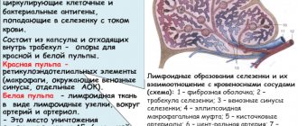
The spleen is made up of several types of tissue. Embryonically, it originates from the middle germ layer, the mesoderm. A certain number of original mesenchymal cells remain in the spleen throughout life, while the rest turn into lymphoid and reticuloendothelial cells. The pulp (pulp) of the spleen consists mainly of the latter, and lymphoid cells are grouped into lymphoid formations, the so-called. Malpighian bodies. The spleen has an abundant blood supply and its color is dull purple. In addition to the peritoneum (serous membrane), it is covered with a dense elastic fibrous capsule mixed with smooth muscle fibers. The capsule continues into the thickness of the organ in the form of crossbars - trabeculae, forming the skeleton of the spleen and dividing it into lobules. The spleen is directly connected to the portal vein system (carrying nutrient-rich blood from the gastrointestinal tract to the liver) and the systemic circulation.
Diseases of the spleen that can be detected by ultrasound
If pathological abnormalities of an organ are detected, the specialist makes the necessary notes in the conclusion.
- Splenomegaly. The organ is increased in size. Possible with cirrhosis, lymphoma, mononucleosis, anemia. A child’s spleen that is larger than normal indicates a viral disease, leukemia. Signs of this pathology are determined in combination with other changes.
- Break. Occurs when there is a blow to the abdomen with a blunt object, a fall from a height, or an accident. Ultrasound examination is practically undetectable, but the presence of a rupture can be assumed by a hematoma under the capsule of an organ or periorgan fluid.
- Heart attack. Formed in the presence of a blood clot or rupture of a vessel. Splenic infarction on ultrasound is defined as an area of tissue with low density and no blood flow.
- Cyst. Possible parasitic, tuberculous, hemorrhagic. The image shows a round formation with a thin capsule that does not reflect ultrasound waves.
- Hemangioma. A benign tumor-like formation consisting of blood vessels. Splenic hemangioma on ultrasound is represented by a dense round formation.
- Abscess. Inflammatory process in the capsule. It appears as a dense area of uneven structure.
The description is deciphered by the attending doctor. An accurate diagnosis is made based on all examination data.
What are the dangers of spleen diseases for a child?
Children's organ parameters differ from those of adults. The organ grows with the child. Up to 1 year, the norm is: 5-5.5 cm X 1.5-2.5 cm. For adolescence: 9-12 cm X 3.5-5 cm. When examined by a pediatrician, the size of the organ must correspond to age and may be acceptable values for its increase:
- in newborns – 30%;
- up to 1.5 years – 15%;
- among younger schoolchildren – 3%.
Splenomegaly in childhood is determined by a table of standards with which specialists compare ultrasound data. Increased processing of red blood cells by an enlarging spleen in a child can lead to anemia and decreased immunity, which will cause frequent pain, weakness and weight loss. Possible digestive tract upset. The child does not eat well due to the pressure of the spleen on the stomach, which creates an imaginary feeling of fullness. Nausea and vomiting, pale skin are possible.
Diseases of the spleen in adults
With tumor-like formations of the pancreas, splenomegaly is formed. The tumor blocks the outflow of venous blood through the splenic vein, and hematopoiesis is disrupted. The enlarged organ captures excess blood cells, at the same time receiving pathological cells. The spleen becomes clogged, enlarges faster, filters blood worse, and damage to healthy cells occurs. The disease affects the full functioning of all organs, connective tissue is damaged, and the lining of the joints is destroyed.
Physiology.
Galen considered the spleen an organ “full of mystery.” The ancient Greeks and Romans are known to have removed runners' spleens to increase their running speed. The functions of the spleen are not fully understood. For a long time it was considered an endocrine (without excretory ducts) gland. Since there is no reliable data on the secretory activity of the spleen, this theory had to be abandoned, although recently it has to some extent received a second life. The spleen is now credited with hormonal regulation of bone marrow function.
Also on topic:
COMPARATIVE ANATOMY
In the early stages of fetal development, the spleen serves as one of the hematopoietic organs. By the ninth month of intrauterine development, the formation of both erythrocytes and leukocytes of the granulocyte series is taken over by the bone marrow, and the spleen, starting from this period, produces lymphocytes and monocytes. In some blood diseases, however, foci of hematopoiesis reappear in the spleen, and in a number of mammals it functions as a hematopoietic organ throughout life.
In an adult, the spleen performs several functions. As part of the reticuloendothelial system, it phagocytizes (destroys) dead blood cells and platelets, and converts hemoglobin into bilirubin and hemosiderin. Because hemoglobin contains iron, the spleen is one of the richest iron reservoirs in the body. As a lymphoid organ, the spleen is a major source of circulating lymphocytes, especially in adolescence and young adults. In addition, it acts as a filter for bacteria, protozoa and foreign particles, and also produces antibodies; People without a spleen, especially young children, are very susceptible to many bacterial infections. Finally, as an organ involved in blood circulation, it serves as a reservoir of red blood cells, which in a critical situation are released back into the bloodstream.
Where is the spleen located and how is it examined?
The small organ is located behind the stomach in the left hypochondrium, so checking its condition is quite difficult. For research, palpation, x-rays, ultrasound and more advanced tomography methods are used. The advantage of MRI is that the diagnosis gives the most complete picture of the location, size, and condition of the organ tissues. In addition, this is the safest method that does not cause any harm to the body. You can undergo examination at least every day if the financial side of the issue is not an obstacle.
But it should be noted that stereotypes about tomography being undemocratic are greatly exaggerated. The price of an MRI of the spleen starts from 2,500 rubles (in Moscow, and even cheaper in the regions). On our website MRT-kliniki.ru you can find a list of all diagnostic centers, compare costs, choose clinics with preferential conditions or promotions and sign up for an examination with a discount of up to 50%.
Splenomegaly,
or enlargement of the spleen is a characteristic response of the organ to many pathological conditions. Splenomegaly may be associated with enlarged lymph nodes, ascites (fluid in the abdomen), jaundice, leukopenia (decreased white blood cell count), fever, enlarged liver, or severe anemia. It is observed in many cardiovascular diseases; for many infectious diseases - malaria, typhoid fever, smallpox, measles, syphilis, meningitis, scarlet fever, etc.; for blood diseases - leukemia, hemolytic jaundice, chronic hemolytic anemia, usually congenital. Sometimes an enlarged spleen occurs in Hodgkin's disease; It reaches enormous sizes in chronic myeloid leukemia. Metabolic disorders, especially fat metabolism, are also often accompanied by splenomegaly. Many liver diseases affect the condition of the spleen. This primarily concerns Banti syndrome, in which cirrhosis of the liver is accompanied by congestive splenomegaly and anemia. With a hereditary disease - Gaucher disease - there is a disorder of fat metabolism and splenomegaly.
Since splenomegaly is only a manifestation of some other disease, treatment should be aimed at the primary cause. Removal of the spleen is indicated in rare cases; sometimes it is performed in diseases associated with increased destruction of red blood cells or platelets, in particular in hemolytic jaundice, thrombocytopenic purpura, Banti syndrome, but even then an improvement in blood counts can be expected only in 30–60% of cases.
See also LYMPHATIC SYSTEM.
Ultrasound of the spleen in adults: what it shows, preparation for the procedure
Ultrasound of the spleen in adults
The spleen is an organ responsible for the protective properties of the body. With the help of the spleen, the immune system and metabolism are formed. If there is a malfunction in its operation, this leads to disruption of many important systems of the human body. In this case, diagnostic procedures are prescribed, namely ultrasonic examination. What does it show?
- Ultrasound of the spleen in adults is one of the safest diagnostic methods.
- It gives an almost complete, and most importantly, objective picture of the health of this organ.
- Ultrasound allows you to determine the size and function of the spleen.
- Both adults and children from an early age can be subject to such examination.
- There is no risk in this procedure.
The spleen is a kind of filter of the body that absorbs bacteria and diseased blood cells, and other pathological particles that enter the body or are produced in it.
An ultrasound is done to determine a number of abnormalities. Reasons to do an ultrasound include:
- Determination of organ size or abnormalities of its development
- Detection of neoplasms
- Pain in the lower back or side
- Pain in the left side of the back
- Pain in the shoulder blade
- Nausea
- Reduced blood pressure readings
- Liver diseases
- Injuries
- Diseases of the blood or lymph nodes
How to prepare for the ultrasound ? Here is the answer:
- An important task is to prevent excess gas formation. A loaded bowel may make examination difficult.
- Three days before the ultrasound you should follow a small diet. Legumes (including peas and beans), sweet foods (confectionery sweets), fresh white bread and vegetables that have not been thermally processed should be excluded from the diet.
- Nine hours before the ultrasound , you should stop eating altogether. Sometimes, to improve the digestion of food, doctors prescribe the evening before the procedure to drink a Filtrum or Smecta . This is necessary in order to neutralize the gases.
The ultrasound examination itself is carried out strictly on an empty stomach, with the exception of diabetics. If you have diabetes, your doctor will recommend how to go about this procedure correctly and what you can eat and when.
Prevention
The spleen, previously considered an insignificant organ, when enlarged can cause damage to the entire functioning of the body. Therefore, it is better to carry out prevention to avoid problems:
- Avoid injuries in the abdominal area;
- Exercise. 10-12 am is the best time to study. At 22:00 the organ’s rest period begins, so it’s better to go to bed earlier;
- a gentle diet without fatty and spicy foods that irritate the liver. The liver and spleen work together;
- If you notice discomfort in the left side, you need to make an appointment with a doctor.