Reception is conducted by:
Krivonosov Denis Sergeevich -
cardiologist, candidate of medical sciences
Make an appointment
Svitova Elena Olegovna —
Obstetrician-gynecologist. Functional diagnostics doctor. Ultrasound diagnostics doctor. Doctor of prenatal ultrasound diagnostics.
Make an appointment
Pelvic varicose veins (PVVD) is a chronic vascular pathology characterized by dilation of the pelvic veins. With the disease, the return flow of blood worsens, causing stagnation and poor circulation. Symptoms of pelvic varicose veins in women are nonspecific and are inherent in many gynecological pathologies, so literally a few decades ago the disease could not be diagnosed in time and only the symptoms were stopped, and their root causes were not eliminated.
According to a study conducted in 1999, in approximately 15-20% of cases in patients with vein disease, the reason for visiting a gynecologist was pain in the lower abdomen. However, the diagnosis was correctly made only in 2% of cases. It’s scary to imagine that about 15% of women had their uterus removed as a result of an incorrect diagnosis.
The Profimedica clinic employs professionals with extensive experience. Modern equipment from the world's best manufacturers allows specialists to diagnose and eliminate pathology at the earliest stages.
Varicose veins of the pelvis in women
Varicose veins, or ectasia (expansion of the lumen of a vessel in a limited area), is a disease that affects the venous network of the pelvic region. Varicose veins of the pelvis lead to disruption of normal blood flow from the internal and external female genital organs. In the vast majority of cases (80%), varicose veins are dilated by the ovarian veins, and extremely rarely (1%) with pelvic varicose veins, the veins of the broad ligament of the uterus are affected. Most often, varicose veins of the pelvis occur in women of fertile age - from 25 to 45 years.
Varicose veins of the small pelvis, associated with the expansion of its vessels, have recently become a fairly common disease in women. Varicose veins of the small pelvis occur in 15-20% of cases. Unfortunately, this pathology in most cases is accompanied by a number of other gynecological diseases, in which their clinical symptoms often hide the manifestations of pelvic varicose veins.
It is precisely because of the difficult diagnosis of pelvic varicose veins that doctors can choose incorrect and ineffective ways to treat varicose veins of the small pelvis, and therefore many patients are not satisfied with the results of standard treatment of veins and intrapelvic organs.
Request a call back Get a free consultation
Our doctors
Drozdov Sergey Alexandrovich
Cardiovascular surgeon, phlebologist, Doctor of Medical Sciences
47 years of experience
Make an appointment
Malakhov Yuri Stanislavovich
Doctor - cardiovascular surgeon, phlebologist, Honored Doctor of the Russian Federation, Doctor of Medical Sciences, doctor of the highest category
Experience 36 years
Make an appointment
Symptoms of pelvic varicose veins
Dilated veins and impaired venous blood flow in the pelvis can lead to quite serious health consequences and also negatively affect women’s self-esteem.
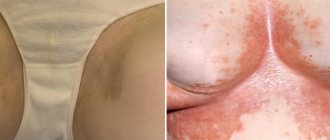
With varicose veins, the venous vessels (perineal and vulvar) dilate in such a way that swollen, enlarged veins become visible and protrude on the patient’s skin; in addition, this process is often accompanied by:
- local edema;
- a feeling of heaviness and bursting pain in the pelvis;
- bleeding;
- mucous discharge.
The undoubted signs of varicose veins of the pelvic veins include:
- Regular long-term pain in the lower abdomen, which manifests itself primarily after physical exertion.
- Increasing discomfort in the second half of menstruation.
- Dysmenorrhea.
- Sharp painful sensations due to hypothermia of the lower extremities, stress;
- Dyspareunia.
If a patient experiences several of the listed symptoms at once, we can with a high degree of probability speak of pelvic varicose veins.
Rice. 2. Cosmetic defects of the external genitalia with pelvic varicose veins (PVVD)
Prices for phlebologist services
0
0 services selected for the amount of 0 rub.
| Total: | from 0 rub. | Sign up |
| Appointment (examination, consultation) with surgeon Globin A.V. (PhD) primary with ultrasound examination of the veins of the lower extremities | from 4000 rub. | Sign up |
| Microsclerotherapy (1 limb) (1 session) surgeon Globin A.V. | from 5400 rub. | Sign up |
| Microsclerotherapy (2 limbs) (1 session) surgeon Globin A.V. | from 7300 rub. | Sign up |
| Microsclerotherapy under ultrasound control or Venolight navigation (1 limb) (1 session) surgeon Globin A.V. | from 6400 rub. | Sign up |
| Microsclerotherapy under ultrasound control or Venolight navigation (2 limbs) (1 session) surgeon Globin A.V. | from 9200 rub. | Sign up |
| Endovenous laser surgery for varicose veins (1 limb) surgeon Globin A.V. | from 48400 rub. | Sign up |
| Endovenous laser surgery for varicose veins. CVI stage 1-2 (2 limbs) surgeon Globin A.V. | from 74600 rub. | Sign up |
| Endovenous laser surgery for varicose veins. CVI stage 3-4 (2 limbs) surgeon Globin A.V. | from 89300 rub. | Sign up |
Causes of varicose veins
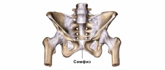
The venous system of the pelvis in women is very complex; varicose veins of the small pelvis are partly provoked by the anatomical and physiological characteristics of the blood supply to this area. In the female body, the uterovaginal, vesical, and rectal plexuses anastomose with each other and carry blood to the internal iliac vein, and the veins of the round and broad ligaments of the uterus, merging with the branches of the uterine vessels, to the hilum of the ovary. The female genital organs and their veins also experience increased stress during pregnancy, which affects the elasticity of the walls of blood vessels.
Typically, pelvic vein pathology results from:
- constant physical overload;
- pregnancy and childbirth;
- age-related changes;
- hormonal imbalances.
Often, patients notice the first manifestations of a disease associated with dilated veins during pregnancy. Please note: with pelvic varicose veins, poorly functioning veins lead, in addition to internal discomfort, to external unaesthetic manifestations on the skin. Since a vein affected by varicose veins makes a depressing impression, it can cause the patient to develop psychological complexes.
Request a call back Get a free consultation
Classification
Pelvic varicose veins can be primary or secondary. The first occurs with congenital insufficiency of the venous valves, and the second, which occurs much more often, as a complication of inflammatory or tumor diseases in the pelvis.
The stage of the disease depends on the diameter of the affected vessels and at the initial stage of varicose veins it does not exceed 5 mm. The second stage of the disease is characterized by an increase in the size of the vessels to 10 mm, and the third stage of the disease and total damage is indicated by a vessel diameter of more than 10 mm.
Varicose veins of the pelvis can be accompanied by varicose veins of the perineum, vulva, pampiniform plexus of the spermatic cord, penis, buttocks, inner thigh and varicose veins of the lower extremities.
Diagnosis of varicose veins of the small pelvis
Specialists who suspect varicose veins in a woman use the following modern methods for diagnosis:
- transabdominal scanning;
- transvaginal scanning;
- Ultrasound angioscanning of the venous system.
It is these methods that make it possible to give an accurate assessment of the condition of the ovarian, inferior vena cava, iliac, and renal veins. During diagnostics in women with possible varicose veins, specialists immediately determine the main parameters of the venous system:
- Vessel diameter.
- Characteristics of blood flow.
- The presence or absence of pathological reflux (reverse flow) of blood in any vein.
- The presence of other venous pathologies.
The second stage of examination of venous vessels involves radionuclide venography and computed tomography with in vivo labeled red blood cells. These procedures allow you to objectively assess the condition of the vessels, detect varicose veins and the presence of signs of pelvic venous congestion. Also an important factor in the successful treatment of venous vessels of the pelvic region is the correct and objective assessment of pain syndrome, which is carried out using the modified McGil visual analogue scale.
| Stage | Characteristic features of affected vessels, diameter in mm | Localization |
| first | less than 5 | any pelvic venous plexus |
| second | from 6 to 10 | ovaries or uterus |
| third | more than 10 | total vascular damage |
Absolutely correct and accurate information about the presence of the disease, the condition of the blood vessels and pelvic organs at the final stage of the examination can be obtained by performing an invasive X-ray contrast study. During selective ovariography and pelvic venography, examining literally every vein, doctors as clearly as possible:
- clarify the anatomical features of the position and structure of the ovarian vessels;
- measure the diameter of the ovarian vessels;
- identify the number and nature of the tributaries of the gonadal vessels;
- identify the presence of reflux characteristic of the internal and ovarian iliac veins;
- detect the connection of subcutaneous and intrapelvic vessels.
Note! If a comprehensive examination confirms the preliminary diagnosis of “varicose veins of the small pelvis”, treatment for this disease should begin immediately!
Diagnostics
Timely detection of the process of vascular dilatation increases the likelihood of its cure and prevention of relapses.
To diagnose pelvic varicose veins, doctors at our clinic use instrumental research methods. One of the most effective ways to detect the disease is ultrasonography. This method is carried out in conjunction with transvaginal and abdominal ultrasound, which allows specialists to obtain the necessary data for diagnosis. During ultrasonography, vascular dilation can be identified by multi-colored spots of irregular shape.
Another research method is laparoscopy. In this case, the presence of pathology is signaled by bluish-colored formations that have a thin, tense wall in the area of the uterine ligament or ovaries.
Recently, a modern diagnostic method – selective oophorography – has been gaining popularity. During the study, a contrast is performed on the patient's blood vessels, thanks to which the doctor is able to see any deviations in their structure.
A CT scan may also be performed. The method helps to see the vessels in the woman’s internal genital organs.
Pronounced discomfort during sexual intercourse and other unpleasant manifestations of the disease contribute to the deterioration of the psychosomatics of patients, which often causes diagnostic errors. The presence of a clear pathognomonic symptom, combined with the absence of signs of pathology of the pelvic organs during a traditional examination and manifestations of clear psychosomatic disorders, often leads specialists to think about the psychoneurological nature of the disease. As a result, instead of treating varicose veins, patients are prescribed consultations with sex therapists and psychotherapists.
How is the treatment carried out?
Pelvic veins affected by varicose veins can be subjected to various medical procedures, in accordance with different methods of treating this disease. Treatment of the disease is carried out using several well-proven techniques. This:
- Operations to restore vein patency.
- Removal of varicose veins. Modern surgery makes it possible to remove varicose veins of the pelvis. However, any operations (even laparoscopic) performed under anesthesia can cause some harm to the patient’s health. Also, with surgical interventions, there is a possibility of relapse and there is always a risk of complications.
- A modern technique of endovascular embolization, which allows preserving the diseased vein.
Request a call back Get a free consultation
Treatment
The main recommendations are aimed at reducing intra-abdominal pressure and improving the outflow of blood from the pelvic veins.
The diet includes vegetables, fruits, and excludes alcohol.
Patients are advised to limit physical activity, stay on their feet for long periods of time, and use a contrast shower on the perineal area.
It is advisable to wear compression tights of compression class 2.
Physical therapy is of great importance. Exercises are performed lying down (“birch tree”, “scissors”, “bicycle”), breathing exercises (slow deep inhalation and exhalation with the involvement of the abdominal wall muscles).
Among medications, a course of phlebotonics (detralex) is indicated in long courses of 2-3 months.
If conservative therapy is ineffective, surgical intervention is performed - resection of the gonadal veins.
Modern method of treating pelvic vein dilatation
Today, treatment of pelvic varicose veins using modern embolotherapy techniques is the most effective and patient-friendly way to correct the venous system in this area.
After conducting a single invasive intravascular examination of varicose veins, specialists at the endovascular surgery center use an innovative method - endovascular embolization of gonadal vessels.
This treatment method involves the introduction of embolization (clogging) coils into a vein through a diagnostic catheter, which allows you to completely block the pathological blood flow of varicose veins. The operation for embolization of varicose veins takes a minimum of time. In this case, the patient can return to her normal life in the near future after embolotherapy on the vessels in the pelvis. The rehabilitation period after the procedure is simple and takes place in a short time.
Diagnosis of the disease
An examination by a phlebologist begins with clarifying the patient’s complaints, collecting anamnesis and visual examination of superficial vessels. Additionally, instrumental studies of blood vessels are performed.
- Ultrasound duplex scanning of the vessels of the pelvic veins solves a complex of diagnostic problems. It allows you to determine the presence of varicose veins, measure the diameter and patency of blood vessels, blood flow speed, choose the optimal method of therapy, etc. The study is carried out in two ways - transvaginally and transabdominally. In the first case, the veins of the parametrium, acrylic plexuses and uterine veins are visualized. Transabdominal examination allows you to evaluate the condition and functionality of the inferior vena cava, ovarian veins and left renal vein.
- Pelvic venography with selective bilateral radiopaque ovariography. Before the examination, a contrast agent is injected into one of the main veins using a catheter and the vessels are examined under the control of an X-ray machine.
- Diagnostic laparoscopy is a minimally invasive surgical procedure. Used when other diagnostic methods are insufficiently informative.
To assess the degree of development of venous pathology and the possibility of relapse, an additional scan of the vessels of the lower extremities, perineum, vulva of the inner thigh and buttocks is performed.
Benefits of using endovascular embolization
Thanks to the endovascular embolization method, varicose veins of the small pelvis can be successfully treated. The main advantages of this method in the fight against dilated veins are:
- High efficiency. Endovascular embolization helps patients whose veins are dilated and pelvic blood flow is impaired in 94.8% of cases.
- High speed of the procedure. Embolization of varicose veins of the pelvic venous system takes only 20-30 minutes.
- No general anesthesia. For pelvic vein embolization, only local anesthesia is used. Any vein is treated using proven local anesthetics, which avoids the long recovery associated with general anesthesia.
- Conducting minimally invasive intervention on an outpatient basis. Women with pelvic varicose veins spend a minimum of time in the hospital after surgery. In some cases, patients can go home immediately after surgery, but usually operated patients remain in the hospital under the supervision of a doctor for several hours.
- Minimum rehabilitation period. Within a few weeks after the intervention, reproductive function will be restored and pain in the lower abdomen will disappear. And after 1-2 months, the patient will be able to completely forget about what varicose veins of the small pelvis are.
Treatment with embolization, carried out by experienced specialists, is truly the most effective and safe. No matter how much the veins are dilated, the operation will not cause discomfort. During the intervention and insertion of emboli into the vein, patients do not experience pain: all unpleasant sensations are relieved by modern medications. In the postoperative period, to fully restore health, the patient will need to meet several easy conditions:
- strengthen the drinking regime (increase the frequency and volume of liquids drunk daily);
- avoid intense physical activity (sports, heavy lifting);
- exclude visiting a pool or bathhouse and taking baths in the first postoperative week, while using a shower is allowed.
Prevention of varicose veins of the small pelvis
Despite the fact that treatment of an altered vein using embolotherapy is accessible and effective, it is better to prevent any disease. In order for each vein to remain in its natural state, it is necessary to ensure normal blood circulation in the body. To do this, you should follow the simplest rules:
- To live an active lifestyle. Even if you have to spend the whole day in the office at the computer, you can always take a leisurely walk in the evening. Exercises for the abdominal area, dance movements of “waggling” and “swaying” with the pelvis, which accelerate blood flow in this part of the body, would be appropriate. Of course, it should be remembered that excessive exercise can provoke the disease, rather than prevent it.
- Be able to relax emotionally. Constant stress often becomes the main cause of problems with blood circulation and stagnant processes. It is very useful to take baths with various aromatic oils, experience positive emotions, sing, and dance.
- It is advisable to have a regular intimate life. Of course, if you have already discovered signs of the disease, you should consult a specialist.
By properly combining work and rest, it is easy to prevent the occurrence of many unpleasant diseases, as well as significantly improve your health.
Request a call back Get a free consultation
Features of therapy at the center of Professor Kapranov
Professor S. A. Kapranov offers modern and effective treatment for vascular pathology. His students and colleagues have accumulated (over more than 10 years) experience in carrying out such minimally invasive interventions, unique for Russian medicine, having operated on several hundred patients. Possessing unique knowledge and skills, they will quickly and effectively help in the treatment of varicose veins.
When treating gynecological diseases (including varicose veins of the small pelvis), the endovascular surgery center uses the latest equipment and innovative instruments. This allows you to treat pelvic varicose veins as carefully and accurately as possible.
An important factor is the optimal cost of treatment for a disease such as pelvic varicose veins.
Sergei Anatolyevich was awarded a high award (Russian Federation Government Prize) for restoring the reproductive health of the fair half of humanity.
The Center for Endovascular Surgery offers an innovative approach to treatment - embolization of pathological veins of the small circle with detachable coils. You can choose the clinic for the intervention yourself. Experienced specialists will do everything so that you can avoid discomfort and numerous risks. You will get rid of varicose veins in the shortest possible time and can return to your normal lifestyle.