The color of the hematoma can be different - from bright red to purple, most often it is heterogeneous - its edges are darker, bluish in color, and the inside of the hematoma is red. Below in the article you will find the causes of the disease; the doctors who treat him; necessary medical procedures for treatment; as well as general information about the disease, its localization, features of diagnosis of diseases and their treatment. However, we advise you to consult a doctor, because self-medication in 90% of cases is fraught with the disease progressing to the chronic stage with extremely unpleasant complications
Make an appointment and consultation
Causes
Hemorrhage usually occurs after injury. This could be a bruise of the skin, internal organs, a concussion or bruise of the brain, an injection with thin (sharp) objects. Sometimes blood leaves the vessels and pours into the skin and internal organs as a result of infections, autoimmune diseases, and poisoning. The occurrence of hemorrhages and bruises is promoted by increased fragility of blood vessels, fasting, lack of vitamins in food, high blood pressure, and congenital bleeding disorders.
At CELT you can get a consultation with a traumatologist-orthopedic specialist.
- Initial consultation – 3,000
- Repeated consultation – 2,000
Make an appointment
Symptoms
A bruise on the skin is always clearly visible. At first it has a purplish-blue color, and then begins to “bloom”, acquiring yellow and green colors. If a sufficiently large amount of blood accumulates under the skin, a protruding lump forms. At first it is very painful to feel, but later the pain goes away.
The outpouring of blood into the internal organs and into the substance of the brain is preceded by trauma. The main symptom is pain. In this case, the hematoma is not visible externally. If bleeding continues, the victim becomes pale, weak, and dizzy. With chronic internal bleeding, anemia comes to the fore. Bleeding in the brain is especially dangerous. Compression of brain structures may occur, sometimes leading to death.
WHEN YOU SHOULD SOUND THE ALARM, THE FIRST SYMPTOMS OF HEMATOMA
A hematoma gives its symptoms and signs almost immediately after injury.
- Firstly, the skin at the site of the hematoma is sharply painful.
- After a short period of time, the site of injury begins to swell, the tumor can spread significantly and interfere with movement (for example, with a hematoma on the ankle, the swelling may be such that it is impossible to move independently or step on the affected leg).
- After swelling, the site of hemorrhage quickly turns red. Patients feel internal tension in the area of the hematoma; it is hard to the touch.
The color of the hematoma can be different - from bright red to purple, most often it is heterogeneous - its edges are darker, bluish in color, and the inside of the hematoma is red.
Predisposing factors
The formation of hematomas occurs after injuries, including pinching, blows, squeezing, and bruises. Subarachnoid hemorrhage does not fall into this category, since it does not appear due to trauma, but due to damage to an unchanged vessel. Often small hematomas appear due to eating large quantities of food or drinking alcoholic beverages. This is due to stretching of the gastrointestinal tract and the appearance of cracks.
The development of pathology is influenced by vascular weakness and problems with blood clotting. Often due to a weakened immune system due to infections or age-related changes, the likelihood of pus accumulating in the affected area increases.
Treatment of hematoma on the head of a child
Due to excessive mobility, children often get injured. A large percentage of childhood injuries are head contusions, which are often accompanied by hematomas and the formation of “bumps.”
First aid involves applying a cold object to the site of the hematoma. This should be done in the first few hours. Important: given the imperfection of thermoregulation in children (high sensitivity to low temperatures), the use of cold in children should be dosed - about half an hour and at intervals.
We recommend visiting our clinic’s website https://www.dobrobut.com/, where you will learn in more detail about the causes and consequences of hematomas in children.
Related services: Departure of an emergency team Departure of a specialized pediatric emergency team
Classification of hematomas
In modern medicine, when classifying hematomas, the following are taken into account:
- Relation to the vessel - pulsating and non-pulsating hematomas.
- Localization - in the cranial cavity, internal organs, under the skin or mucous membrane.
- The state of the blood in the affected area is suppurated, clotted, fresh, infected.
- Symptoms – limited, encysted, diffuse.
There are hematomas that do not fall into this classification. For example, intracerebral, intracranial, intraventricular. They are of the epidural or subdural type and cause serious complications.
Soft tissue hematomas
Soft tissue hematomas are divided into 3 types:
- Lungs - appear 24 hours after injury and are accompanied by mild pain. No special treatment is required.
- Medium - appear within 5-6 hours and are accompanied by pain and swelling. The motor function of the limb deteriorates. Consultation with a traumatologist is required.
- Heavy - formed within 2 hours after tissue damage. The function of the limb is impaired, acute pain and diffuse swelling are observed. You should immediately consult a doctor to determine a treatment strategy.
Immediately after the injury, swelling appears, and the skin acquires a purplish-bluish tint. After 5 days, the skin takes on a green tint as hemoglobin breaks down. Gradually, the hematoma resolves and “flows” down.
If there are no complications, the hematoma will resolve on its own. In the worst case, a hard area appears that causes discomfort and impairs motor function. When an intramuscular lump forms, external symptoms are rarely observed, but the limb swells significantly and an area forms inside, the touch of which causes severe pain.
Note! For chronic intramuscular hematomas, an MRI is prescribed to determine the location and extent of tissue damage.
When large lumps form, surgical intervention is required. Treatment is carried out by a traumatologist. The opening of infected seals is performed by a surgeon after a comprehensive diagnosis. The operation is performed on an outpatient basis, but for large hematomas hospitalization is required. An autopsy is performed, during which blood clots are removed and washing is carried out. Drainage and suturing are required. Sutures are not applied only for infected hematomas. Antibiotics are often prescribed in combination to eliminate the infection.
Using ointments for bruises
It is important that if there is a bruise or a hematoma, try to apply ice or a heating pad with cold water to the damaged area. This will prevent the development of severe swelling. Only after this is it recommended to use anti-bruise products.
On the first day after injury, it is better to use ointments containing heparin. The drugs increase the speed of blood flow, thin the blood and prevent the expansion of the hematoma.
If there is pain, you can apply an ointment containing non-steroidal anti-inflammatory drugs to the damaged area. The most well-known drugs are diclofenac and ibuprofen. They can be applied to the damaged area a couple of hours after injury.
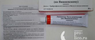
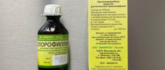
In the future, to speed up the resorption of the bruise, you need to use ointments based on badyagi or comfrey. You can also use other ointments with a warming effect to increase blood circulation. They often contain snake and bee venom.
With proper and timely treatment of bruises, the occurrence of large hematomas can be prevented. But if bruises do appear, it is necessary to use special ointments to speed up their resorption and tissue restoration. When you cannot get rid of hematomas on your own, and the bruises increase in size, you should urgently consult a doctor.
Intracranial hematomas
Intracranial hematomas are divided into the following types:
- Epidural.
- Subdural.
- Intracerebral.
- Intraventricular.
Epidurals appear in 1-3% of cases and are due to injury to the middle meningeal artery. Pathology is often observed with skull fractures or depressed fractures. A hematoma develops in 2-3 hours or within 24 hours. Lack of treatment leads to coma. The first symptoms are confusion and weakness. Children rarely lose consciousness after a severe blow. Significant swelling of the brain does not lead to the detection of a light gap (which is rare in adults).
Subdurals appear in 1-7% of cases and pose a threat to human life, since death occurs in 60% of cases. There is an acute, subacute and chronic form of the pathology. Bleeding occurs due to a rupture of a vein or artery in the damaged area. People report nausea and severe headaches. Symptoms characteristic of compression of the brain stem are often observed. Lack of treatment and worsening symptoms lead to coma.
Intracerebral are observed extremely rarely with severe traumatic brain injuries. The light gap is not visible, the development of pathology occurs quickly. Hemiplegia or hemiparesis often occurs, as well as extrapyramidal symptoms.
Intraventricular diseases are rarely diagnosed due to the serious condition of patients. There are acute disturbances of consciousness, an increase in body temperature, a decrease in heart rate, and an increase in blood pressure. To establish a diagnosis, a survey of close people is carried out, since the patient is unconscious. To establish the location of the hematoma, MRI is used. In the most severe cases, lombal puncture is used.
Products based on comfrey and badyagi
Preparations based on hematomas based on comfrey root and freshwater sponge badyagi have good resorption properties. Such products are presented by manufacturers in a huge variety. You can buy them at the pharmacy at an affordable price.
The effectiveness of comfrey root, which is also known as larkspur, against bruises has long been known. The plant was dried and crushed, and after that infusions were prepared, which were applied to bruises in the form of compresses. Ointments, gels and balms based on larkspur are presented in pharmacies in a wide range of multi-component formulations. Any remedy for bruises can quickly reduce swelling. The drugs have an analgesic effect and promote tissue restoration. Their peculiarity is the absence of contraindications.
The skeleton of the freshwater sponge badyaga consists of silicon oxide. The product is crushed and a powder is obtained, consisting of microscopic needles of silicon oxide. It is the basis of an ointment, which, when applied to damaged skin, has a local irritating effect. This allows you to increase blood circulation and speed up the processes of healing and tissue regeneration. When using it, it is important to exclude individual intolerance to the active substance.
Mani based on comfrey and badyagi is applied to damaged areas 3-4 times a day. It is not recommended to use the products during pregnancy and lactation, as well as before reaching the age of 12 years.
Diagnostics
A hematoma is diagnosed by visual examination. If the hemorrhage is located deep under the skin, in internal organs, or in a joint, it is often very difficult to assess its size and possible consequences.
Patients are prescribed an examination, which may include:
- Ultrasound of internal organs, joints;
- computed tomography and magnetic resonance imaging;
- puncture (puncture with a needle): for example, a puncture of the knee joint is often done if there is a suspicion that blood has accumulated in it after an injury.
Treatment
Minor bruises can be treated conservatively: physiotherapeutic procedures and medications are prescribed.
In case of large accumulations of blood, the hematoma is treated surgically: it is opened, the blood or pus is evacuated, washed with antiseptics and drainage is installed. Antibiotics are prescribed if necessary.
When hemorrhaging into internal organs, it is often necessary to perform surgical intervention, during which it is necessary not only to remove the spilled blood, but also to stop the bleeding.
The multidisciplinary CELT clinic employs experienced traumatologists and surgeons who perform operations on hematomas of various locations. Modern techniques used in our clinic help provide effective treatment and minimize the risk of complications.
Drainage of hematoma and other methods of its treatment
The human body is riddled with blood vessels.
During domestic, industrial or sports injuries, the integrity of the wall of a particular vessel is compromised. This can lead to bleeding or hematoma formation. Hematoma is an accumulation of liquid or coagulated blood in the thickness of soft tissue. This is often a harmless phenomenon, but in some cases doctors are often faced with the need to remove a large hematoma, which can interfere with the functioning of tissues and neighboring organs. Hematomas can form:
- for all types of injuries – closed and open;
- in any part of the human body;
- for injuries of any degree - from minor (in this case, the role is played not by physical effort, but by the fragility of the vascular wall) to severe.
Hematomas can occur not only when physical force is applied to the tissues, but also several hours and days after that. An example is an internal hematoma after surgery - when sutures placed on blood vessels damaged during surgery fail. The possibility of such tissue hemorrhages must be constantly remembered. The timeliness of diagnosis, and therefore the success of hematoma treatment, depends on this.
Orthopedics and traumatology services at CELT
The administration of CELT JSC regularly updates the price list posted on the clinic’s website. However, in order to avoid possible misunderstandings, we ask you to clarify the cost of services by phone: +7
| Service name | Price in rubles |
| Appointment with a surgical doctor (primary, for complex programs) | 3 000 |
| X-ray of the chest organs (survey) | 2 500 |
| Ultrasound of soft tissues, lymph nodes (one anatomical zone) | 2 300 |
All services
Make an appointment through the application or by calling +7 +7 We work every day:
- Monday—Friday: 8.00—20.00
- Saturday: 8.00–18.00
- Sunday is a day off
The nearest metro and MCC stations to the clinic:
- Highway of Enthusiasts or Perovo
- Partisan
- Enthusiast Highway
Driving directions