Autoimmune hemolytic anemia
With the acute onset of autoimmune hemolytic anemia, patients experience rapidly increasing weakness, shortness of breath and palpitations, pain in the heart, sometimes in the lower back, fever and vomiting, intense jaundice. In the chronic course of the process, a relatively satisfactory state of health of patients is noted even with deep anemia, often pronounced jaundice, in most cases an enlargement of the spleen, sometimes the liver, alternating periods of exacerbation and remission. Anemia is normochromic, sometimes hyperchromic; during hemolytic crises, severe or moderate reticulocytosis is usually observed. Macrocytosis and microspherocytosis of erythrocytes are detected in the peripheral blood, and normoblasts may appear. ESR is increased in most cases. The content of leukocytes in the chronic form is normal; in the acute form, leukocytosis occurs, sometimes reaching high numbers with a significant shift in the leukocyte formula to the left. The platelet count is usually normal. In Fisher-Evens syndrome, autoimmune hemolytic anemia is combined with autoimmune thrombocytopenia. In the bone marrow, erythroposis is enhanced, and megaloblasts are rarely detected. In most patients, the osmotic resistance of erythrocytes is reduced, which is due to a significant number of microspherocytes in the peripheral blood. The bilirubin content is increased due to the free fraction, and the content of stercobilin in the feces is also increased.
Incomplete heat agglutinins are detected using a direct Coombs test with polyvalent antiglobulin serum. With a positive test, using antisera to IgG, IgM, etc., it is clarified which class of immunoglobulins the detected antibodies belong to. If there are less than 500 fixed IgG molecules on the surface of red blood cells, the Coombs test is negative. A similar phenomenon is usually observed in patients with a chronic form of autoimmune hemolytic anemia or those who have suffered acute hemolysis. Cases when antibodies belonging to IgA or IgM (against which polyvalent antiglobulin serum is less active) are fixed on red blood cells are also Coombs-negative. In approximately 50% of cases of idiopathic autoimmune hemolytic anemia, antibodies to one’s own lymphocytes are detected simultaneously with the appearance of immunoglobulins fixed on the surface of red blood cells.
Hemolytic anemia due to warm hemolysins is rare. It is characterized by hemoglobinuria with black urine, alternating periods of acute hemolytic crisis and remissions. Hemolytic crisis is accompanied by the development of anemia, reticulocytosis (in some cases thrombocytosis) and an enlarged spleen. There is an increase in the level of free bilirubin fraction and hemosiderinuria. When treating donor red blood cells with papain, it is possible to detect monophasic hemolysins in patients. Some patients have a positive Coombs test.
Hemolytic anemia caused by cold agglutinins (cold hemagglutinin disease) has a chronic course. It develops with a sharp increase in the titer of Cold hemagglutinins. There are idiopathic and symptomatic forms of the disease. The leading symptom of the disease is excessive sensitivity to cold, which manifests itself in the form of blueness and whiteness of the fingers and toes, ears, and tip of the nose. Peripheral circulatory disorders lead to the development of Raynaud's syndrome, thrombophlebitis, thrombosis and trophic changes up to acrogangrene, sometimes cold urticaria. The occurrence of vasomotor disorders is associated with the formation of large intravascular conglomerates of agglutinated erythrocytes during cooling, followed by spasm of the vascular wall. These changes are combined with increased predominantly intracellular hemolysis. In some patients, enlargement of the liver and spleen occurs. Moderately expressed normochromic or hyperchromic anemia, reticulocytosis, a normal number of leukocytes and platelets, an increase in ESR, a slight increase in the level of the free fraction of bilirubin, a high titer of complete cold agglutinins (detected by agglutination in a saline medium), and sometimes signs of hemoglobinuria are observed. Characteristic is the agglutination of erythrocytes in vitro, which occurs at room temperature and disappears when heated. If it is impossible to perform immunological tests, a provocative test with cooling takes on diagnostic significance (in the blood serum obtained from a finger tied with a tourniquet after lowering it into ice water, an increased content of free hemoglobin is determined).
With cold hemagglutinin disease, in contrast to paroxysmal cold hemoglobinuria, hemolytic crisis and vasomotor disturbances arise only from hypothermia of the body and hemoglobinuria, which began in cold conditions, ceases when the patient moves to a warm room.
The symptom complex characteristic of cold hemagglutinin disease can occur against the background of various acute infections and some forms of hemoblastosis. In idiopathic forms of the disease, complete recovery is not observed; in symptomatic forms, the prognosis depends mainly on the severity of the underlying process.
Paroxysmal cold hemoglobinuria is one of the rare forms of hemolytic anemia. It affects people of both sexes, most often children.
Patients with paroxysmal cold hemoglobinuria may experience general malaise, headache, body aches and other unpleasant sensations after exposure to cold. Following this, chills begin, the temperature rises, nausea and vomiting are noted. Urine turns black. At the same time, jaundice, enlarged spleen and vasomotor disturbances are sometimes detected. Against the background of a hemolytic crisis, patients exhibit moderate anemia, reticulocytosis, increased content of the free fraction of bilirubin, hemosiderinuria and proteinuria.
The final diagnosis of paroxysmal cold hemoglobinuria is established on the basis of the detected biphasic hemolysins using the Donath-Landsteiner method. It is not characterized by autoagglutination of erythrocytes, which is constantly observed in cold hemagglutination disease.
Hemolytic anemia caused by erythropsonins. The existence of autoopsonins to blood cells is generally accepted. In acquired idiopathic hemolytic anemia, liver cirrhosis, hypoplastic anemia with a hemolytic component and leukemia, the phenomenon of autoerythrophagocytosis was discovered.
Acquired idiopathic hemolytic anemia, accompanied by the positive phenomenon of autoerythrophagocytosis, has a chronic course. Periods of remission, sometimes lasting a considerable time, are replaced by a hemolytic crisis, characterized by icterus of the visible mucous membranes, darkening of the urine, anemia, reticulocytosis and an increase in the indirect fraction of bilirubin, sometimes an enlargement of the spleen and liver.
In idiopathic and symptomatic hemolytic anemia, the detection of autoerythrophagocytosis in the absence of data indicating the presence of other forms of autoimmune hemolytic anemia gives grounds to classify them as hemolytic anemia caused by erythropsonins. The diagnostic test of autoerythrophagocytosis is carried out in direct and indirect versions.
Immunohemolytic anemia caused by drug use. Various medicinal drugs (quinine, dopegit, sulfonamides, tetracycline, ceporin, etc.), capable of causing hemolysis, form complexes with specific heteroantibodies, then settle on erythrocytes and add complement, which leads to disruption of the erythrocyte membrane. This mechanism of drug-induced hemolytic anemia is confirmed by the detection of complement on the erythrocytes of patients in the absence of immunoglobulins on them. Anemia is characterized by an acute onset with signs of intravascular hemolysis (hemoglobinuria, reticulocytosis, increased content of the free bilirubin fraction, increased erythropoiesis). Acute renal failure sometimes develops against the background of a hemolytic crisis.
Hemolytic anemias that develop when penicillin and methyldopa are prescribed proceed somewhat differently. Administration of 15,000 or more units of penicillin per day can lead to the development of hemolytic anemia, characterized by intracellular hyperhemolysis. Along with the general clinical and laboratory signs of hemolytic syndrome, a positive direct Coombs test is also detected (the detected antibodies are classified as IgG). Penicillin, by binding to the red blood cell membrane antigen, forms a complex against which antibodies are produced in the body.
With long-term use of methyldopa, some patients develop hemolytic syndrome, which has features of the idiopathic form of autoimmune hemolytic anemia. The detected antibodies are identical to warm agglutinins and belong to IgG.
Hemolytic anemia caused by mechanical factors is associated with the destruction of red blood cells as they pass through altered vessels or through artificial valves. The vascular endothelium changes in vasculitis, malignant arterial hypertension; At the same time, platelet adhesion and aggregation are activated, as is the blood coagulation system and thrombin formation. Widespread blood stasis and thrombosis of small blood vessels (DIC syndrome) develop with traumatization of red blood cells, as a result of which they fragment; Numerous fragments of red blood cells (schistocytes) are found in the blood smear. Red blood cells are also destroyed when they pass through artificial valves (more often during multivalve correction); Hemolytic anemia has been described in the setting of a senile calcified aortic valve. The diagnosis is based on signs of anemia, an increase in the concentration of free bilirubin in the blood serum, the presence of schistocytes in a peripheral blood smear and symptoms of the underlying disease that caused mechanical hemolysis.
Less common in clinical practice is hemolytic anemia caused by exposure to lead, poisoning with acids, snake venoms or vitamin E deficiency, as well as intracellular parasites. Hemolytic anemia develops, for example, after a snake bite, accidental or intentional (suicide) ingestion of acetic acid, upon contact with lead vapor, against the background of malaria. Anemia is normocytic, normochromic, regenerative in nature; in the blood serum the content of the free fraction of bilirubin and iron is increased.
Hemolytic-uremic syndrome (Moschkovich's disease, Gasser's syndrome) can complicate the course of autoimmune hemolytic anemia. A disease of an autoimmune nature is characterized by hemolytic anemia, thrombocytopenia, and kidney damage. Disseminated damage to blood vessels and capillaries is noted, involving almost all organs and systems, and pronounced changes in the coagulogram characteristic of DIC syndrome.
Publications in the media
Hemolytic anemias are a large group of anemias characterized by a decrease in the average lifespan of red blood cells (normally 120 days). Hemolysis (destruction of red blood cells) can be extravascular (in the spleen, liver or bone marrow) and intravascular. General signs are severe intoxication with chills and fever, pain in the lower back and abdomen, possible shock as a result of impaired microcirculation, jaundice, splenomegaly, hemoglobinuria.
Etiology. Hemolytic anemia occurs due to defects in red blood cells (intracellular factors) or under the influence of causes external to the red blood cells (extracellular factors). Typically, intracellular factors are inherited, and extracellular factors are acquired.
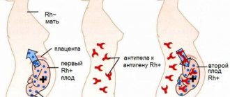
• Extracellular factors. The microenvironment of erythrocytes is represented by plasma and vascular endothelium. The presence of toxic substances or infectious agents in the plasma causes changes in the erythrocyte wall, which leads to: (1) the production of autoantibodies to the altered erythrocyte (a classic example is autoimmune hemolytic anemia), (2) direct destruction of the erythrocyte •• Isoimmune hemolytic anemias are observed in erythroblastosis fetalis; this also includes hemolytic transfusion reactions •• Defects in the vascular endothelium (microangiopathy) can also damage red blood cells - hemolytic microangiopathic anemia. In children, it can occur acutely in the form of hemolytic-uremic syndrome •• Paroxysmal cold hemoglobinuria •• Prescription of certain drugs (for example, sulfonamides, antimalarial drugs) leads to a hemolytic crisis.
• Intracellular factors. Intracellular defects include abnormalities of red blood cell membranes, Hb, or enzymes. These defects are inherited (excluding paroxysmal nocturnal hemoglobinuria) •• Membrane defects ••• Inherited spherocytosis ••• Inherited elliptocytosis ••• Paroxysmal nocturnal hemoglobinuria •• Hemoglobinopathies (eg, sickle cell anemia). More than 300 diseases are known to be caused by point mutations in globin genes. A defect in the globin molecule contributes to the disruption of its polymerization. The membrane and shape of the red blood cell change, susceptibility to hemolysis increases •• Enzymopathies.
Some hemolytic anemias caused by enzyme deficiency
• Anemia due to deficiency of glucose-6-phosphate dehydrogenase (G-6-PD, see also appendix to this article). Clinical picture: acute episodes of hemolytic anemia, usually provoked by drugs, infections (also with neonatal jaundice and favism). Treatment • Replacement therapy (blood transfusions) • Splenectomy for G-6-PD deficiency is useless • Patients with variants of G-6-PD deficiency associated with acute hemolytic crises should avoid taking drugs that cause hemolysis. ICD-10 D55.0 Anemia due to G-6-PD deficiency.
• Anemia due to pyruvate kinase deficiency. Pyruvate kinase deficiency is the second most common (after G6PD deficiency) inherited erythrocyte enzymopathy. Pyruvate kinase catalyzes the final step of glycolysis; the consequence of its deficiency is inadequate production of adenosine triphosphoric acid (ATP), which negatively affects the functions of red blood cells, incl. at the work of Na+-K+-ATPase - defective cells lose K+. Reticulocytes (mainly in the spleen) undergo especially intensive destruction. Clinical picture • Possible manifestations of hemolysis of varying severity; in newborns - jaundice, chronic hemolysis, splenomegaly • Symptoms of pyruvate kinase deficiency are identical to those in other chronic hemolytic anemias • Due to the selective destruction of reticulocytes, their number in the peripheral blood may not reflect the severity of anemia. Treatment: splenectomy is effective. ICD-10 D55.2 Anemia due to disorders of glycolytic enzymes.
• Hemolytic anemia also develops with Norum's disease, as well as due to deficiency of the erythrocyte form of adenylate kinase (*103000, EC 2.7.4.3, AK1 gene, 9q34.1, r), adenosine triphosphatase (*102800, ATPase, EC 3.6.1.3, r, also B), d-aminolevulinate dehydratase (*125270, porphobilinogen synthetase, EC 4.2.1.24, 9q34, ALAD gene, at least 7 mutant alleles inherited by both r and B), hexokinase 1 (EC 2.7.1.1, *142600 , locus 10q22, gene HK1, r), g-glutamylcysteine synthetase (*230450, EC 6.3.2.2, r], glutathione peroxidase 1 (EC 1.11.1.9, *138320, GPX1, 3q11–q12, r), glutathione reductase ( *138300, EC 1.6.4.2, 8p21.1, GSR gene, r), glucose-6-phosphate isomerase (*172400, EC 5.3.1.9, 19q13.1, GPI gene, Â, r), 2,3-diphosphoglycerate mutases (222800, EC 5.4.2.4, 7q31–q34, BPGM gene, r), enolases (*172430, EC 4.2.1.11, ENO1 gene), coproporphyrinogen oxidase (*121300, EC 1.3.3.3, 3q12, CPO gene, В ), pyrimidine nucleotidase (*266120, EC 3.1.3.5, gene P5N, r), triosephosphate isomerase (*190450, EC 5.3.1.1, 12p13, TPI1 gene), ferrochelatase (*177000, EC 4.99.1.1, 18q21.3, FECH gene, Â, r), phosphoglycerate kinase (*311800, EC 2.7.2.3, À [Xq13, PGKA gene]; also  [arch. 19, PGKB gene]), 6-phosphogluconolactonase (*172150, EC 3.1.1.31), phosphofructokinase (*171860, EC 2.7.1.11, 21q22.3, PFKL gene). Treatment is individualized depending on the type of hemolytic anemia (for example, splenectomy or iron replacement therapy).
ICD-10 • D55 Anemia due to enzyme disorders • D56 Thalassemia • D57 Sickle cell disorders • D58 Other hereditary hemolytic anemias • D59 Acquired hemolytic anemia
APPLICATIONS
Glucose-6-phosphate dehydrogenase (G-6-PD, EC 1.1.1.49, *305900, Xq28) is important for maintaining the intracellular content of reduced nucleotides; inherited enzyme deficiency (A, extreme polymorphism, more than three hundred allelic variants) leads to the development of anemia, favism, and chronic granulomatous disease. There are variants of the enzyme: variant A and Mediterranean • Option A is found in African Americans (rapidly degrading isoenzyme, half-life - 13 days) • Mediterranean variant is found mainly in Greeks and Italians (extreme instability of the enzyme, half-life - several hours), in fact, the activity of the isoenzyme absent • In variant A or the Mediterranean variant of G-6-PD deficiency, taking drugs that exhibit oxidative properties (sulfonamides, salicylates, phenacetin) causes an acute hemolytic crisis after 1–3 days of the incubation period. The subsequent course of the disease is different for the two variants of the enzyme •• Option A. Hemolysis of the pre-existing erythrocyte population occurs. The hemolytic crisis resolves when new populations of red blood cells with high G-6-PD activity are formed in the bone marrow •• Mediterranean variant. Hemolysis leads to the destruction of almost all red blood cells. Transfusion is indicated until the drug is completely eliminated from the body.
Favism (primaquine anemia) is an acute condition that occurs when eating certain types of legumes (for example, fava beans Vicia fava), as a result of inhalation of pollen from their flowers, as well as after taking certain drugs (primaquine, sulfonamides, salicylates, nitrofurans, vitamin derivatives K, etc.). It is found everywhere, but more often in areas where malaria is endemic. Possible in some people with hereditary G6PD deficiency (305900, G6PD gene, Xq28). Clinical picture: high body temperature, headache, abdominal pain, hemolytic anemia, eosinophilia, jaundice, diarrhea, vomiting, loss of strength and coma. Treatment is supportive • blood transfusions • folic acid • maintaining adequate diuresis • urine alkalinization. ICD-10. D55.0 Anemia due to deficiency of glucose-6-phosphate dehydrogenase. Favism.
Paroxysmal nocturnal hemoglobinuria is a chronic disease with episodes of hemolytic anemia and hemoglobinuria (mainly at night). Characteristic features include yellowness of the skin or its bronze coloration, moderate enlargement of the spleen, and sometimes the liver, macrocytosis, and anisocytosis. Rarely observed (2/1 million population). Predisposition to developing the disease (*311770, Xq22.1, PIGA gene, À). Diagnosis: episodes of morning black urine; when table sugar is added to the patient's blood, hemolysis occurs. Treatment. GCs (eg, prednisolone 20–40 mg/day) cause stable remission in 50% of patients. Plasma transfusions are not recommended, but washed red blood cells can be transfused during crises. Heparin should be used with caution, because increased hemolysis is possible. Iron supplements orally. Synonyms: Marchiafava–Miceli syndrome, Marchiafava–Miceli disease. ICD-10. D59.5 Paroxysmal nocturnal hemoglobinuria. Cold is a rare disease characterized by the occurrence of hemolysis after cold exposure (atmospheric air, cold water). Usually occurs after a viral infection. Hemolysis is induced by Donath–Landsteiner hemolysins fixed on erythrocytes. The course can be acute, progressive or recurrent. Synonyms: Donath-Landsteiner hemolytic anemia, Dressler syndrome, Harley syndrome. ICD-10. D59.6 Hemoglobinuria due to hemolysis caused by other external causes.
Stomatocytosis • I (#185020, 9q34.1, EPB72 gene [133090], Â), mutation of the stomatin gene (internal protein of erythrocyte membranes). Clinically: hemolytic anemia, stomatocytosis. Laboratory: shortening of the circulation time of erythrocytes, their increased osmotic resistance, increase in the intracellular content of sodium ions • Stomatocytosis II (*185010, Â). Clinically: hemolytic anemia, stomatocytosis, cholelithiasis, periodic jaundice. Laboratory: decreased osmotic resistance of erythrocytes, increased intracellular sodium • Cold-sensitive stomatocytosis (185020, Â). Clinically: hemolytic anemia, stomatocytosis. Laboratory: Increased autohemolysis and increased osmotic resistance of erythrocytes at 5 °C, cold hemolysis prevented by a decrease in pH or an increase in ATP content.
Aldolase deficiency. Fructose-1,6-diphosphate aldolase (triosephosphate lyase, EC 4.1.2.13, 3 isoenzymes: aldolases 1, 2 and 3, or A, B and C) is a glycolytic enzyme that catalyzes the reversible conversion of fructose-1,6-bisphosphate to glyceraldehyde 3 -phosphate and dihydroxyacetone phosphate. Aldolase A is expressed in fetal tissues and skeletal muscles (5% of total muscle protein), aldolase B in the liver, kidneys, and intestines, and aldolase A and C in nervous tissue. Hereditary diseases are known that develop as a result of deficiency of various aldolases. Aldolase A (103850, 16q22–q24, ALDOA gene, r). With enzyme deficiency, congenital non-spherocytic hemolytic anemia develops (jaundice, splenomegaly, cholelithiasis, possible short stature, delayed mental development and puberty). Aldolase B (229600, 9q22, ALDOB gene, r). When the gene is mutated, congenital fructose intolerance (fructosemia) develops. Clinically: sluggish sucking, delayed growth and development, hypoglycemia, metabolic acidosis, vomiting, hepatomegaly, liver cirrhosis, gastrointestinal bleeding, seizures, proximal tubular acidosis. Laboratory: fructosemia, hyperbilirubinemia, hyperuricemia, glucosuria, hypophosphatemia and phosphaturia, high urine pH values. ICD-10. D55.2 Anemia due to disorders of glycolytic enzymes.
Anemia, pathology of hemostasis, oncohematology
Materials are presented from the RUDN textbook
Anemia. Clinic, diagnosis and treatment / Stuklov N.I., Alpidovsky V.K., Ogurtsov P.P. – M.: Medical Information Agency LLC, 2013. – 264 p.
Copying and reproducing materials without indicating the authors is prohibited and is punishable by law.
Erythrocyte enzymopathies are hereditary disorders of enzyme activity. A decrease in enzymatic activity in most cases does not mean the absence of enzymes in the erythrocyte, but is the result of the presence of a pathological low-active form in the patient.
Hereditary deficiency in the activity of erythrocyte enzymes due to impaired energy production, as well as as a result of a decrease in the ability to withstand the effects of oxidizing agents, can lead to the development of so-called congenital, non-spherocytic hemolytic anemia, which combines the following symptoms:
- absence of spherocytosis or other characteristic changes in the shape of erythrocytes;
- normal or increased osmotic resistance of erythrocytes;
- frequent occurrence of hemolysis when taking certain medications;
— ineffectiveness of splenectomy;
- recessive type of inheritance.
Depending on the level of disturbance in erythrocyte glucose metabolism and related systems in enzymopathies, several types of congenital non-spherocytic hemolytic anemia are distinguished:
I. hemolytic anemia caused by a deficiency in the activity of enzymes of the pentose phosphate cycle (G-6-FDG);
II. hemolytic anemia caused by a deficiency in the activity of glycolytic enzymes (pyruvate kinase, triosephosphate isomerase, hexokinase, etc.);
III. hemolytic anemia associated with deficiency of enzymes involved in glutathione metabolism (reductase synthetase and glutathione peroxidase);
hemolytic anemia caused by a deficiency in the activity of enzymes involved in the use of ATP (ATPase, adenylate cyclase).
Hemolytic anemia due to glucose-6-phosphate dehydrogenase (G-6-PDH) deficiency
Among congenital enzymopathic hemolytic anemias, the most common is hemolytic anemia, which develops as a result of deficiency of glucose-6-phosphate dehydrogenase activity. According to WHO, there are about 200 million people around the globe with a genetic defect of G-6-FDG in red blood cells, mainly in countries located around the Mediterranean Sea, in Africa, the Middle East, the Indian subcontinent and in the countries of Southeast Asia. Asia. The prevalence of G6PD deficiency in these areas appears to have been significantly influenced by the high incidence of malaria in the population. The greater resistance of carriers of G-6-FDG deficiency to malaria served as a selective factor in the spread of deficiency of this enzyme.
Currently, more than 200 pathological variants of G-6-FDG are known, differing in biochemical and kinetic properties and intracellular stability. The most studied are the African (A⁻) and Mediterranean variants of G-6-FDG deficiency.
The A⁻-variant of G-6-FDG deficiency is characterized by mild enzyme deficiency (average enzymatic activity - 8 - 20% of normal) and high electrophoretic mobility. The structural features of the A⁻ variant of G-6-FDG cause a rapid decrease in enzyme activity as cells age, while in young erythrocytes the activity of G-6-FDG is not significantly changed. During the period of a hemolytic crisis, only old red blood cells are destroyed, so hemolysis with the A⁻-variant of G-6-FDG is self-limiting and not so significant.
The Mediterranean variant of G-6-FDG deficiency is characterized by a pronounced decrease in enzyme activity (0 - 4% of normal activity) with its normal electrophoretic mobility. G-6-FDG deficiency of this type is present even in young erythrocytes and reticulocytes, so the hemolytic process is more severe, sometimes threatening the patient’s life.
In the countries of East and Southeast Asia, there are other, less studied variants of G-6-FDG deficiency, which differ from the Mediterranean and A⁻ variants.
The gene responsible for the structure of G-6-PDG is located on the X chromosome, so the inheritance of G-6-PDG deficiency is associated with gender. Hemolytic anemia, caused by a deficiency of G-6-FDG activity, is observed mainly in men who inherited this pathology from their mothers, and in homozygous women to whom this enzymopathy was transmitted by both parents. In women who are heterozygotes with pathology of the gene responsible for the structure of G-6-FDG, only on one chromosome-X in the blood are there two populations of erythrocytes: normal and pathological with G-6-FDG deficiency. In most cases, due to the low content of pathological erythrocytes in the blood, heterozygous carriage of G-6-FDG deficiency is asymptomatic, however, in 1/3 of heterozygous women with a fairly high proportion of G-6-FDG-deficient erythrocytes, clinical signs of hemolysis are observed.
Clinic
The most typical clinical manifestation of G-6-FDG deficiency is hemolytic crises. Hemolysis usually develops in patients whose red blood cells contain less than a quarter of the normal G-6-FDG activity. Hemolytic crises develop after taking antimalarial drugs (quinine, primaquine), sulfonamides, nitrofurans, nevigramon, isonicotinic acid group of drugs, salicylates, PAS and vikasol.
All these drugs, being active reducing agents, catalyze the oxidative denaturation of hemoglobin by molecular oxygen, which, in case of G-6-PDG deficiency, occurs due to impaired glutathione metabolism. Precipitated globin chains, interacting with erythrocyte membrane proteins, form Heinz bodies, which disrupt the permeability of the cell membrane and cause intravascular lysis of erythrocytes, and also promote their phagocytosis by spleen macrophages.
Drug-induced hemolysis develops with any type of G-6-FDG deficiency. However, the most severe hemolytic crises with a sharp drop in hemoglobin levels and black urine, and sometimes with the development of shock and acute renal failure, are observed with the Mediterranean variant of G-6-FDG deficiency.
In typical cases, drug-induced hemolysis with G-6-FDG deficiency occurs 2-3 days after taking the drug, and its severity depends on the dose taken.
However, in each specific case, the activity of hemolysis is influenced by the degree of G-6-FDG deficiency, the peculiarities of the interaction of the hemolyzing drug and this variant of the pathological enzyme, individual differences in the metabolism and excretion of the drug from the patient’s body.
In some patients with G-6-FDG deficiency, the development of anemia may be due to an infectious process unrelated to the use of any medications. Hemolysis in these cases occurs several days after the onset of febrile fever and is usually not significant. Jaundice is moderate, and reticulocytosis is absent while the infectious process persists.
In patients with the Mediterranean variant of G-6-FDG deficiency, hemolysis can be caused by consumption of Fava beans (favism). This complication, however, is not observed with the A⁻ variant of G-6-FDG deficiency. Consumption of Fava beans usually provokes acute hemolytic anemia, accompanied by severe hemoglobinemia and hemoglobinuria, which can result in acute renal failure and shock.
Although all patients with favism suffer from G-6-FDG deficiency, not every case of deficiency of this enzyme causes favism. Since the distribution of favism appears to be limited to individual families, it is assumed that additional genetic factors, the nature of which is unknown, are necessary for the development of this complication. The hemolyzing agent found in Fava beans has also not yet been identified.
G-6-FDG deficiency may be responsible for the development of jaundice in newborns in the absence of any group incompatibility between mother and fetus. Neonatal hyperbilirubinemia often develops in the Mediterranean variant of G-6-FDG deficiency and in people of Chinese origin with a deficiency of this enzyme. In severe cases of enzymopathic disease of newborns, kernicterus with severe neurological symptoms is observed.
Some patients with G-6-FDG deficiency have chronic hemolytic anemia, which is detected in early childhood. The reasons for persistent hemolysis in these cases are unclear, and splenectomy is ineffective. Chronic hemolysis due to G-6-FDG deficiency increases after taking certain medications and during infectious diseases.
Laboratory data
Outside of a hemolytic crisis, red blood cells with G-6-FDG deficiency do not differ from normal ones. Immediately before the development of drug-induced hemolysis or in its early phase, Heinz bodies are found in erythrocytes with G-6-FDG deficiency. In severe hemolysis, spherocytes and fragmented red blood cells appear in the blood smear. A decrease in hemoglobin levels is accompanied by the appearance of reticulocytosis and polychromasia. In plasma, hyperbilirubinemia is determined due to the unconjugated fraction and hemoglobinemia, and in urine - an increase in the content of urobilin and free hemoglobin.
Diagnostics
G-6-FDG deficiency is established based on the quantitative determination of the activity of this enzyme in erythrocytes using biochemical or cytochemical methods, as well as using screening tests: the brilliant cresyl blue test, the tetrazolium spot test and the GSU (glutathione) reduction-inhibitory test.
Treatment
The need for therapy for G-6-FDG deficiency arises only when hemolytic crises occur. For episodes of mild hemolysis, only discontinuation of the provoking drug is indicated. In severe hemolytic crises occurring with severe hemoglobinuria, the main therapeutic measures should be aimed at the prevention and treatment of acute renal failure. If there is a significant drop in hemoglobin levels and a sharp decrease in hematocrit, a red blood cell transfusion is necessary. For neonatal jaundice caused by G-6-FDG deficiency, exchange transfusions are performed. Splenectomy for anemia caused by G-6-FDG deficiency is usually ineffective.
Anemia is a condition in which the number of red blood cells and/or hemoglobin in the blood becomes lower than normal.
Red blood cells and the hemoglobin they contain are involved in the transport of oxygen from the lungs to the tissues. Without oxygen, many tissues and organs may experience hypoxia (oxygen starvation), which has an extremely negative effect on their work. Depending on the degree of decrease in red blood cell and/or hemoglobin levels, anemia may be mild, moderate, or severe.
The most common cause of anemia is iron deficiency.
Synonyms Russian
Anemia.
English synonyms
Anemia, Anaemia.
Symptoms
Despite the fact that different types of anemia are caused by different reasons, their symptoms are very similar:
- fatigue,
- weakness,
- fast fatiguability,
- dizziness,
- pallor,
- headache,
- feeling cold,
- numbness of the limbs,
- dyspnea,
- feeling of lack of air,
- increased heart rate,
- chest pain.
Who is at risk?
Anemia is very common and occurs in both women and men of all ages. However, there are groups of people who are more susceptible to developing this disease than others:
- those who consume little iron and vitamins in their diet,
- patients with chronic kidney disease, diabetes, cancer, inflammatory bowel disease,
- people whose relatives had a form of anemia that can be inherited,
- patients with chronic infections, such as tuberculosis or HIV,
- patients who have lost a lot of blood after injury or surgery.
General information about the disease
There are two main mechanisms for the development of anemia:
- decreased or impaired production of red blood cells, as in iron deficiency or aplastic anemia,
- decreased lifespan of red blood cells in the vascular bed, excessive destruction of red blood cells, as in hemolytic anemia.
Mechanisms of development of the most common types of anemia
| Type of anemia | Mechanism | Possible reasons | Recommended tests |
| Iron deficiency | A decrease in the amount of iron in the body leads to a decrease in hemoglobin and, as a consequence, a decrease in the number of red blood cells. | Blood loss, low iron diet, impaired iron absorption. | Complete blood count (without leukocyte count and ESR), serum iron, serum iron-binding capacity, transferrin, reticulocytes. |
| B12-deficient | A lack of vitamin B12 interferes with the growth and development of red blood cells, which in turn causes a decrease in the number of red blood cells. | Lack of intrinsic factor, diet low in vitamin B12, impaired absorption of vitamin B12. | Complete blood count (without leukocyte formula and ESR), vitamin B12. |
| Aplastic | Reduced production of all types of cells in the bone marrow, including red blood cells. | Antitumor therapy, exposure to toxins, autoimmune diseases, viral infections. | Complete blood count (without leukocyte formula and ESR), reticulocytes, erythropoietin, erythrocyte sedimentation rate (ESR). |
| Hemolytic | The lifespan of red blood cells, which is normally about 120 days, decreases due to their destruction in the bloodstream. | Congenital disorders of red blood cell development, blood transfusion reactions, autoimmune diseases, taking certain medications. | Complete blood count (without leukocyte count and ESR), reticulocytes, bilirubin. |
| Anemia in chronic diseases | Various long-term diseases lead to a decrease in the formation of red blood cells. | Kidney diseases, diabetes, tuberculosis, HIV. | Complete blood count (without leukocyte count and ESR), erythropoietin, erythrocyte sedimentation rate (ESR). |
All anemia can be divided into 2 groups: acute and chronic. Chronic develops slowly, without obvious symptoms, over a long period of time, for example in chronic kidney disease or oncology. In this case, the symptoms of anemia can be smoothed out by the manifestations of the underlying disease. Therefore, this species may remain unrecognized for a long time.
Acute anemia occurs as a result of sudden, heavy blood loss, for example after damage to blood vessels due to injury or gastrointestinal bleeding. Its symptoms appear immediately: severe dizziness, weakness, pallor, tinnitus.
Diagnostics
The main method for detecting anemia is a complete blood count, which shows the number and ratio of various cells in the bloodstream. It allows the doctor to assess the size, shape and maturity of blood cells. Other tests may be prescribed, such as a test for ferritin, serum iron, etc. If anemia is already diagnosed, a biochemical blood test is used to determine its type.
Treatment
The treatment of the most common forms of mild anemia is most often carried out by a general practitioner. Based on the results of a general blood test, he may refer the patient to another specialist, such as a hematologist.
Prevention
To prevent anemia, a balanced diet is necessary, and most importantly, it should be varied, because this disease can be caused by a lack of various substances: iron, proteins, B vitamins, folic, ascorbic acid, copper, cobalt, etc. In addition For example, to satisfy a person’s need for iron, the diet should, in addition to iron itself, also contain substances that promote its absorption, primarily vitamin C.
Also recommended
- General blood analysis
- Reticulocytes
- Vitamin B12 (cyanocobalamin)
- Serum iron
- Erythropoietin
- Erythrocyte sedimentation rate (ESR)
Anemia: symptoms, treatment, prevention
Probably no one likes feeling lethargic, tired and overwhelmed. It’s one thing when such a condition appears periodically - after a hard day at work, severe stress, or after an illness. And it’s completely different when the state of “brokenness” becomes permanent: fatigue and weakness do not go away even after proper rest. And suddenly the skin becomes pale, the hands and feet are constantly freezing, the heart jumps out of the chest, the hair falls out and there is a ringing in the ears. This is a reason to sound the alarm and consult a doctor, since there can be a great many reasons for this condition. We will talk about one of them in our article. Today we will talk about anemia. What kind of disease is this? How dangerous is she? How to treat and prevent anemia? These, and more, questions will be answered by the therapist at the SMITRA Clinic, a doctor with more than 30 years of experience, Svetlana Aleksandrovna Marchenko.
What is anemia?
Erythrocytes (red blood cells) contain a complex protein - hemoglobin
, which is synthesized by the bone marrow with the help of iron. Its main function is to carry oxygen throughout the body: after delivering oxygen to the tissues, hemoglobin picks up carbon dioxide and delivers it to the lungs.
Anemia (or anemia)
is a condition in which there is a decrease in the amount of hemoglobin, sometimes with a decrease in the number of red blood cells. Because of this, the transfer of oxygen to tissues deteriorates and hypoxia occurs, i.e. oxygen starvation of tissues.
Causes and types of anemia
Anemia can be either an independent disease or a symptom or manifestation of a complication of another disease or syndrome.
Among the main causes of anemia are the following: 1.
Anemia that occurs when blood formation is impaired
.
These are, first of all, iron deficiency anemia (occurs due to a lack of iron in the body), anemia of pregnant and lactating women (during pregnancy and lactation, the volume of circulating blood in a woman’s body increases, which leads to its “dilution” and a drop in the proportion of hemoglobin in the total blood volume) . 2. Iron-saturated anemia
.
With such anemia, iron in erythrocytes is contained in sufficient quantities, but for various reasons cannot be used by the bone marrow for the synthesis of hemoglobin. Iron-saturated anemias include B-12 deficiency anemia, cancer anemia, helminthic anemia; anemia that occurs during infections. Anemia may occur when exposed to chemical reagents, radiation, medications, as well as due to immune disorders, gastritis, and enteropathies. 3. Anemia that occurs with increased blood destruction (hemolytic).
With this type of anemia, the lifespan of red blood cells is significantly shortened.
Hemolytic anemia can be either hereditary or acquired. In the hereditary form, the problem lies in genetic abnormalities in the structure or function of red blood cells. Acquired hemolytic anemia develops due to excessive destruction of red blood cells due to exposure to antibodies, toxins and other factors. 4. Anemia that occurs during acute or chronic blood loss.
Severe acute or chronic bleeding depletes the body's iron stores, leading to anemia.
Different forms of the disease differ in symptoms and complications. Without adequate treatment, anemia can be complicated by pathology of the cardiovascular system, premature birth, or even death.
What is the danger of long-term anemia?
As we said above, anemia leads to hypoxia - oxygen starvation of tissues. Therefore, even with a mild course, long-term anemia can cause serious harm to health. Prolonged hypoxia leads to metabolic disorders, accumulation of toxic metabolic products, and excessive load on the life-support organs - heart, lungs, liver, kidneys, brain. Against the background of chronic anemia, any acute disease - sore throat, viral infection, etc. – are much more severe and more difficult to treat.
Symptoms of anemia
Each type of anemia has its own manifestations, but a number of general signs of anemia can still be identified:
- weakness;
- noise in ears;
- dizziness;
- dyspnea;
- darkening of the eyes;
- heartbeat;
- decreased appetite;
- difficulty swallowing;
- pallor and dryness of the skin, as well as visible mucous membranes;
- hair loss;
- fragility, “striations” of the nails, sometimes their spoon-shaped concavity;
- cracks in the corners of the mouth;
- decreased blood pressure;
- from the gastrointestinal tract, glossitis (inflammation of the tissues of the tongue), atrophy of the mucous membranes of the esophagus, atrophic gastritis, pain in the left hypochondrium are noted.
Iron deficiency anemia is characterized by a perversion of taste (addiction to chalk, lime, coal, earth, tooth powder, ice). Hemolytic anemia can be manifested by yellowness of the skin and sclera, enlargement of the liver and spleen, dark urine and feces.
As we can see, the manifestations of anemia can be completely diverse. Therefore, if your health worsens or if the symptoms described above or any other symptoms appear, we recommend that you immediately consult a doctor.
How to diagnose anemia?
Laboratory tests help diagnose the disease, because it is impossible to determine the disease by eye.
Only correct diagnosis will help choose the right tactics for further treatment and, accordingly, guarantee a successful outcome for the patient. If you suspect one or another type of anemia, your doctor will definitely prescribe blood tests: general and biochemical. Changes in indicators (hemoglobin, iron, red blood cells, ESR, vitamin B12, etc.) will not only answer the question: is there anemia, but will also help the doctor choose the appropriate treatment path.
In some cases, laboratory blood tests alone are not enough, so the patient is prescribed a myelogram - a bone marrow test. A myelogram helps in the differential diagnosis of blood diseases.
Also, the doctor will definitely advise you to conduct a stool test for occult blood, stool for worm eggs and protozoa, caprogram, and, if necessary, stool for calprotectin (a specific marker of intestinal inflammation).
To exclude acute and chronic blood loss, fibrogastroduodenoscopy (FGDS) and colonoscopy are performed; if these procedures are impossible, irrigoscopy is prescribed. Consultation with a proctologist, ultrasound of the abdominal organs and kidneys are also indicated. Women are recommended to consult a gynecologist.
At the initial stages, anemia is treated by a general practitioner. Then, if necessary, the patient can be referred to a hematologist - a specialist who deals with the prevention, diagnosis and treatment of blood and hematopoietic organs.
Treatment of anemia
There are a huge number of types of anemia.
And they are all treated differently. That is why when treating an illness it is so important to take into account the cause that caused it. In case of bleeding, this is, first of all, the fight against bleeding; for inflammatory diseases - antibiotics, hormones; for chronic renal failure - hemodialysis; for iron deficiency anemia - iron supplements, sometimes even blood transfusions; for B12-deficiency anemia - injections of vitamin B12; if there is a lack of vitamin B9 (folic acid), use folic acid supplements. Treatment of any anemia can only be prescribed by a qualified doctor after a thorough examination!
Otherwise, all attempts to independently increase hemoglobin will be ineffective, in vain, or even dangerous.
Is it possible to treat anemia with folk remedies?
To treat anemia, first of all, you need to contact a specialist and receive a course of drug therapy.
Folk remedies can be used as auxiliaries when undergoing the main course of treatment prescribed by the doctor. For example, rose hips contain a huge amount of vitamins and other useful substances - iron, copper, magnesium. Rosehip decoctions and infusions are useful for iron deficiency anemia. Garlic is also an effective aid for anemia, and if you don’t want to eat it because of the smell, you can take garlic infusion.
Folk remedies cannot be the main treatment for anemia! If you suspect a disease, be sure to consult a doctor!
Nutrition for anemia
Pay close attention to the foods in your daily diet.
Do they have enough iron? To prevent a deficiency of this important element in the body, it is necessary to eat foods rich in iron. Both animal and plant products are rich in iron. Animal iron is found in meat, organ meats, poultry and fish. Moreover, the darker the meat, the more iron it contains. This type is absorbed most efficiently (from 15 to 30%). Among plant products, the richest in iron are legumes, spinach, apples, cereals, nuts, and dried fruits. Iron from plant foods is absorbed less efficiently by the body. To improve the absorption of iron from plant foods, they should be taken with foods rich in vitamin C (citrus fruits, greens, tomatoes, bell peppers, rose hips, cabbage). Folic acid also goes well with iron. It can be found in grain bread, corn, avocado, rice, oatmeal, barley, and pearl barley. It is not recommended to simultaneously consume foods rich in iron and calcium (for example, buckwheat with milk), since these two elements make it difficult to absorb each other. In addition, it is not recommended to drink tea and coffee with foods rich in iron. The tannin contained in drinks prevents iron from being absorbed.
When trying to boost your iron levels through food, don't overdo it! Large amounts of iron-rich foods can lead to an increase in iron levels, which can lead to new problems.
Anemia is a common and serious disease that should not be left to chance. In order to avoid the development of anemia and other diseases, it is necessary to undergo an annual preventive examination: take a general blood and urine test, feces for occult blood, undergo fluorography, visit a therapist, urologist, dentist for women - a gynecologist.
If there are signs of chronic bleeding and inflammation, as well as any symptoms of anemia, consult a doctor immediately! The therapist will prescribe the necessary examination and, based on its results, adequate treatment, which will help you return to a healthy, fulfilling life as soon as possible!
Be healthy!
The material was prepared with the participation of the physician-therapist of the SMITRA Clinic, Svetlana Aleksandrovna Marchenko.
© 2010-2021 SMITRA. All rights reserved. No material on this site may be copied or used without written permission except for private, non-commercial viewing.

