Cor pulmonale is dilatation of the right ventricle, which occurs against the background of lung diseases in which pulmonary artery hypertension develops. Right ventricular failure develops. Symptoms include swelling of the jugular veins, peripheral edema, bulging in the sternum and hepatomegaly. Diagnosis is based on symptoms and ECG data. Therapy consists, first of all, in eliminating the cause.
- Causes and pathogenesis
- Classification
- Symptoms
- Diagnostics
- Treatment
- Forecast
- Prevention
Cor pulmonale is a consequence of lung disease, but this condition does not include congenital heart defects, dilatation of the right ventricle secondary to left ventricular failure, or acquired valve pathology. The disease in question can be acute, chronic, or reversible.
Causes and pathogenesis
Lung diseases can lead to pulmonary arterial hypertension if the following mechanisms occur:
- vasoconstriction caused by hypercapnia, hypoxia, or both
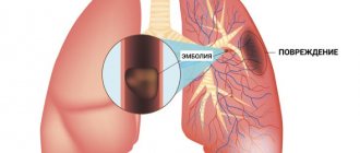
- loss of capillary bed (including due to pulmonary thromboembolism or chronic obstructive pulmonary disease)
- hypertrophy of the middle layer of the arteriolar wall
- artificial ventilation
The bronchopulmonary form of cor pulmonale may have the following causes:
- bronchial asthma
- chronic obstructive bronchitis
- emphysema
- bronchiolitis
- polycystic lung disease
- diffuse pneumosclerosis of various origins
- tuberculosis
- bronchiectasis
- pneumoconiosis
- sarcoidosis
- Hamman-Rich syndrome, etc.
The thoracodiaphragmatic form of the disease can have the following causes:
- ankylosing spondylitis
- kyphoscoliosis
- pleural pathologies
- polio
- pathology of the diaphragm (with paresis of the diaphragm, pneumosclerosis, etc.)
The vascular form can be caused by the following reasons:
- pulmonary vasculitis
- primary pulmonary hypertension
- compression of the pulmonary trunk by an aortic aneurysm
- thromboembolism of the branches of the pulmonary artery
- mediastinal tumors
- pulmonary artery atherosclerosis
Pulmonary hypertension increases afterload on the right ventricle. Therefore, the same pathogenesis develops as in heart failure. End-diastolic and central venous pressure increases, as does ventricular dilation and hypertrophy. The load on the right ventricle may increase due to increased blood viscosity. The latter process is associated with hypoxia-induced polycythemia. In some cases, failure of the right ventricle can cause pathology of the left ventricle, when the interventricular septum protruding into the cavity of the left ventricle makes it difficult to fill the left ventricle, causing diastolic dysfunction.
Acute cor pulmonale is often formed during artificial ventilation (which is prescribed for the diagnosis of “acute respiratory distress syndrome”) or massive pulmonary embolism. Cor pulmonale in chronic form most often develops with COPD (emphysema, chronic bronchitis). Less commonly, development can occur with extensive loss of lung tissue, which occurs with trauma and surgery. Among the causes of chronic pulmonary heart disease:
- kyphoscoliosis
- interstitial pulmonary fibrosis
- neuromuscular disorders
- obesity with alveolar hypoventilation
- idiopathic alveolar hypoventilation
In patients with chronic obstructive pulmonary disease, infection in this organ or exacerbation of the disease can lead to overload of the right ventricle. In chronic cor pulmonale, the risk of venous thromboembolism increases.
Causes of cardiopulmonary failure
Cardiopulmonary failure never occurs without a reason. As a rule, several reasons lead to its development. There are three main groups of causes that can provoke pathology:
1. Bronchopulmonary diseases. According to statistics, they act as a provoking factor in more than 70% of all cases. Among the most common diseases are bronchial asthma, pulmonary tuberculosis, chronic bronchitis and other severe disorders of the respiratory system (for example, cystic fibrosis).
2. Thoradiaphragmatic diseases. They are directly related to pronounced deformation of the chest and limited mobility of the diaphragm. The appearance is caused by severe curvatures of the spinal column (in particular, kyphoscoliosis), ankylosing spondylitis, and restrictions on the expansion of the lungs (pleuritis).
3. Vascular diseases. This group includes diseases that affect lesions of the pulmonary bed. These include sickle cell anemia, pulmonary vasculitis, compression of the pulmonary veins and arteries by neoplasms.
4. Vascular diseases. Often, the development of cardiopulmonary failure is provoked by aneurysms, atherosclerosis and blood clots in the arteries of the lungs.
Classification
Today there are several classifications, including such authors as B. E. Votchal (pulmonary heart disease) and N. R. Paleev (pulmonary hypertension in COPD). They are successfully combined, and three stages are distinguished.
- Stage I - transient - an increase in pulmonary arterial pressure is observed during physical activity, often the condition is caused by an exacerbation of the inflammatory process in the lungs or worsening bronchial obstruction
- Stage II - stable - pulmonary arterial hypertension is now observed not only during exercise, but also at rest, and is not associated with exacerbations of pulmonary pathology
- Stage III - pulmonary hypertension is stable, circulatory failure also develops
Symptoms
Initially, the disease does not manifest symptoms. But patients often have pronounced manifestations of the underlying lung disease. This may be fatigue during physical activity, shortness of breath.
Gradually, as the pressure in the right ventricle increases, physical symptoms are manifested by systolic pulsation in the sternum, a loud pulmonary component of the second heart sound, murmurs of functional insufficiency of the 3-leaf valve and the pulmonary valve. A little later, the gallop rhythm of the right ventricle (third and fourth heart sounds), which intensifies with inspiration, hepatomegaly, swelling of the jugular veins (with a dominant wave a, in the event that there is no blood regurgitation due to tricuspid valve insufficiency), swelling of the legs are also noted.
Functional classes
During the examination, it is necessary to establish the functional class of manifestations of cor pulmonale.
- Class 1 - the main symptoms are diseases of the bronchi and lungs, hypertension in the pulmonary circle is detected only with instrumental examination and stress tests;
- Class 2 - in addition to the listed symptoms, there is respiratory failure due to narrowing of the bronchi;
- Class 3 - respiratory failure is severe, followed by cardiac failure. Constant shortness of breath, tachycardia, dilatation of the neck veins, cyanosis. Studies reveal persistent hypertension in the pulmonary circulation;
- Class 4 - decompensation, all clinical manifestations are pronounced, there are congestion, respiratory and heart failure of the third degree.
Doppler examination allows you to quantify the pressure in the pulmonary artery, measure the reverse flow of blood (regurgitation) from the right ventricle into the atrium
Diagnostics
For diagnosis, it is important not only to collect anamnesis and take into account symptoms, but also to laboratory and instrumental diagnostics. An examination for cor pulmonale is carried out for everyone who has at least one reason (factor) for the development of this pathology. Chest radiographs show proximal dilatation of the pulmonary artery with distal weakening of the vascular pattern, as well as an increase in the size of the right ventricle.
An ECG diagnosis is performed, which reveals typical signs of right ventricular hypertrophy. This may be a QR wave in lead V, a deviation of the electrical axis to the right, a dominant R wave in leads V1-V3. Signs are dependent on the degree of pulmonary hypertension. But, because pulmonary hyperventilation and bullae in chronic obstructive pulmonary disease cause cardiac remodeling, methods such as radiography, physical examination and electrocardiography may be partially insensitive.
To assess the function of the right and left ventricles, cardiac imaging methods are used: radionuclide scanning or echocardiography. Echocardiography is needed to assess right ventricular systolic pressure. But often the possibility of performing it is technically limited if the patient has lung diseases. Confirmation of the diagnosis in some cases is carried out using catheterization of the right heart.
Treatment
Cor pulmonale is difficult to treat. The main measure is to eliminate the cause, thereby slowing down the progression or reducing hypoxia. If the patient has peripheral edema, the doctor prescribes diuretics. But they have the desired effect only if there is insufficiency of the left ventricle and, at the same time, overload of the lungs with fluid.
Diuretics can cause worsening of the condition, because even a slight decrease in preload can cause worsening of the manifestations of cor pulmonale. Pulmonary vasodilators do not produce results in cor pulmonale , although they are relevant for the treatment of primary pulmonary hypertension (calcium channel blockers, hydralazine, prostacyclin and dinitrogen oxide).
Digoxin gives the desired effect only if there is concomitant left ventricular dysfunction. This medication is prescribed with caution because patients diagnosed with COPD are extremely sensitive to the effects of digoxin. With hypoxic cor pulmonale, some doctors have suggested performing a venotomy, but the effect of reducing blood viscosity cannot cancel out the negative consequences of reducing the volume of blood that carries oxygen, with the exception of cases of polycythemia major. In patients with chronic cor pulmonale, long-term use of anticoagulants reduces the risk of venous thromboembolism.
Symptomatic treatment includes the use of:
- mucolytic agents
- bronchodilators
- oxygen therapy
- respiratory analeptics
If cor pulmonale is decompensated against the background of bronchial obstruction, the patient is prescribed constant use of drugs from the glucocorticoid group (for example, prednisolone may be effective). If, in the chronic form of cor pulmonale, the patient experiences a persistent increase in blood pressure, aminophylline can be prescribed (orally, intravenously or rectally); at the onset of the disease, nifedipine (Corinfar, Adalat) can be prescribed. In case of decompensated course, nitrates (nitroglycerin, nitrosorbide) are prescribed; monitoring of blood gas composition is mandatory, because there is a danger of worsening hypoxemia.
For symptoms of heart failure with cor pulmonale, cardiac glycosides and diuretics are prescribed. Caution must be exercised because glycosides have a highly toxic effect on the myocardium, especially under conditions of hypoxia and hypokalemia. Hypokalemia can be corrected with potassium supplements, such as potassium chloride or panangin. If diuretics are prescribed, first of all consider potassium-sparing drugs, including aldactone, triampur, etc.
In cases of severe erythrocytosis, bloodletting of 200-250 ml of blood is performed, after which low-viscosity infusion solutions, for example, rheopolyglucin, are administered intravenously. Also, therapy for cor pulmonale will include prostaglandins, especially those with antiproliferative, cytoprotective, and antiaggregation effects.
In some cases, endothelin receptor antagonists are relevant. Endothelin is a powerful vasoconstrictor of endothelial origin, the level of which increases in various forms of cor pulmonale. If acidosis develops, intravenous infusion of sodium bicarbonate solution is necessary. Treatment of circulatory failure of the right ventricular type is carried out with potassium-sparing diuretics, for example, veroshpiron or triamterene. Cardiac glycosides are effective for the treatment of left ventricular failure. In order to improve the metabolism of the heart muscle in cor pulmonale, it is recommended to prescribe mildronate (0.25 g 2 times a day orally), as well as potassium orotate or panangin (asparkam).
Therapy for cor pulmonale, as already noted, must be comprehensive (not only medications are used). The following methods are effective:
- massage
- physiotherapy
- hyperbaric oxygen therapy
- breathing exercises
Forecast
The mortality rate of patients from chronic pulmonary heart disease remains high: 45% of patients survive in the decompensation stage for about two years. Even with intensive therapy, their life expectancy is no more than four years. Lung transplantation gives 60% of patients survival over the next two years.
The disease is very difficult to treat. Any person has the opportunity to rid himself of bad habits and take care of his health in a timely manner. The appearance of cough, shortness of breath and other symptoms requires immediate medical attention.