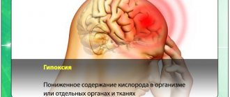
A description is given of the forms of hydrocephalus that can be incidental findings on MRI/CT of the brain - primary hydrocephalus and conditions with which a differential diagnosis must be made. Information about benign intracranial hypertension (BIH), normotensive and replacement hydrocephalus is presented to the extent that is necessary for a neurologist in outpatient practice to make decisions and explain to the patient the essence of his condition. The structure of the document reflects the tasks facing it. You can get acquainted with the question in more detail using the links in the relevant sections of the text. The clinical picture of intracranial hypertension consists of general cerebral and focal symptoms. Intracranial hypertension is characterized by the absence of pathognomonic disorders. In a typical clinical picture, a triad stands out: headache, vomiting, congestion in the fundus. Increased intracranial pressure is often associated with hydrocephalus, a disorder involving the accumulation of excess cerebrospinal fluid (CSF) in the cerebral ventricles and/or subarachnoid spaces, due to an imbalance between secretion and absorption, which is accompanied by dilation of the ventricles and/or subarachnoid spaces. The set of disorders in patients may be similar to the clinical picture of other lesions of the nervous system: - disseminated demyelinating focal lesions of the brain substance and cranial nerves, - focal lesions of the brain stem with involvement of the medial longitudinal fasciculus and the development of disorders of consciousness - space-occupying formations of the brain with general cerebral changes, focal symptoms and symptoms of damage to the cranial nerves, - conditions with diffuse brain damage - encephalopathy. — neuroinfections (congenital syphilis, cytomegalovirus infection, mumps, etc.) — thrombosis of the sinuses and veins of the brain. Asymptomatic cases of intracranial hypertension (ICH) are accompanied by nonspecific complaints - dizziness, fatigue. The variety of variants of the clinical picture, both in severity and in the set of symptoms, is the reason for the overdiagnosis of hypertensive-hydrocephalic syndrome, both in adult and pediatric neurological practice. An additional harmful effect is the high rate of false-positive data on dilation of the ventricular system when using echoencephaloscopy. Doctors can claim that a patient has ICH if rheoencephalography, duplex scanning of the brachiocephalic arteries, or transcranial Doppler sonography reveals venous drainage from the cranial cavity.
Imaging signs of hydrocephalus with increased ICP include: An increase in the size of the inferior horns of the lateral ventricles by more than 2 cm with the absence of visualization of the subarachnoid spaces of the convexital areas, interhemispheric and lateral fissures of the brain; Balloon-shaped dilatation of the anterior horns of the lateral ventricles (Mickey Mouse symptom) and the third ventricle; Periventricular decrease in tissue density seen on CT or increased T2 signal seen on magnetic resonance imaging (MRI) as a result of transependymal infiltration or migration of cerebrospinal fluid. In addition to structural changes, the clinical manifestations of hydrocephalus are taken into account. The last clarification is necessary, since taking into account only structural changes does not allow us to classify disorders of liquorodynamics without changing the configuration of the ventricles as hydrocephalus, for example, benign intracranial hypertension (BIH).
Types of hydrocephalus
There are several variants of the disease depending on the cause of occurrence and localization. By origin, there is a congenital form, which develops as a result of problems suffered by the baby in the prenatal state, and an acquired form, provoked after birth by external factors (injuries, infections).
According to the type of location, they are distinguished:
- external hydrocephalus, when in a child cerebrospinal fluid accumulates mainly in the space under the membranes of the brain;
- internal form, in which cerebrospinal fluid flows into the ventricles of the brain, causing them to expand;
- mixed or general hydrocephalus, which is characterized by a combination of external and internal forms of the disease.
There are also open (communicating) and closed (obstructive) types of pathology: in the first case, the communication between the ventricles and the subarachnoid space is not broken, in the second it is absent. According to the speed of flow, acute, subacute and chronic dropsy of the brain are distinguished.
Pathogenesis
The disease provokes expansion of the cerebral ventricles for various reasons. The main provocateur is a violation of the outflow of cerebrospinal fluid.
The process of cerebrospinal fluid circulation
The choroid plexuses produce approximately 600–700 ml of cerebrospinal fluid per day. It enters the central canal through special openings, washing the brain and then the spinal cord. Reabsorption occurs through the cerebral hemispheres through the venous sinuses.
Brain damage process
Excessive pressure of the cerebrospinal fluid provokes its leakage into the surrounding substance, which causes the development of edema. The dilated ventricles begin to compress the brain tissue.
The white matter takes the brunt of the attack, causing destruction of the myelin sheaths that transmit nerve impulses to the body.
Up to a certain point, such changes are reversible—after timely surgery, it is possible to almost completely restore neurological functions. Otherwise, atrophy of the brain substance develops, causing spastic paralysis, and the person ceases to control the functioning of the pelvic organs.
In infants, under the influence of such excess pressure, there is a rupture of adhesions inside the subarachnoid space, which opens the way for the outflow of cerebrospinal fluid and self-healing. In advanced situations, brain tissue ruptures. Timely treatment can prevent the development of neurological disorders.
Symptoms of hydrocephalus of the brain in a child
Classic signs of hydrocephalus in children over 1–3 years of age are symptoms of increased pressure on the brain, which include:
- intense headache;
- nausea and vomiting;
- spastic paresis, when muscles involuntarily tense and limb movements become sluggish;
- constant fatigue;
- urinary incontinence;
- gait disturbances;
- convulsions;
- strabismus;
- nystagmus;
- developmental delay.
Hydrocephalus, which occurs as a consequence of a brain injury or infection, can cause in an older child a deterioration in memory and attention, decreased visual acuity, irritability, dizziness and head pain that does not go away after taking painkillers.
The following symptoms help to suspect hydrocephalus in infants:
- bulging and pulsation of the fontanel;
- divergence of the sutures of the skull;
- deformation of the head with a predominance of the cerebral region over the facial region;
- rapid increase in head circumference with a significant deviation from the norm;
- increased excitability, moodiness;
- poor appetite;
- frequent volumetric regurgitation, similar to vomiting;
- exotropia;
- Graefe syndrome;
- a distinct pattern of saphenous veins on the head.
In some cases, an additional sign may be a slow heartbeat, tremor (trembling) of the chin and limbs, which persists for a long time.
Participation in care
Children with implanted shunts should be monitored by a neurosurgeon throughout their lives. Most patients suffering from hydrocephalus, after implantation of a shunt system, can lead a normal life, but constant monitoring by loved ones in collaboration with a neurosurgeon is necessary.
The neurosurgeon monitors each patient to prevent drainage failure. The first time after implantation or re-operation - regularly, with a gradual transition to examinations once a year.
Relatives are advised to be able to recognize early signs of complications. Quick and accurate assessment of health problems is very important. Flu symptoms may mask symptoms of a blocked shunt. Quickly identifying them will allow you to plan a repeat operation and avoid an emergency.
Patients and their loved ones should pay close attention to signs and symptoms of complications. The main causes are: blockage, infection and excessive drainage.
Reasons for the development of hydrocephalus in children
Factors that can lead to the internal form of the disease are:
- anomalies and malformations of the fetus during the period of intrauterine formation, associated with infections, injuries, and the influence of bad habits of the mother;
- various birth injuries;
- prematurity and premature birth with low body weight.
The causes of acquired hydrocephalus in a child can be infectious diseases of the brain and its structures: meningitis, malignant and benign neoplasms, encephalitis. In some cases, the starting mechanism is traumatic brain injuries and bruises resulting from falls, blows, accidents and other injuries.
Diagnosis of dropsy of the brain
In infancy, a visual examination is sometimes sufficient to determine the nature of the pathology: the doctor, by examining the baby’s physiological parameters and listening to the parents’ complaints, can make a preliminary diagnosis at the initial appointment.
To clarify the disease in children with an open fontanel, including to identify pathology during intrauterine development, ultrasound is used to assess the degree of enlargement of the ventricles. Additional methods are:
- fluoroscopy;
- computed and magnetic resonance imaging;
- lumbar puncture, necessary for a detailed analysis of the cerebrospinal fluid.
A child with suspected hydrocephalus must be examined by an ophthalmologist or pediatrician; if necessary, concomitant diseases may require consultation with a cardiologist, surgeon, oncologist and other specialists.
Emotional support
The physical side of hydrocephalus is only part of the problem of this disease. The patient and his relatives need to take into account his emotional factors.
Although surgery should resolve your hydrocephalus to some extent, you may feel fearful, depressed, irritable, or doubtful. If the patient is a child, it should be taken into account that he has the same feelings as an adult. If your child is feeling out of shape or uncomfortable because they need to visit the doctor frequently or have repeated tests, it is best to reassure them with simple explanations. If he knows what awaits him, he will be more willing to cooperate with you. Children, like adults, as a rule, do not like unpleasant surprises. A calm atmosphere among loving loved ones is the best environment for children. It is advisable to explain the phenomenon of hydrocephalus in words that the child can understand.
It is very important to know how the child feels and be able to explain to him what he is experiencing. The needles hurt. It is natural to cry and want to get rid of them. Being admitted to hospital is a new challenge for your child. You must tell him the truth in order to gain and secure his trust. Sincerity is the best way to maintain your child's trust.
Children over ten years of age are generally able to comprehend more complex concepts. They may associate signs and symptoms with their illness. Restrictions associated with the disease are easier for them to bear. Tell your doctor about your feelings and give him the right to guide you. Some people share their feelings with close friends, others need professional help. The health professionals treating you or your child are interested in your well-being and their goal is to do what is best for you and your loved ones.
Patients and the patient's parents should communicate with their doctor quite frequently. It is important to actively participate in this communication so that your doctor can better understand your needs and the needs of your loved ones.
Treatment of hydrocephalus in children
Depending on the type and nature of the pathology, treatment for hydrocele can be conservative or surgical. Conservative tactics are used only in cases with a non-progressive open form of the disease, which is caused by external factors. It involves medication to relieve symptoms and eliminate the cause.
In all other situations, it is rational to use surgical treatment methods, which are aimed primarily at eliminating the obstacle that impedes the outflow of cerebrospinal fluid (tumor, abscess, intracranial hematoma, developmental anomalies). In cases where it is not possible to eliminate the cause of the pathology, a special operation is performed - bypass surgery. The technique involves introducing a system of tubes and valves into the brain, which will drain excess cerebrospinal fluid to other parts of the body.
In situations that threaten the child’s life, when immediate assistance is required, external ventricular drainage is performed - an operation during which excess cerebrospinal fluid is quickly pumped out of the ventricles of the brain using the drainage system.
With timely diagnosis and adequate treatment, a child after hydrocephalus grows and develops in accordance with the norms; the disease does not affect the mental or mental state in any way.
Infection
Infection is the second type of complication. It poses a significant risk for any surgical procedure, most often when implanting a foreign body.
It manifests itself in the form of redness or suppuration along the edges of the suture or along the path of the drainage system under the skin. The surgeon fixes his attention on these signs. If left untreated, the wound may erode or open, and in more serious cases, the infection may cause chills and fever. Typically, the drain needs to be removed. Sometimes antibiotic therapy can be administered without removing the system.
Since the shunt is a foreign body, the patient may experience an allergic or inflammatory reaction. Inflammation at one of the drainage sites should be immediately shown to a neurosurgeon.
Disease prevention
The list of measures that can prevent the development of pathology includes:
- pregnancy planning;
- prevention of birth injuries;
- giving up bad habits before conceiving a baby and during pregnancy;
- vaccine prevention of infectious diseases of the brain (meningitis, encephalitis);
- early perinatal diagnosis;
- regular monitoring of children at risk by a pediatrician and neurologist;
- timely completion of preventive examinations up to a year;
- providing the child with safe living conditions to prevent household traumatic brain injuries.
Parents need to closely monitor the development and condition of young children, promptly contacting doctors in case of infectious diseases, falls and injuries affecting the head and neck area.
Specialists at the SM-Doctor clinic will conduct a detailed diagnosis if hydrocephalus is suspected, plan treatment and monitor the child throughout therapy and rehabilitation.