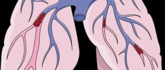
Pulmonary embolism (PE) is an emergency condition that is accompanied by occlusion (blockage) of the pulmonary artery or its branches by a thrombus formed in the systemic circulation. This pathology causes a generalized spasm of the arterioles of the pulmonary circulation and a sharp limitation of pulmonary blood flow.
To confirm the diagnosis, the following diagnostic methods are used:
- ECG;
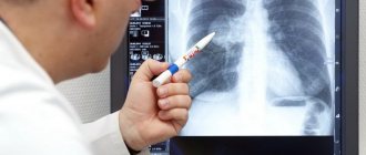
- radiography;
- Echocardiography;
- lung scintigraphy;
- angiopulmonography;
- computed tomography, etc.
This allows you to accurately establish a diagnosis in the early stages of the disease and make its treatment more effective.
Treatment tactics
Treatment of pulmonary artery diseases depends on the severity of the process. As for stenosis, in the absence of pronounced manifestations, there is no need for surgical intervention, but clinical observation and symptomatic therapy are required.
As the pathology progresses, the question of surgical treatment arises. When the pressure gradient increases above 50 mmHg, the question of surgery is not discussed - it must be performed immediately. Operation options are different. Valvuloplasty can be open, closed, endoscopic - balloon. Indications for a specific intervention are determined by a specialist.
The main directions in the treatment of pulmonary hypertension are the prevention of thrombosis, elimination of spasm of vascular smooth muscle fibers, stopping the proliferation of connective tissue structures of the vascular wall. In this case, the following application is indicated:
· drugs that normalize blood clotting;
· diuretics;
cardiac glycosides;
· vasodilators;
· oxygen therapy.
In case of hypertension, called secondary, complex measures should correct the underlying pathology - the root cause of the increase in pressure in the artery. The list often includes bronchodilators and corticosteroids.
Surgical treatment for hypertension: embolectomy, lung transplant, pulmonary-cardiac complex transplant.
Pulmonary embolism
Diagnosis of pulmonary embolism
Clinical manifestations of pulmonary emboli depend on the location of emboli, the degree of pulmonary blood flow impairment and concomitant diseases. Clinical signs, although not specific, give reason to suspect the disease and roughly judge the location of the lesion. With embolism of the distal branches of the pulmonary arteries, most patients develop symptoms of infarction pneumonia: sharp “pleural” chest pain associated with breathing, shortness of breath, cough with scanty sputum, fever. Hemoptysis is observed only in 1/3 of cases. An objective examination reveals moist rales and pleural friction noise. It should be taken into account, firstly, that in 60% of patients, infarction pneumonia does not develop (and then there are no symptoms), and secondly, it takes 2-3 days after the embolism for the formation of a heart attack. In the presence of concomitant pathology of the cardiovascular system, distal embolism can manifest itself as collapse and symptoms of right ventricular failure. With massive pulmonary emboli, emboli are localized in the pulmonary trunk or main pulmonary arteries. It usually manifests itself with symptoms of acute cardiopulmonary failure: collapse, severe shortness of breath, tachycardia, chest pain. If more than 60% of the arterial bed of the lungs is excluded from the blood circulation, an enlarged liver and swelling of the neck veins appear. If PE is suspected, the following studies are required: - electrocardiography - echocardiography - chest radiography - perfusion (perfusion-ventilation) lung scintigraphy or spiral computed tomography or angiopulmonography - ultrasound examination of the main veins of the legs.
On the ECG, the most typical signs are the appearance of Q in lead III, deep S in lead I and negative T in lead III (McGinn-White syndrome), as well as right bundle branch block. Negative symmetrical T waves may appear in leads V1-3(4); ST elevation in III, aVF, aVR and V1-3(4); displacement of the transition zone to the left chest leads. Only a third of patients show signs of right heart overload on the ECG. In 20% of patients with pulmonary embolism, there are no changes on the ECG.
An x-ray can reveal dilation of the superior vena cava, enlargement of the right heart, bulging of the conus of the pulmonary artery, high standing of the dome of the diaphragm on the affected side, disc-shaped atelectasis, pleural effusion - however, all these symptoms are not very specific. The only characteristic of PE is the Westermarck symptom: expansion of the root of the lung and depletion of the pulmonary pattern in the affected area, but it is observed only in 5% of cases. However, X-ray data are important for excluding pneumonia, pneumothorax, myocardial infarction, and pericarditis.
Echocardiography can confirm the diagnosis of PE and differentiate it from other acute heart diseases. An echocardiogram reveals hypokinesia and dilatation of the right ventricle; paradoxical movement of the interventricular septum; tricuspid regurgitation; absence or reduction of respiratory collapse of the inferior vena cava; pulmonary artery dilatation; signs of pulmonary hypertension.
Scintigraphy is informative in 87% of cases. It demonstrates perfusion defects of embolic origin - with a clear delineation, triangular shape and location corresponding to the blood supply zone of the affected vessel (lobe, segment). When small branches of the pulmonary artery are occluded, the diagnostic value decreases.
Multislice CT with vascular contrast allows visualization of blood clots in the pulmonary artery, as well as changes in the lungs caused by other diseases manifested by perfusion or filling defects. The sensitivity of this method is high when emboli are localized in large pulmonary arteries and is significantly reduced when subsegmental and smaller arteries are affected.
Pulmonary angiography is recognized as the “gold standard” in the diagnosis of pulmonary embolism. Signs of embolism in this study are: amputation of a vessel or a filling defect in its lumen. A laboratory method for determining D-dimer is used to exclude PE. Its normal plasma level allows us to reject with 90% accuracy the assumption of the presence of pulmonary embolism in patients with low or moderate clinical probability. The diagnosis of pulmonary embolism is established by analyzing the results of clinical, instrumental and laboratory studies. However, during life the diagnosis is correctly established only in 34% of patients. At the same time, in 9% of cases it is overdiagnosed.
Statistics on morbidity and mortality of pulmonary embolism
Statistics show that about 0.1% of the world's population dies every year from pulmonary embolism. In terms of the frequency of deaths, this pathology is second only to coronary heart disease, strokes and some cancers.
The main cause of mortality in pulmonary embolism is late diagnosis. About 90% of patients do not receive the necessary treatment in the early stages of this acute condition.
The difficulty in diagnosing thromboembolism is that this condition can masquerade as a large number of heart and lung diseases. Many patients begin receiving therapy for heart attack or asthma. As a result, valuable time is lost.
Another danger of pulmonary embolism is suddenness. It affects not only patients with cardiovascular diseases, but also women during childbirth, and practically healthy people after injuries. Most often, after a diagnosis of pulmonary embolism is made, only 70% of patients can be saved. But if the pathology was diagnosed in a timely manner and optimal treatment was started, then this figure can increase to 98% percent.
How to prevent pulmonary embolism?
To avoid pulmonary embolism, it is necessary to prevent and promptly treat chronic diseases. One of the causes of venous thrombosis is varicose veins. Any person who suspects he has this disease should visit a phlebologist and have the venous system of the lower extremities diagnosed. After diagnosis, you need to follow the doctor's recommendations. Also, patients planning major surgery should be assessed for the risk of deep vein thrombosis, and people at high risk of DVT may require prophylactic doses of anticoagulants before and after surgery.
Who develops pulmonary embolism?
Almost all cases of pulmonary embolism are caused by deep vein thrombosis. Thus, people who are more likely to get pulmonary embolism are people who are prone to deep vein thrombosis. Let's look at some risk factors for pulmonary embolism. Important factors are immobility, severe somatic pathologies and major surgical intervention (especially gynecological operations, operations on the pelvis and lower extremities). The risk of developing deep vein thrombosis or PE in hospital can be significantly reduced by activating the patient to walk as soon as possible. Medicines to help prevent deep vein thrombosis and PE are also prescribed to those patients who are at high risk.
What causes pulmonary embolism?
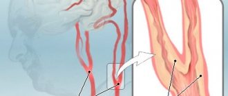
The usual cause of pulmonary embolism is deep vein thrombosis (DVT).
In almost all cases, the cause is a blood clot (thrombosis) that originally formed in a deep vein of the lower limb (known as deep vein thrombosis). This clot travels through the circulation and eventually becomes lodged in one of the lung's blood vessels. The broken thrombus is now called an embolus (and therefore can cause an embolism). Most deep vein thromboses arise from the veins of the legs or pelvis. Sometimes PE can occur from a blood clot in a vein in the arm or from a blood clot that forms in the heart. However, these situations are quite rare.
Other causes of pulmonary embolism
In rare cases, a blockage of a blood vessel in the lung may be caused by an embolus, which is not a blood clot. It could be:
- Fatty tissue from the bone marrow of a broken bone (if a large long bone such as a femur is broken).
- Foreign material from unsterile injection - for example, due to drug abuse.
- Amniotic fluid during pregnancy or childbirth (rare).
- Entry of a significant volume of air into the vein (rare).
- A small fragment of cancerous material (tumor) that has broken off from a larger tumor in a different location.
- Mycotic emboli are material from the focus of a fungal infection.
Treatment of pulmonary hypertension
Therapy depends on the cause that led to the development of pulmonary hypertension. It must be remembered that the pressure in the pulmonary artery may exceed normal due to the following factors:
- physical exercise
- hypothermia
- pregnancy
- stay in the highlands
The condition of a person with pulmonary hypertension can be alleviated by inhaling oxygen. But it is contraindicated for those who have right-to-left shunting and arterial hypertension.
In chronic nonspecific lung diseases, pulmonary hypertension can be reduced with the help of such medications6
- slow calcium channel blockers
- aminophylline
- nitrates
But they do not affect the life expectancy of people with PH, which was proven by studies no more than 20-30 years ago. It is possible to reduce PH and increase life expectancy with long-term low-flow oxygen therapy.
Taking diuretics reduces the severity of right ventricular failure. But when prescribing, some caution is needed, because in patients with pulmonary hypertension, large diuresis can reduce the preload on the right ventricle and cardiac output will decrease. A relevant drug is furosemide, which is prescribed at a dose of 20-40 mg per day. Taken orally. Hydrochlorothiazide is also effective at a dose of 50 mg/day orally.
Slow calcium channel blockers are used to reduce pulmonary artery pressure in patients with established pulmonary hypertension. The effect is exerted by nifedipine at a dose of 30-240 mg/day or diltiazem at a dose of 120-900 mg/day.
Cardiac glycosides are not effective drugs for the treatment of patients with PH. They can be used in small doses for atrial fibrillation to slow the heart rate. It should be taken into account that in patients with PH, glycoside intoxication develops faster as a result of hypoxemia and hypokalemia during diuretic therapy.
If drug treatment does not produce results, lung or heart-lung transplantation is recommended.
What are the prospects for pulmonary embolism?
This depends on the type of pulmonary embolism and the presence of concomitant diseases.
If PE is treated correctly and promptly, the prognosis is good and most patients can make a full recovery.
The prognosis is worse if there is a serious disease that contributed to the embolism, such as advanced cancer. Massive PE is more difficult to treat and is life-threatening. PE is a serious condition and can have a high risk of death, but this is greatly reduced with early treatment.
The most dangerous time for complications or death is the first few hours after an embolism. In addition, there is a high risk of developing a new pulmonary embolism within six weeks of the first one. Therefore, treatment must be started immediately and continued for at least six months.
Complications of pulmonary embolism
Most people with PE are treated successfully and have no serious complications. However, serious complications are possible, including:
- Collapse - due to the effect of a blood clot on the heart and circulation. This can cause cardiac arrest and can be fatal. PE can cause heart strain. This can lead to a condition called heart failure, where the heart pumps less than normal.
- Recurrence of pulmonary embolism. Blood clots may occur again later (called recurrent PE). Treatment with anticoagulants helps prevent this.
- Bleeding. Complications due to treatment. Treatment with anticoagulants may have side effects. The main one is bleeding in various locations, for example, from a stomach ulcer. Approximately 3 in 100 patients will have significant bleeding due to treatment of PE with anticoagulants. This type of bleeding can usually be treated successfully. However, it is almost always safer to take anticoagulants to prevent recurrent PE, which can be even more severe.
- Chronic pulmonary hypertension. If small PEs recur, they may contribute to the development of a condition in which there is increased pressure in the blood vessels of the lungs (pulmonary hypertension).
Causes and pathogenesis
Pulmonary hypertension is divided into congenital and acquired. Otherwise, these forms are called primary and secondary. Among the causes of increased pressure in the pulmonary artery are the following:
- chronic lung diseases (bronchial asthma, COPD, sarcoidosis, interstitial pulmonary fibrosis, tuberculosis, silicosis, asbestosis)
- heart diseases (heart failure, acquired and congenital heart defects)
- stay in the highlands
- vasculitis
- effects of drugs on the body
- TELA
Pulmonary hypertension without a known cause is called primary hypertension. The cause of PH may be an increase in pressure in the pulmonary veins. Growth is provoked by the following factors:
- left ventricular failure
- mitral valve defects
- pulmonary vein compression
- left atrium myxomas
Increased pulmonary blood flow can also trigger pulmonary hypertension:
- patent ductus arteriosus
- ventricular septal defect
- atrial septal defect
Among the causes of the disease in question is an increase in pulmonary vascular resistance, which can be caused by:
- taking drugs that reduce appetite (fenfluramine) or chemotherapy drugs
- obstruction of the pulmonary artery and its branches
- inflammatory changes in the lung parenchyma
- destruction of the pulmonary vascular bed (with chronic destructive lung diseases, pulmonary emphysema)
- chronic alveolar hypoxia (for example, when staying high in the mountains)
Valvular defects lead to the occurrence of passive PH as a result of the transfer of pressure from the left atrium to the pulmonary veins and then to the pulmonary artery system. With mitral orifice stenosis, there can sometimes be a reflex spasm of the pulmonary arterioles due to increased pressure at the mouths of the pulmonary veins and in the left atrium. Long-term development of PH leads to irreversible sclerotic changes in arterioles and their obliteration.
If a person has congenital heart defects, PH results from increased blood flow in the pulmonary artery system and resistance to blood flow in the pulmonary vessels. Left ventricular failure, which occurs with dilated cardiomyopathy, coronary heart disease, and hypertension, can also cause pulmonary hypertension because the left atrium and pulmonary veins become higher than normal.
In chronic diseases of the respiratory organs, PH for a long period of time has a compensatory nature, because hypoxic pulmonary vasoconstriction turns off non-ventilated areas of the lungs from perfusion, which eliminates the shunting of blood in them, and hypoxemia decreases. At this stage, the use of a drug such as aminophylline reduces pressure in the pulmonary circulation, reduces compensatory vasostriction, and reduces intrapulmonary shunting of blood with deterioration of oxygenation. As lung disease progresses, PH becomes a factor in the pathogenesis of the formation of chronic “pulmonary heart.”