Skin structure
The skin consists of 3 layers: epidermis, dermis and subcutaneous fat (SAT) (Fig. 1). The epidermis is the thinnest of them and is a stratified keratinizing epithelium. The dermis is the middle layer of the skin. Mainly composed of fibrils of the structural protein collagen. PFA contains fat cells - adipocytes. The thickness of these layers can vary significantly depending on the anatomical location.
Fig.1. Skin structure
Skin is a living organ. Structure. Upper layer. Why can't you wash your face with soap?
The most important thing is that you need to know about it and remember that the skin is a living organ , and the largest organ in our body. This is not just a mechanical protective barrier from external influences, it is an indicator that shows us the state of health of the body as a whole. Because each organ is reflected on the skin and it is like a map, following which we will go in the right direction. And the structure of the skin is unique. This is its own microanatomy. That is, it is not what we see on the surface, like skin and skin. No! These are three full layers, each of which consists of its own cells, its own structures, which are not interchangeable. So let's figure out what is what, why and where?
3 layers of leather
For ease of understanding, if we imagine these three layers in the form of a house, where the “roof” we have is the epidermis, the main part of the “house” is the dermis and under it there will be a “basement” called “hypodermis”, then we will get all three layers of skin. The roof is the epidermis, which literally translates as “above the skin”, under it there will be the dermis, the skin itself, and under it lies the subcutaneous fatty tissue.
Epidermis - the top layer of skin
Today we are talking about the “roof”, about our epidermis. Our “roof” consists of five layers and each layer consists of several more rows. All this lies on a wavy line - on the basement membrane. It’s like a border that separates our roof from the main part of the house, and under the wave of this “roof” there are vessels that feed our “roof.” To better understand, you can look at your fingertips. Fingerprints are the imprints of this wavy basement membrane. On top of the basement membrane lies the germ layer, which gives rise to all skin cells. Skin cells are called keratinocytes. From here, from the germ layer, they make their way up. But in addition to skin cells, there are also special “workers”, some “workers” are responsible for the color of the skin, others for protecting the skin, and others for its sensitivity.
Basement membrane and germinal layer
What happens if we violate the integrity of the skin? Some kind of injury or damage occurs. Due to this sprout layer, the “roof hole” is sealed. And the skin healed, we forgot and that’s it, nothing really happened. But if we destroy this wavy line, then for the body - for our home this is a big defect, and here the question is not about quality, not about high-quality healing, but about rapid healing. But since the basement membrane is destroyed, and, accordingly, the germ layer, how can it be repaired? Due to this main part, due to the skin itself, due to its connective tissue. That is, the “hole” of the connective tissue is sealed not with cells, but with tissue, and this formation is called a “scar”.
The germ layer that lay on the basement membrane, this layer gives rise to new skin cells, new keratinocytes. During their lifetime, these cells must pass through all five layers to the top. These are not just cells that divided and went who where? No, they have their own life plan and motto, which can be expressed: “by serving others, I burn myself.” That is, during life, and the life of a cell is 28-30 days, the cell must go through, synthesize special proteins, special lipids and become a cancerous scale. And by the way! When we say that we don’t see the effect of peeling, scrubs, or serums, one of the reasons is that the cells have not yet had time to go through their life cycle, that is, we acted on cells that were already at the end of the cycle, in the middle of the cycle , they will peel off soon or have already peeled off. That is, these cells did not have time to receive the beneficial substances that we provided them. And in order to see the effect, two, three, four cell cycles must occur and somewhere there will be new cells with these beneficial substances obtained and then we will see the effect.
"Bricks in Cement"
And so our cells, as they move upward, they synthesize special granules: special granules with fats and special granules with proteins. And as they move upward, that is, starting from the germ layer in the next three layers, they specifically synthesize these granules and when they approach the top layer, to the end of our “roof,” as it were, they open, granules with fats come out, granules with proteins, the cell loses its nucleus, it is no longer a cell, it is now a scale. And the result was a structure called “Bricks in Cement,” where “bricks” are our scales, and “cement” are synthesized substances. Lipids were used to form a hydrolipidic barrier - protective, and proteins began to break down into special amino acids. That is, these substances have moisture-retaining properties.
Hydro-lipid mantle and PH
And so our skin cell has passed its life path from the germ layer to the stratum corneum. And this is such a precise mechanism, where everything is thought out. The life plan of the cells has been thought out, what should they synthesize, when, where, why, why? Sometimes we unknowingly or deliberately interfere with these processes for the wrong reasons. The whole chain is broken. Let's remember the simplest example. What do we wash with? If we wash our face with soap, soap is an alkaline product, alkali kills our PH, changes our PH. And we need an acidic PH in order for skin cells to exfoliate normally. If they exfoliate normally, then the cells of the germ layer receive information that new cells need to be synthesized. We change the pH, we wash out the hydrolipid film, then our skin “thinks”: “Do I really synthesize so little fat that I’m drying out? We need to synthesize even more.” And it synthesizes fat even more. It seems to us, what a horror, the skin is oily again, I’ll wash it all off again. It lasts for a while, half an hour, after half an hour all the skin is oily again. This is a pathological circle and if we interfere with these processes, then some part of the skin, its link, will invariably suffer.
Summary
And so, to summarize today’s conversation, we have dismantled the “roof”, which lies on a special basement membrane, which consists of skin cells, the life cycle of which is 28 - 30 days, during their life they must synthesize special lipids, special proteins that They will create a protective layer for us that will not allow foreign organisms to penetrate inside, which will protect the body from loss of moisture, which will create normal microflora for us and will maintain normal, imperceptible exfoliation.
Epidermis
Rice.
2. Epidermis The epidermis is a constantly exfoliating epithelial layer of the skin, which consists mainly of 2 types of cells - keratinocytes and dendritic cells. Melanocytes, Langerhans cells, Merkel cells, and intraepidermal T lymphocytes are present in small quantities in the epidermis. Structurally, the epidermis is divided into 5 layers: basal, spinous, granular, shiny and horny, differing in the position and degree of differentiation of keratinocytes, the main cell population of the epidermis (Fig. 2).
Keratinization. As keratinocytes differentiate and move from the basal layer to the stratum corneum, their keratinization (keratinization) occurs - a process that begins with the phase of keratin synthesis by keratinocytes and ends with their cellular degradation. Keratin serves as a building block for intermediate filaments. Bundles of these filaments, reaching the cytoplasmic membrane, form desmosomes necessary for the formation of strong contacts between neighboring cells. Further, as epithelial differentiation progresses, epidermal cells enter the degradation phase. The nuclei and cytoplasmic organelles are destroyed and disappear, metabolism stops, and cell apoptosis occurs when it is completely keratinized (turns into horny scales).
The basal layer of the epidermis consists of a single row of mitotically active keratinocytes, which divide on average every 24 hours and give rise to new cells in the overlying epidermal layers. They are activated only in special cases, such as when a wound occurs. Next, a new cell, a keratinocyte, is pushed into the spinous layer, where it spends up to 2 weeks, gradually approaching the granular layer. The movement of the cell to the stratum corneum takes another 14 days. Thus, the lifespan of a keratinocyte is about 28 days.
It should be noted that not all cells of the basal layer divide at the same rate as keratinocytes. Epidermal stem cells under normal conditions form a long-lived population with a slow proliferation cycle.
The spinous layer of the epidermis consists of 5-10 layers of keratinocytes, differing in shape, structure and intracellular content, which is determined by the position of the cell. Thus, closer to the basal layer, the cells have a polyhedral shape and a round nucleus, but as the cells approach the granular layer, they become larger, acquire a flatter shape, and lamellar granules appear in them, containing various hydrolytic enzymes in abundance. Cells intensively synthesize keratin filaments, which, gathering into intermediate filaments, remain unconnected on the nuclear side, but participate in the formation of multiple desmosomes on the membrane side, forming connections with neighboring cells. The presence of a large number of desmosomes gives this layer a spiny appearance, for which it received the name “spinous.”
The granular layer of the epidermis is composed of still living keratinocytes, distinguished by their flattened shape and a large number of keratohyalin granules. The latter are responsible for the synthesis and modification of proteins involved in keratinization. The granular layer is the most keratogenic layer of the epidermis. In addition to keratohyalin granules, keratinocytes of this layer contain large quantities of lysosomal granules. Their enzymes break down cellular organelles during the transition of the keratinocyte to the phase of terminal differentiation and subsequent apoptosis. The thickness of the granular layer can vary; its value, proportional to the thickness of the overlying stratum corneum, is maximum in the skin of the palms and soles of the feet.
The shiny layer of the epidermis (so named for its special shine when viewing skin preparations on a light microscope) is thin, consists of flat keratinocytes, in which the nuclei and organelles are completely destroyed. The cells are filled with eleidin, an intermediate form of keratin. It is well developed only in some parts of the body - on the palms and soles.
The stratum corneum of the epidermis is composed of corneocytes (dead, terminally differentiated keratinocytes) with a high protein content. The cells are surrounded by a waterproof lipid matrix, the components of which contain compounds necessary for exfoliation of the stratum corneum (Fig. 3). The physical and biochemical properties of cells in the stratum corneum vary depending on the position of the cell within the layer, directing the exfoliation process outward. For example, cells in the middle layers of the stratum corneum have stronger water-binding properties due to the high concentration of free amino acids in their cytoplasm.
Rice. 3. Schematic representation of the stratum corneum with the underlying granular layer of the epidermis.
Regulation of proliferation and differentiation of epidermal keratinocytes. Being a continuously renewing tissue, the epidermis contains a relatively constant number of cells and regulates all interactions and contacts between them: adhesion between keratinocytes, interaction between keratinocytes and migrating cells, adhesion with the basement membrane and underlying dermis, the process of terminal differentiation into corneocytes. The main mechanism for regulating homeostasis in the epidermis is supported by a number of signaling molecules - hormones, growth factors and cytokines. In addition, epidermal morphogenesis and differentiation are partially regulated by the underlying dermis, which plays a critical role in maintaining postnatal skin structure and function.
Leather
Skin is the largest human organ by area. The skin forms the outer covering that separates internal organs and tissues from the environment.
The skin consists of the epidermis (from the Greek epi - above and derma - skin) - the outer layer, and the dermis (the skin itself) - the inner connective tissue layer. Below the skin is the hypodermis (Greek hypo - down), represented by adipose tissue.
Epidermis
The epidermis of the skin is represented by stratified keratinizing epithelium. In the epidermis there are (from bottom to top) 5 layers: basal, spinous, granular, shiny and horny. In the basal layer, cells rapidly divide by mitosis; as cells move to the surface, they die and become keratinized. Keratinization is associated with the accumulation of a special substance by cells - keratin.
The horny (uppermost) layer of the epidermis is completely renewed in 7-11 days. Thanks to this renewal, the epidermis is very resistant to mechanical and chemical factors, is a barrier to microbes - bacteria, and is impenetrable to water.
In the basal layer there are melanocytes (from the Greek melanos - black) - cells that accumulate the black pigment - melanin. The synthesis of this pigment increases with prolonged exposure to the sun, which is the reason for the appearance of “tanning” on the skin.
In fact, tanning represents the body's protective reaction to the harmful effects of ultraviolet rays, which prevents them from passing through the skin into internal tissues and organs.
Dermis
Under the epidermis is the dermis (the skin itself), in which you can find sweat and sebaceous glands, as well as hair follicles (lat. folliculus - pouch). The dermis contains blood and lymphatic vessels, nerves, and muscle fibers.
The dermis has two layers:
- Papillary
- Reticulate
Formed by loose connective tissue in the form of papillae, protruding into the lower layers of the epidermis. It is the papillary layer that determines the unique pattern of human skin. Blood and lymphatic vessels and nerve endings are located here.
Formed by dense fibrous connective tissue. Structural proteins - collagen and elastin (along with hyaluronic acid) - give this layer (and the skin in general) strength and elasticity. Sweat and sebaceous glands and hair follicles are localized in the reticular layer.
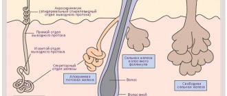
We begin to study the appendages of the skin: sebaceous, sweat glands, hair and nails. The term appendages in no way downplays the significance of these formations; it only emphasizes that all of them are a derivative (formed from) the epidermis of the skin.
Sweat glands are tubular exocrine glands whose ducts open into pores on the surface of the skin. A secretion is secreted - sweat, which contains water, urea, uric acid, and salts. Sweat glands are found almost throughout the surface of the skin.
Functions of sweat glands:
- Excretory - removes urea and uric acid from the body
- Participation in water and salt metabolism - water and salts are released with sweat to maintain homeostasis
- Thermoregulation - when sweat evaporates, the skin cools, getting rid of excess heat
The sebaceous glands are located, unlike the sweat glands, more superficially. Their excretory ducts can open both into the hair follicle and onto the surface of the skin. The secret of the sebaceous glands is sebum, which prevents the development of microbes on the skin, prevents the skin from drying out, softens its surface and is a lubricant for the skin appendages - hair.
Hair is a derivative of the epidermis, consisting of a root and a shaft. The hair root ends in a hair follicle, into which the hair papilla with vessels and nerves enters from below. Hair growth occurs due to cell division of the hair follicle. Outside, the hair root is surrounded by a hair follicle, to which the levator pili muscle is attached.
The duct of the sebaceous gland opens into the hair funnel - the place where the hair root passes into the shaft. The rod consists of medulla and cortex, represented by keratinized cells. With old age, the amount of pigment in keratinized cells (scales) decreases, and the number of gas bubbles increases, which is the cause of graying of hair.
Human hair, compared to many other animals, is tiny and cannot serve as thermal insulation. Eyelashes, eyebrows, nose and ear hairs perform a protective function. Eyebrows serve to prevent sweat, an irritant, from getting into the eyes.
Nails are derivatives of the epidermis, which are convex horny plates located in the nail bed. The nail bed consists of germinal epithelium and connective tissue, rich in nerve endings and blood vessels. Nail growth occurs due to the division of germ epithelial cells.
In the lower part, the nail bed is surrounded by a dense leathery cushion - the cuticle, which protects the growth zone of the nail from bacteria and foreign particles. The function of the nail is to protect the sensitive part of the finger from mechanical damage and create support for it.
Skin is an organ of thermoregulation
You already know that due to the evaporation of sweat, the skin can cool, thereby performing a thermoregulatory function. However, this is not the only mechanism of thermoregulation. The skin contains networks of blood vessels.
During the heat, blood vessels dilate, blood fills them - heat transfer increases, thus the body gives off excess heat to the environment.
During cold weather, the blood vessels narrow, there is less blood in them (heat transfer decreases), it rushes to the internal organs (liver) so that the body can maintain the optimal temperature for as long as possible.
Skin is the organ of touch
The skin contains nerve endings (receptors) that perceive various stimuli: cold, heat, pressure, pain. Cold receptors are located at the surface of the skin, while thermal receptors are located in the dermis (the skin itself). Pain is perceived through free nerve endings.
Skin is the site of vitamin D synthesis
The skin is actively involved in the synthesis of vitamin D. It contains a substance that is a precursor to vitamin D - ergosterol, which is converted into vitamin D under ultraviolet rays (which is why it is useful to be in the sun).
In children, with a lack of solar radiation (insolation), rickets can develop - softening of bone tissue, since vitamin D is involved in the absorption of calcium.
Skin functions
At the end of the article, we will collect all the functions of the skin together into a system to make it easier to memorize. So, the functions of the skin:
- Protective
- Immune
- Thermoregulatory
- excretory
- Blood depot
- Receptor
- Participation in metabolism
Protects internal organs and tissues from mechanical damage, covered with sebum, which prevents the development of pathogenic microorganisms.
When foreign substances (antigens) enter the skin, they are recognized, destroyed and removed. Inflammation of the skin is called dermatitis (from ancient Greek δέρμα, δέρματος - skin + lat. itis - inflammation).
Thermoregulation is carried out by sweat glands, blood vessels and subcutaneous fat, which insulates internal organs and tissues.
Thanks to the work of the sweat glands, uric acid and urea, metabolic by-products, are removed from the body.
When the skin vessels are filled, up to 1 liter of blood can be deposited in them.
The skin contains temperature, cold, pain, and pressure receptors. All of them provide the tactile function of the skin.
Due to the work of the sweat glands, the skin takes part in water-salt metabolism, and due to the formation of vitamin D during insolation (sun exposure).
Diseases
The branch of medicine that studies the skin is called dermatology. A severe hereditary skin disease is known - ichthyosis (Greek "ichthys" - fish). It is characterized by a violation of keratinization of the skin: scales are formed that resemble fish scales. Sometimes keratinization is so severe that it is incompatible with life.
© Bellevich Yuri Sergeevich 2018-2021
This article was written by Yuri Sergeevich Bellevich and is his intellectual property. Copying, distribution (including by copying to other sites and resources on the Internet) or any other use of information and objects without the prior consent of the copyright holder is punishable by law. To obtain article materials and permission to use them, please contact Yuri Bellevich
.
Dermis
The dermis is a complex, loose connective tissue consisting of individual fibers, cells, a network of blood vessels and nerve endings, as well as epidermal outgrowths surrounding the hair follicles and sebaceous glands. The cellular elements of the dermis are represented by fibroblasts, macrophages and mast cells. Lymphocytes, leukocytes, and other cells are capable of migrating into the dermis in response to various stimuli.
The dermis, making up the main volume of the skin, performs primarily trophic and supporting functions, providing the skin with such mechanical properties as plasticity, elasticity and strength, which it needs to protect the internal organs of the body from mechanical damage. The dermis also retains water, participates in thermoregulation and contains mechanoreceptors. Finally, its interaction with the epidermis supports the normal functioning of these layers of skin.
In the dermis there is no such directed and structured process of cell differentiation as in the epidermis, however, it also exhibits a clear structural organization of elements depending on the depth of their occurrence. Both the cells and the extracellular matrix of the dermis also undergo constant renewal and remodeling.
The extracellular matrix (ECM) of the dermis, or intercellular substance, which consists of various proteins (mainly collagen, elastin), glycosaminoglycans, the most famous of which is hyaluronic acid, and proteoglycans (fibronectin, laminin, decorin, versican, fibrillin). All these substances are secreted by dermal fibroblasts. The ECM is not a random accumulation of all components, but a complexly organized network, the composition and architectonics of which determine such biomechanical properties of the skin as rigidity, extensibility and elasticity. Epidermal keratinocytes, which are closely connected to each other, are attached to the ECM proteins. They form a dense protective layer of the skin. The structure of the ECM is also capable of exerting a regulatory effect on the cells immersed in it. Regulation can be either direct or indirect. In the first case, ECM proteins and glycosaminoglycans directly interact with cell receptors and initiate specific signal transduction pathways in them. Indirect regulation is carried out through the action of cytokines and growth factors retained in the cells of the ECM network and released at a certain moment to interact with cell receptors. The ECM structural network is subject to remodeling by enzymes from the matrix metalloproteinase (MMP) family. In particular, MMP-1 and MMP-13 initiate the degradation of collagen types I and III. The density of the ECM network of the dermis is uneven - in the papillary layer it is looser, in the reticular layer it is much denser, both due to the closer arrangement of the fibers of structural proteins and due to the increase in the diameter of these fibers.
Collagen is one of the main components of the ECM of the dermis. Synthesized by fibroblasts. The process of its biosynthesis is complex and multi-stage, as a result of which the fibroblast secretes procollagen into the extracellular space, consisting of three polypeptide α-chains folded into one triple helix. Procollagen monomers are then enzymatically assembled into extended fibrillar structures of various types. In total, there are at least 15 types of collagen in the skin; in the dermis, types I, III and V of this protein are most abundant: 88, 10 and 2%, respectively. Type IV collagen is localized in the basement membrane zone, and type VII collagen, secreted by keratinocytes, plays the role of an adapter protein for attaching ECM fibrils to the basement membrane (Fig. 4). Fibers of structural collagens of types I, III and V serve as a framework to which other ECM proteins are attached, in particular collagens of types XII and XIV. It is believed that these minor collagens, as well as small proteoglycans (decorin, fibromodulin and lumican), regulate the formation of structural collagen fibers, their diameter and the density of the formed network. The interaction of oligomeric and polymeric collagen complexes with other proteins, ECM polysaccharides, various growth factors and cytokines leads to the formation of a special network with certain biological activity, stability and biophysical characteristics important for the normal functioning of the skin. In the papillary layer of the dermis, collagen fibers are located loosely and more freely, while its reticular layer contains larger strands of collagen fibers.
Rice. 4. Schematic representation of skin layers and distribution of different types of collagens.
Collagen is constantly renewed, degrading under the action of proteolytic enzymes collagenases and being replaced by newly synthesized fibers. This protein makes up 70% of the skin's dry weight. It is the collagen fibers that “withstand the blow” under mechanical influence on it.
Elastin forms another network of fibers in the dermis, giving the skin such qualities as firmness and elasticity. Compared to collagen, elastin fibers are less rigid and curl around collagen fibers. It is with elastin fibers that proteins such as fibulins and fibrillins bind, to which, in turn, latent TGF-β-binding protein (LTBP) binds. Dissociation of this complex results in the release and activation of TGF-β, the most potent of all growth factors. It controls the expression, deposition and distribution of collagens and other matrix proteins in the skin. Thus, the intact network of elastin fibers serves as a depot for TGF-β.
Hyaluronic acid (HA) is a linear polysaccharide consisting of repeating dimers of D-glucuronic acid and N-acetylglucosamine. The number of dimers in the polymer varies, which leads to the formation of HA molecules of different molecular weights and lengths - 1x10⁵-107 Da (2-25 μm), which, accordingly, have different biological effects.
HA is a highly hydrophilic substance that affects the movement and distribution of water in the dermal matrix. Thanks to this property, our skin, normally and in youth, has high turgor and resistance to mechanical pressure.
HA easily forms secondary hydrogen bonds both within one molecule and between neighboring molecules. In the first case, they ensure the formation of relatively rigid spiral structures. In the second, association with other HA molecules and nonspecific interaction with cell membranes occurs, which leads to the formation of a network of polysaccharide polymers with fibroblasts included in it. Shorter molecules of proteoglycans (versican, lumican, decorin, etc.) “sit” on a long HA molecule, like on a thread, forming aggregates of enormous sizes. Extended in all directions, they create a scaffold, contributing to the stabilization of the ECM protein network and fixing fibroblasts in a specific matrix environment. Taken together, all these properties of HA endow the matrix with certain chemical characteristics - viscosity, density of “cells” and stability. However, the ECM network is a dynamic structure that depends on the state of the organism. For example, under conditions of inflammation, HA aggregates with proteoglycans dissociate, and the formation of new aggregates between newly synthesized HA molecules (renewed every 3 days) and proteoglycans is blocked. This leads to a change in the spatial structure of the matrix: the size of its cells increases, the distribution of all fibers changes, the structure becomes looser, the cells change their shape and functional activity. All this affects the condition of the skin, leading to a decrease in its tone.
In addition to regulating water balance and stabilizing the ECM, HA plays an important regulatory role in maintaining epidermal and dermal homeostasis. HA actively regulates dynamic processes in the epidermis, including keratinocyte proliferation and differentiation, oxidative stress and inflammatory response, maintenance of the epidermal barrier and wound healing. In the dermis, HA also regulates fibroblast activity and collagen synthesis. By remodeling the matrix, HA controls the functioning of cells in the matrix, influencing their availability to various growth factors and changing their functional activity. Cell migration and the immune response in tissue depend on the action of GC. Thus, changes in the distribution, organization, molecular weight and metabolism of GCs have significant physiological consequences.
Fibroblasts are the main type of cellular elements of the dermis. It is these cells that are responsible for the production of HA, collagen, elastin, fibronectin and many other intercellular matrix proteins necessary for the formation of connective tissue. Fibroblasts in different layers of the dermis differ both morphologically and functionally. Not only the amount of collagen they synthesize depends on the depth of their location in the dermis, but also the ratio of the types of this collagen, for example types I and III, as well as the synthesis of collagenase: fibroblasts in the deeper layers of the dermis produce less of it. In general, fibroblasts are very plastic cells, capable of changing their functions and physiological response and even differentiating into another cell type depending on the stimulus received. The role of the latter can be played by signaling molecules synthesized by neighboring cells and the restructuring of the surrounding ECM.
The structure of human skin and its main functions
Skin is one of the types of tissue of the human body, which has a number of inherent properties such as: elasticity, strength, some porosity, waterproofness, sensitivity and antibacteriality.
The skin protects the body from the negative effects of the environment (from excess solar radiation, for example), it is capable of secreting fat and producing odorous substances, selectively absorbing some chemicals useful to the body and rejecting others. In addition, the skin has such an important function as regeneration, which gives it the ability to independently recover from damage.
On average, there are about 5 million hairs on the surface of the skin of an adult, with about 200 receptors and 100 pores located on each square centimeter.
Which layers of skin are affected by cosmetics?
In accordance with the legislation of most countries, cosmetic products can only affect the outer layers of the skin; their deep penetration and impact on the living layers of the skin is unacceptable. That is, cosmetics should have exclusively external use, which is its fundamental difference from medications.
It should be noted here that there is no barrier in the lower part of the epidermis that could effectively prevent the penetration of cosmetics into the lymphatic and blood vessels, and, consequently, the distribution of the substances they contain throughout the human body. The possibility of effective exchange between the dermis and epidermis has been proven experimentally; accordingly, the risk of substances crossing the transepidermal barrier is quite high.
What substances are able to overcome the transepidermal barrier and enter the dermis?
It was experimentally established that substances such as caffeine, nicotine, hyaluronic acid, essential oils, vitamin E (it accumulates at the border of the dermis and epidermis). Nanoparticles contained in sunscreens penetrate deep into the skin, as well as liposomes, which easily penetrate into the deep layers of the skin and deliver the necessary nutrients there.
The structure of human skin
The versatility and multifunctionality of the skin is based on the characteristics of its structure. Our skin consists of 3 important layers, each of which performs its own function:
|
Epidermis The epidermis is the outer layer of skin, which consists of keratinized (dead) skin cells. In layers of the epidermis distant from the surface, the cells are living. They actively divide and make a slow movement to the surface, where over time they are replaced by keratinized ones, and then peel off.
The epidermis is an effective barrier to water and aqueous solutions . Fat-soluble substances penetrate the skin through the epidermis much better. This is due to the fact that cell membranes themselves contain a significant amount of fats and fat-soluble substances dissolve in them naturally.
Almost all epidermal cells produce keratin , which is why they are called keratinocytes. These keratinocytes are in constant motion, as a result of which the keratinocyte loses its own nucleus and main organelles, turning into a kind of flat “bag” filled with keratin - a corneocyte. Corneocytes form the stratum corneum of dead epidermal cells and look like flat scales. They are responsible for the barrier function of the epidermis. Corneocytes are held together by a double layer of lipids (ceramides), a kind of plastic “cement.”
The basal layer of the skin contains melanocytes, which produce melanin . Melanin is a pigment that gives our skin color and reduces the effects of external radiation. So, thanks to its presence, the skin completely blocks infrared rays and is partially capable of blocking ultraviolet rays. Often, the formation of pigment spots on the skin will depend on the condition of the basement membrane. The epidermis also contains Langerhans cells, which protect the body from microbes and foreign bodies .
The thickness of the epidermis and the rate of its renewal As a rule, the thickness of the epidermis layer varies from 0.07 to 0.12 millimeters (this is the usual thickness of a sheet of paper or plastic film). Especially rough skin can be much thicker - up to 2 mm. (on the soles of your feet, for example). The process of skin cell renewal is continuous. During our lifetime, we get rid of approximately 18 kg of dead skin, while losing about 10 billion cells every day.
The rate of complete renewal of the outer layer of the epidermis slows down with age. So, if in childhood and adolescence this process takes 21-28 days, then already at 25 years old 35-45, and after 50 years 56-72 days.
The process of division and advancement of mature skin cells is not only slower, but also heterogeneous in different areas, which also affects the aesthetic appearance of the skin. If dead skin cells become layered, the cell division process occurs more slowly, leading to faster skin aging. In addition, the layer of dead cells makes it difficult for oxygen and nutrients to penetrate the skin. Therefore, the older a person is, the more often peelings are required.
Dermis The dermis is the inner layer of the skin, which also contains functional skin glands, thanks to which excess salts and moisture are removed from the body: sweat and sebaceous glands, producing sweat and sebum, respectively.
Sebum protects the skin from the aggressive effects of the external environment and gives the skin such properties as bactericidal (due to the acidic environment it creates and then has a detrimental effect on microorganisms) and water resistance. Sweat glands, in addition, participate in the natural thermoregulation of the body.
Hypodermis The hypodermis is a subcutaneous fat layer that protects us from both excess cold and excess heat, and in addition significantly mitigates damage from impacts.
Subcutaneous fatty tissue is capable of storing fat-soluble vitamins such as A, E, F, and K. An important function of the hypodermis is that it serves as the basis for the outer layers of the skin. Skin whose hypodermis layer is weakly expressed has more folds and wrinkles and is subject to faster aging. Another important function of the hypodermis is the hormone-producing function. Adipose tissue is not only capable of accumulating estrogens, but also stimulating their synthesis.
The hypodermis layer also contains leptin, a unique hormone responsible for the feeling of fullness during meals. Thanks to it, the body is able to regulate our appetite and, as a direct consequence, the amount of fat in the subcutaneous layer.
Subcutaneous fat
Subcutaneous fat, or hypodermis, is the lowest layer of skin, located under the dermis. It consists of fat lobules separated from each other by connective tissue septa containing collagen and penetrated by large vessels. The main cells of fat lobules are adipocytes, the number of which varies in different areas of the body. Currently, PFA is considered not only as an energy depot, but also as an endocrine organ, the adipocytes of which are involved in the production of a number of hormones (leptin, adiponectin, resistin), cytokines and mediators that affect metabolism, insulin sensitivity, reproductive and immune functional activity. systems