Causes
In many cases, the formation of ulcers in the facial area contributes to the appearance of inflammatory processes. Staphylococci and streptococci are among the most common pathogens. In accordance with the cause of development, specific and nonspecific lymphadenitis are distinguished. The development of the former can be caused by serious diseases such as tuberculosis. The second form develops due to direct penetration of infection into the lymph node. Entry may occur through lesions in the neck. Those people who are at risk include: patients with weak immunity; people whose field of activity is related to animals, dirty waters, etc. As a rule, people over 18 years of age experience this disease.
Lymphadenitis in children
As a rule, lymphadenitis in children affects the lymph nodes in the face and neck (most often - submandibular and cervical on one or both sides, less often - parotid, buccal, occipital, behind-the-ear), in some cases - axillary, inguinal.
The serous stage of acute nonspecific lymphadenitis in children (days 1-3 of the disease) is manifested by painful, noticeably enlarged regional lymph nodes that are densely elastic to the touch, without loss of their mobility and the development of a local skin reaction. The child's general condition is not affected; the temperature ranges from normal to subfebrile values.
The transition of acute lymphadenitis to the purulent stage (days 3-6 of the disease) occurs with pronounced local signs and a sharp deterioration in the well-being of children. Signs of intoxication are observed: chills, high temperature (up to 40ºC), headache, severe weakness, lack of appetite and sleep disturbance. In the area of the affected lymph nodes, intense nagging or shooting pains, periadenitis, local hyperemia and swelling of the skin occur. The development of adenophlegmon with the appearance of foci of fluctuation and the release of purulent inflammation beyond the lymph node is possible. Adenophlegmons of the maxillofacial area can be complicated by thrombosis of the cavernous sinus, mediastinitis, and sepsis.
The chronic form of lymphadenitis in children can develop primarily if the causative agents are weakly virulent microorganisms, or become a continuation of the acute form of the disease. In a chronic course without exacerbation, the lymph nodes are enlarged, limited in mobility, quite dense, but painless; the child’s well-being is not impaired; suppuration develops rarely. If a child has a focus of chronic infection that maintains sluggish lymphadenitis for a long time, the lymph nodes are gradually destroyed and replaced by granulation tissue.
Tuberculous lymphadenitis in children has a long course and is usually limited to the cervical group of lymph nodes, collected in a dense, painless, large package, similar to a collar. Tuberculous lymphadenitis in children can be complicated by caseous decay, the formation of fistulous tracts, and cicatricial changes in the skin.
Specific lymphadenitis associated with vaccination against tuberculosis (“betsejeitis”) can develop in children with low and altered general reactivity, manifested by chronic calcific caseous lesions of the axillary lymph nodes. Generalized lymphadenitis in children is observed with disseminated pulmonary tuberculosis and chronic tuberculosis intoxication; accompanied by characteristic fibrosis of nodes (“pebble glands”).
What contributes to the development of the disease?
There are several factors that contribute to the spread of lymphadenitis in children and adults:
- infection of the nasal and oral cavities,
- dysfunction of the endocrine system,
- HIV,
- severe allergies,
- metabolic disorder,
- excessive consumption of alcoholic beverages.
Cervical lymphadenitis is not transmitted from one person to another, since it is only an additional process that appears as a complication. In accordance with the diseases occurring against the background of this pathology, treatment is prescribed by a certain specialist in his field (surgeon, infectious disease specialist, and so on). Initially, lymphadenitis occurs in an acute form, gradually turning into a chronic form.
causes of cervical lymphadenitis
Varieties
Types of cervical lymphadenitis:
- Penetration of infection into the lymph node (quickly cured, practically does not lead to complications).
- Presence of serious illnesses. In this situation, diagnosis is carried out in the acute form of the pathology.
There are several stages of the disease in acute form:
- Serous. Does not cause intoxication or severe fever. The initial stage of penetration of harmful organisms into the lymph node.
- Purulent. Indicates bacterial infection. Promotes the appearance of high temperature. In this case, surgical intervention is a necessary condition.
- Complicated. In this situation, we are talking about immediate surgical intervention, since the disease can cause infection of the entire body. The disease occurs along with the spread of infections through the lymph nodes. It is worth noting that this form is highly curable and does not cause complications. The development of pathology in other nodes can cause the spread of a serious disease, which is called generalized lymphadenitis.
Symptoms
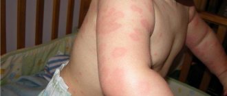
The main symptoms that indicate the presence of lymphadenitis are the following: increased body temperature, insomnia, fatigue, nervous system disorders; indifference to life, poisoning, etc.
In the initial stage of acute lymphadenitis, thickening of the lymph nodes can be observed. When pressed, discomfort and pain occur. Such signs are considered dangerous, so patients should immediately consult a doctor for advice. Otherwise, further development of a dangerous disease may occur.
Symptoms of the chronic form of the studied lymphadenitis in adults and children include: hardening of the lymph nodes, high body temperature, weakness and fatigue, insomnia, pain when pressing the lymph nodes.
At the stage of the chronic form of the disease, the symptoms become somewhat muted. This is due to the fact that the body reduces the number of mechanisms spent on fighting pathology. As a result, the body is poisoned. Purulent inflammation leads to an increase in the number of external symptoms of the disease, which leads to exacerbation. Purulent inflammation is indicated by intense pain and swelling of the lymph nodes. A similar condition is life-threatening and requires urgent intervention.
lymphadenitis symptoms
Publications in the media
Sarcoidosis is a systemic, relatively benign granulomatosis of unknown etiology, characterized by the accumulation of activated T-lymphocytes (CD4+) and mononuclear phagocytes, the formation of non-secreting epithelioid cell non-caseating granulomas. Intrathoracic manifestations of this disease predominate; damage to all organs and systems except the adrenal gland has been described. Modern ideas about diagnosis, observation and treatment currently reflect the international document “ATS/ERS/WASOG Statement on sarcoidosis” (1999).
Classification. According to the course - acute, subacute, chronic. According to intrathoracic radiographic changes, sarcoidosis is divided into stages • Stage 0. No changes on chest x-ray • Stage I. Hilar lymphadenopathy. The lung parenchyma is not changed • Stage II. Lymphadenopathy of the roots of the lungs and mediastinum. Pathological changes in the lung parenchyma • Stage III. Pathology of the pulmonary parenchyma without lymphadenopathy • Stage IV. Irreversible pulmonary fibrosis • Extrathoracic manifestations of sarcoidosis (damage to the eyes, skin, bones, etc.) are described separately.
Frequency. Newly identified cases are most often registered at the age of 20–50 years with a peak at 30–39 years, 2/3 of patients are women. In the United States, the prevalence of sarcoidosis in different states and different ethnic groups ranges from 5 to 100 per 100 thousand population. Sarcoidosis is most common in Scandinavian countries - up to 64 per 100,000 population. In Russia, the incidence of sarcoidosis in Russia is 3.0 per 100,000 population. The prevalence of sarcoidosis in the Republic of Tatarstan in 2000 was 14.8 per 100 thousand population. Sarcoidosis also occurs in childhood (intrathoracic lesions are rare in children under 4 years of age).
Etiology. There are 3 groups of factors that lead to the formation of granulomas: 1) bacteria, fungi and parasites; 2) products of plants and animals (pollen, spores, proteins); 3) metal compounds. Chlamydia pneumoniae, Borrelia burgdorferi, Propionibacterium acnes, as well as a number of viruses, including HSV and adenoviruses, are considered potential triggers for the development of sarcoidosis. The role of M. tuberculosis is debated, but a direct link to tuberculosis has not been established.
Genetic features. Cases of familial sarcoidosis were reported in Ireland in 2.4%, in Japan - 4.3%, in Finland - 4.7%, in France - about 5%, in Germany - 7.5%, in Holland - 16.3%, among African Americans - 17%.
Pathogenesis. CD4+ T-lymphocytes accumulate in the affected organ. Activated macrophages produce increased amounts of IL-1, IL-2, IL-12, TNF-a. TNF-a is considered a key cytokine involved in granuloma formation in sarcoidosis. In sarcoidosis, uncontrolled production of 1a-hydroxylase by activated alveolar macrophages (normally produced in the kidneys) with high affinity for 1,25-dihydroxycalciferol has been proven, which can serve as a marker of the activity of the process and sometimes leads to hypercalcemia and nephrolithiasis. The 1a-hydroxylation reaction mediated by pulmonary alveolar macrophages is stimulated by g-interferon and inhibited by GC.
Pathomorphology. Discrete, compact, noncaseating epithelioid cell granuloma, consisting of highly differentiated mononuclear (mononuclear) phagocytes (epithelioid and giant cells) and lymphocytes. Giant cells may contain cytoplasmic inclusions such as asteroid bodies and Schaumann bodies. The central part of the granuloma consists predominantly of CD4+ lymphocytes, while CD8+ lymphocytes are present in the peripheral zone.
Clinical manifestations • In the acute course (usually stages I–II), Löfgren's syndrome is characteristic (up to 30%): fever, bilateral lymphadenopathy of the roots of the lungs, polyarthralgia and erythema nodosum, much less often Heerfordt-Waldenström syndrome: fever, enlarged parotid lymph nodes, anterior uveitis and paralysis of the facial nerve (Bell's palsy) • In a chronic course (usually stages II–IV), manifestations are variable • Complaints: fatigue, weakness, fatigue (up to 90%), nonproductive cough, discomfort and pain in the chest, joint pain, decreased vision, mixed or inspiratory shortness of breath • Physical examination: skin changes in the form of erhytema nodosum (as a non-specific sign), rashes or lupus pernio (as a manifestation of severe cutaneous sarcoidosis); pulmonary manifestations are scanty and atypical (hard breathing, dry wheezing); an increase in the percussion determined size of the liver and spleen. Facial nerve paralysis • Laboratory data: leukopenia, lymphopenia, increased ESR, hypergammaglobulinemia, hypercalciuria, hypercalcemia; increased ACE activity in blood serum and lavage fluid • Kveim-Siltzbach test: intradermal injection of a pasteurized suspension of the spleen affected by sarcoidosis. A biopsy of the papule that forms at the injection site after 4–6 weeks reveals characteristic granulomas. Standard Ag Kveim is missing • Instrumental data. X-ray of the chest organs at the detection stage, high-resolution CT at the stage of primary and dynamic examination: hilar lymphadenopathy, ground glass symptom, pulmonary dissemination syndrome, rarely - local shadows, in later stages signs of fibrosis and bullae, pleural compaction. X-ray of the hands: bone cystic formations. CT scan of the abdominal cavity: hepato- and/or splenomegaly. FVD study: in the early stages, obstruction syndrome of the distal part of the bronchial tree (decrease in MOS50 and MOS75), later decrease in VC, TEL. The ECG shows rhythm and conduction disturbances. MRI for sarcoidosis of the central nervous system, liver, heart: identification of foci, clusters of granulomas. When bronchoscopy in lavage fluid, the ratio of subpopulations of CD4+/CD8+ lymphocytes >3.5 allows diagnosing sarcoidosis with a probability of 94%. Transbronchial or transbronchial videothoracoscopic biopsy of the lungs or intrathoracic lymph nodes, biopsy of the skin, liver, and peripheral lymph nodes reveal characteristic granulomas.
Features of the course of the disease in children. It is rare, 0.1–0.3 per 100 thousand children. In children aged 5 years and older, the disease has manifestations similar to sarcoidosis in adults. At a younger age, the triad is more common - arthritis, uveitis and skin rashes without intrathoracic lesions.
Diagnostic tactics and differential diagnosis: disseminated processes in the lungs (tuberculosis, tumor disseminations, occupational diseases, alveolitis, etc.), hilar lymphadenopathy (tuberculosis, lymphomas and other tumors of the lungs and mediastinum), splenomegaly of various origins, rheumatoid arthritis. The primary diagnostic stage in 70% of cases is carried out in anti-tuberculosis institutions.
Concomitant diseases do not have any features in comparison with healthy ones. Iatrogenic conditions may develop during treatment with GCs and cytostatics (Cushing's syndrome, arterial hypertension, diabetes, secondary immunodeficiency).
Treatment in most cases is carried out on an outpatient basis (pulmonologist or general practitioner), inpatient - during the period of invasive diagnosis or the development of complications • Diet requires correction in case of hypercalcemia, during treatment with steroids • If detected: 3-6 months of observation, use of vitamins E, C, pentoxifylline • Drug treatment is started only when the process progresses in any localization, or when the eyes, heart, or nervous system are affected. The process is stabilized and leads to remission by adrenal hormones (0.5 mg/kg for at least 6 months with a decrease), methotrexate, cyclophosphamide and other cytostatics. Inhaled steroids (budesonide, flunisolide, fluticasone) at a dose of at least 1000 mcg/day are indicated for cough syndrome and as a means of reducing the dose of systemic steroids • Surgical treatment consists of lung transplantation for IV radiation stage of sarcoidosis • Sarcoidosis is not a reason for termination of pregnancy, pregnancy depends on the presence of sarcoidosis of the female genital area, the development of internal organ failure and on the therapy administered (methotrexate and other cytostatics are teratogenic).
Complications. Progression of fibrosis, increasing respiratory and pulmonary heart failure. Sudden cardiac arrest (damage to the conduction system). Severe neurological disorders (CNS sarcoidosis). Renal failure (kidney damage, nephrolithiasis).
Prevention has not been developed because the etiology is unknown.
Course and prognosis of the disease. The course is benign. In 60–70% of cases, spontaneous remission is possible. The cause of death in sarcoidosis (up to 5%) is damage to the heart, central nervous system, stage IV sarcoidosis with decompensated respiratory failure.
Synonyms • Beck's disease • Besnier-Beck-Schaumann syndrome • Besnier-Beck-Schaumann disease • Schaumann syndrome • Benign granulomatosis • Benign lymphogranulomatosis • Epithelioid cell chronic reticuloendotheliosis • Schaumann benign lymphogranulomatosis.
ICD-10 • D86 Sarcoidosis
Notes. MOV - instantaneous volumetric velocity, TEL - total lung capacity /
Diagnostic methods
neck lymphadenitis detected ? During the examination, the doctor presses on the lymph nodes to determine the cause. A general blood test will provide the necessary information about the course of pathological processes, which are characterized by an increase in the number of lymphocytes.
If the diagnosis is established without the presence of certain complications, the doctor immediately prescribes therapy. However, in cases where the doctor notes disturbances in other systems, it is necessary to conduct additional examinations, which include:
- general and biochemical blood test,
- histology examination,
- x-ray of the chest area,
- Ultrasound of the abdominal cavity,
- immune system examinations and blood tests to check for hepatitis.
In any case, consultation with a specialist is mandatory. An exacerbation of the disease can occur at any time.
What situations can lead to a reaction of the inguinal lymph nodes? First of all, these are various inflammatory diseases of the pelvic organs (mainly the reproductive and urinary systems), external genitalia, perineum and thighs. This may be infection of skin lesions, local inflammation in the form of a boil or carbuncle, erysipelas of the lower extremities, fungal infection, syphilis or herpetic infection, bacterial or viral disease of the genitourinary tract - chlamydia, ureaplasmosis, tuberculosis and others, primary damage to the lymph nodes with tularemia, plague, as well as generalized infections – HIV infection, tick-borne rickettsiosis and others.
There are other inflammatory diseases that lead to enlargement of the lymph nodes, but they are all accompanied by a different clinical picture, where inguinal lymphadenopathy is far from the first place. Palpable lymph nodes should always alert the doctor, because often malignant tumors metastasize to the lymph nodes, and generalized blood tumors (leukemia) and local tumors of the lymphatic system (lymphomas and lymphogranulomatosis) are always accompanied by damage to the lymph nodes. Metastases to the inguinal lymph nodes can give rise to tumors of the external and internal genital organs: uterus, ovaries, prostate, bladder; the skin itself (melanoma), rectum. The main manifestation, as the name suggests, is enlarged lymph nodes. However, it is especially important to identify additional symptoms, which can provide necessary information for further diagnosis. Detection of visible inflammatory changes in the skin or external genitalia, urination disorders, and pathological discharge from the urinary or genital tract most likely indicate an infectious etiology. Frequent changes of sexual partners and neglect of barrier methods of contraception indicate the possibility of contracting sexually transmitted infections (syphilis, HIV, chlamydia, trichomoniasis, gonorrhea).
A tumor etiology can be suspected in the presence of symptoms of chronic intoxication - prolonged low-grade fever, sweating, weight loss, weakness, coagulation disorders with multiple hemorrhages and many others, as well as most often dense, painless lymph nodes. Diagnosis and treatment of inflammatory diseases associated with inguinal lymphadenitis in men is carried out by a urologist and andrologist, and in women by a gynecologist and urologist, and for specific infections, consultation with an infectious disease specialist may be indicated. Suspicion of tumor processes requires a consultation with an oncologist.
For diagnosis, it is especially important to identify additional symptoms during a conversation and physical examination. Additional methods of laboratory and instrumental diagnostics include a general analysis of urine, blood, biochemical blood test, taking samples for testing for bacterial, viral and fungal infections, cystoscopy (examination of the bladder cavity), vaginal examination in women, rectal examination, ultrasound, MRI , CT and other imaging methods of the pelvic organs and other areas to search for the primary tumor, the use of cytological and histological (biopsy) methods to verify the diagnosis (if a tumor is suspected).
Treatment of inguinal lymphadenitis itself is possible when the nature of the underlying disease is established. For inflammatory diseases, most often this is drug therapy - antibacterial drugs, antiviral and antifungicidal drugs are used to treat inflammation. The prognosis for inguinal lymphadenitis is favorable in most cases.
Possible complications
This diagnosis is a good reason to consult a doctor and carry out urgent treatment. In the worst case, dangerous consequences may occur through:
- purulent pimples,
- thrombophlebitis,
- septicopyemia.
When purulent fluid is released into the esophagus or bronchi, then there is a high risk of the spread of dangerous complications (for example, fistulas in the esophagus).
As a result of the chronic form of the disease, inflammatory processes in the form of sepsis or adenophlegmon are often observed.
Diagnostics
The difficulties of differential diagnosis for local lymphadenopathy lie in identifying a local inflammatory process of an infectious (usually) or non-infectious nature. Lymphatic vessels supplying the upper extremities of the body, the chest wall and the mammary glands pass through the axillary lymph nodes, and as a result, the inflammatory process in this part of the body can provoke enlargement of the lymph nodes. Much less often, the pathogen enters the lymph node and directly through the skin of the axillary region when its integrity is violated. Possible causes of axillary lymphadenitis:
- panaritiums;
- erysipelas of the upper limb;
- phlegmon of soft tissues of the hand, forearm and shoulder;
- osteomyelitis of the bones of the upper limb;
- boils of the same localization;
- purulent mastitis;
- cat scratch disease;
- lymphoma;
- mammary cancer;
- melanoma.
Bacterial axillary lymphadenitis is caused by the following infectious agents:
- staphylococci and streptococci;
- Proteus wand;
- coli;
- enterobacteria
In order to establish the correct diagnosis, it is necessary to take into account the anamnesis and all clinical indications. Diagnosis is usually made using puncture biopsy of lymph nodes; if necessary, lymph node excision can be performed, followed by histological analysis. Before making a diagnosis, a number of examinations are carried out:
- lymphoscintigraphy;
- computed tomography;
- radiopaque lymphography.
Often a patient has to go through several specialists at once to verify the diagnosis, from a general practitioner to an infectious disease specialist, a venereologist, an oncologist and a therapist. The examination begins with palpation (palpation) of the areas where the nodes are located. If primary signs of lymphadenitis are detected, an ultrasound examination is first prescribed. Differential diagnosis should be carried out with systemic blood diseases (leukemia, lymphogranulomatosis), storage diseases (Gaucher, Niemann-Pick), immunopathological diseases (chronic granulomatous disease, juvenile rheumatoid arthritis, systemic lupus erythematosus, dermatomyositis, etc.), tumor metastases. Sometimes it is necessary to carry out a differential diagnosis with other surgical diseases, for example, breast cancer can metastasize to nearby (axillary lymph nodes) and then treatment will be aimed at the underlying disease.
Therapy
Lymphadenitis is treated in an adult using traditional methods. These include:
- bed rest,
- treatment with antibiotics,
- UHF,
- electrophoresis,
- galvanization,
- no impact on damaged areas of the body,
- vitamin complexes.
Among other things, they may also prescribe special products and ointments.
Importance is attached to lymphadenitis treatment with antibiotics . Before prescribing this type of treatment, the specialist is obliged to conduct an examination to determine whether harmful organisms have reacted to such drugs. It is clear that the doctor prescribes to the patient those medications to which the pathogens give a reaction.
When it comes to urticaria lymphadenitis with purulent processes, then to cure it is necessary to open the purulent pimples and remove all the liquid from them.
If a tuberculosis bacillus is detected, the patient is transferred to hospital treatment. Within the clinical center, strict monitoring of the patient’s condition is carried out, as well as his isolation from healthy people to prevent the development of infection.
treatment of lymphadenitis
To treat chronic lymphadenitis, it is necessary to detect and eliminate the main factor of lymph node compaction. Only this will allow you to get rid of the pathology 100% forever.