Through the placenta, the baby in the womb eats and breathes. The anatomical structure protects the fetus from negative influences. Adverse effects include disruption of the maternal body and changes in environmental factors. Given the importance of this natural barrier, even partial placental abruption is dangerous for the condition of the fetus if rejection occurs prematurely. In the normal course of pregnancy, it occurs at 40 obstetric weeks, after the birth of the child.
1.What is placental abruption and its causes?
Placental abruption
is one of the problems that can arise during pregnancy.
When an abruption occurs, the placenta separates from the wall of the uterus too early, and this can cause very serious consequences, and in rare cases, be fatal. The main threats with placental abruption
are premature birth and too much blood loss from the mother.
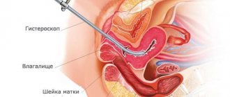
Placenta
is a disc-shaped, flat organ that forms in the uterus during pregnancy. The placenta provides nutrition to the growing fetus and supplies it with oxygen from the mother's body. During a normal pregnancy, the placenta remains firmly attached to the inner wall of the uterus until delivery. With premature placental abruption, its separation begins too early, even before the baby is born.
According to statistics, placental abruption most often occurs in the third trimester, but can begin earlier, after the 20th week of pregnancy.
Causes of placental abruption
Doctors cannot always accurately pinpoint the causes of placental abruption in the early stages, but there are several common risk factors
that may cause this problem:
- High blood pressure (140/90 and above). This is the most significant risk factor and cause of placental abruption, but during pregnancy, doctors always carefully monitor pressure readings;
- Cases of placental abruption in the early stages of a previous pregnancy;
- Smoking during pregnancy.
Less common risk factors are:
- Drug use (not because it is less dangerous, but the factor itself is less common);
- The presence of a scar on the uterus from surgery or due to uterine fibroids in the place where the placenta is attached to the wall of the uterus;
- Injury to the uterus (for example, due to an accident, fall, or physical abuse) can also cause placental abruption in the early stages;
- Premature rupture of membranes, especially when there is an infection in the uterus.
A must read! Help with treatment and hospitalization!
Premature abruption of a normally located placenta
Ultrasound machine HS40
Top seller in high class.
21.5″ high-definition monitor, advanced cardio package (Strain+, Stress Echo), expert capabilities for 3D ultrasound in obstetrics and gynecology practice (STIC, Crystal Vue, 5D Follicle), high-density sensors.
Premature abruption of a normally located placenta is a complication that is manifested by untimely separation of the placenta, which does not occur after the birth of the fetus, as it should normally be, but during pregnancy or during labor. This complication occurs with a frequency of 0.5 - 1.5% of cases. In 1/3 of cases, premature placental abruption is accompanied by heavy bleeding, with the development of corresponding complications in the form of hemorrhagic shock and DIC syndrome (disseminated intravascular coagulation).
Causes of premature placental abruption
Premature abruption of a normally located placenta most often develops in primiparous women. With premature birth, placental abruption is observed 3 times more often than with timely birth. The reasons that lead to premature abruption of a normally located placenta can be divided into two groups.
First group
- these are the reasons that directly contribute to the development of this complication. These include: gestosis (nephropathy, late toxicosis), most often long-term, untreated or insufficiently treated; various diseases, among which are diseases with an increase or decrease in blood pressure, heart defects, kidney diseases, diabetes mellitus, thyroid diseases, diseases of the adrenal cortex; incompatibility of the blood of mother and fetus by Rh factor or blood group; antiphospholipid syndrome; systemic lupus erythematosus; blood diseases; inflammatory diseases of the uterus; operations performed on the uterus; malformations of the uterus; location of the placenta in the projection of the myomatous node; post-term pregnancy.
Second group of reasons
- these are factors that provoke the occurrence of premature placental abruption against the background of existing disorders. These include: overstretching of the uterine walls due to polyhydramnios, multiple pregnancies, the presence of a large fetus; sudden, rapid and abundant rupture of amniotic fluid with polyhydramnios; injury (fall, blow to the stomach); discoordination of contractile activity of the uterus; improper use of uterotonic drugs during childbirth.
These factors lead to disruption of the connections between the placenta and the uterine wall, rupture of blood vessels with the formation of hemorrhage (retroplacental hematoma).
Symptoms of placental abruption, uterine bleeding
If the area of placental abruption is small, then after the formation of a retroplacental hematoma, thrombosis of the uterine vessels is possible, and further placental abruption stops. With significant placental abruption, heavy bleeding and extensive retroplacental hematoma, escaping blood can saturate the wall of the uterus, which leads to disruption of its contractility. This condition was called “Couvelaire’s uterus” after A. Couvelaire, who first described a similar picture.
If placental abruption forms closer to its edge, then blood, penetrating between the membranes and the wall of the uterus, pours into the vagina, which is manifested by external bleeding. When bleeding occurs immediately after placental abruption, the blood flowing from the vagina is usually scarlet in color; dark blood with clots is noted if some time has passed from the moment of abruption to the appearance of bleeding.
Premature placental abruption can occur in a mild form, the patient’s condition is most often satisfactory, the uterus is in normal tone or somewhat tense, the fetal heartbeat is not affected, and there is a small amount of bloody discharge from the vagina.
A severe form of placental abruption is usually characterized by severe bleeding and significant pain. However, there may not be bleeding if blood accumulates between the placenta and the uterine wall. In the area of the uterus where the placenta is located, due to the formation of a retroplacental hematoma, a local swelling forms and pain occurs, which quickly intensifies and gradually spreads to the rest of the uterus.
When the placenta is located on the posterior wall, the pain is diffuse and unclear. Local pain may be mild or not expressed at all when blood leaks out. The uterus becomes tense, painful, and takes on an asymmetrical shape. The abdomen is distended, the patient experiences weakness, dizziness, and vomiting. The skin is cold, damp and pale. Breathing is rapid, pulse is rapid, blood pressure is reduced.
Simultaneously with the detachment, signs of an increasing lack of oxygen in the fetus appear. When the size of the retroplacental hematoma is 500 ml or more and/or the area of detachment is more than 1/3, the probability of fetal death is highest.
With progressive bleeding and an increase in the time interval from the moment of placental abruption to delivery, the phenomena of disruption of the blood coagulation system increase, which ultimately manifests itself in the fact that the blood stops clotting altogether.
Diagnosis of premature placental abruption
Diagnosis of premature abruption of a normally located placenta is based on the detection of blood discharge from the genital tract during pregnancy or childbirth against the background of increased tone and changes in the shape of the uterus, as well as abdominal pain in combination with signs of increasing oxygen deficiency of the fetus. The patient’s complaints, medical history, clinical course of the complication, as well as the results of objective, instrumental and laboratory studies should be taken into account.
Women with gestosis deserve special attention. If premature placental abruption occurs during childbirth, contractions weaken, become irregular, and the uterus does not relax between contractions. Ultrasound examination provides significant assistance in diagnosing premature placental abruption, which allows one to determine the location and volume of the retroplacental hematoma.
Delivery with premature placental abruption
In case of progressive premature abruption of the placenta, its severe course, and the absence of conditions for urgent delivery through the birth canal (during pregnancy, regardless of the term, or during childbirth), it is necessary to perform only an emergency cesarean section, ensuring immediate delivery. In the absence of labor, the amniotic sac should not be opened, since a decrease in intrauterine pressure can worsen the onset of premature placental abruption.
If there is a slight non-progressive placental abruption during pregnancy, the patient’s condition is satisfactory, there is no anemia and signs of fetal impairment, it is possible to use expectant management in a maternity hospital. In this case, careful monitoring of the condition of the fetus and placenta is necessary. For this purpose, ultrasound, Doppler, and cardiotocography are regularly performed. The state of the blood coagulation system is also assessed. Concomitant diseases and complications are treated.
If repeated, even minor, bleeding appears, indicating the progression of placental abruption, even if the condition of the pregnant woman is satisfactory, then expectant management should be abandoned and the issue should be resolved in favor of an emergency cesarean section for health reasons, both on the part of the mother and the fetus.
With a mild form of premature placental abruption, delivery through the natural birth canal is possible only in a favorable obstetric situation, when there is a cephalic presentation of the fetus, a mature cervix, full proportionality of the fetal head and maternal pelvis, and normal labor. In the process of conducting childbirth through the natural birth canal, it is necessary to conduct constant monitoring of the condition of the fetus and the contractile activity of the uterus and organize careful medical supervision.
When regular labor has developed, it is advisable to open the amniotic sac. At the same time, a decrease in the volume of the uterus after the rupture of amniotic fluid reduces the tone of the uterus and helps reduce bleeding. Labor induction and labor stimulation in case of premature placental abruption are contraindicated. If abruption worsens during childbirth, bleeding intensity increases, uterine hypertonicity develops, and fetal condition deteriorates, a cesarean section is indicated.
Immediately after extraction of the fetus, in the case of vaginal delivery, it is necessary to manually separate the placenta and release the placenta. It is also necessary to examine the cervix and vaginal walls using mirrors to exclude possible damage and eliminate them if detected.
Preventive actions
Among the preventive measures aimed at preventing premature placental abruption, the most important are the following. From the early stages of pregnancy, it is necessary to identify possible risk factors in a pregnant woman that can lead to premature abruption of a normally located placenta. These pregnant women undergo a thorough examination and treatment of concomitant diseases and complications, with monitoring of the effectiveness of the treatment.
Particular attention should be paid to pregnant women with gestosis. It is necessary to promptly hospitalize a pregnant woman in a maternity hospital if there is no effect from the treatment on an outpatient basis. Prenatal hospitalization is mandatory at 38 weeks of pregnancy. The issue of timing and method of delivery is decided on an individual basis.
Ultrasound machine HS40
Top seller in high class.
21.5″ high-definition monitor, advanced cardio package (Strain+, Stress Echo), expert capabilities for 3D ultrasound in obstetrics and gynecology practice (STIC, Crystal Vue, 5D Follicle), high-density sensors.
2.What are the symptoms of placental abruption in the early stages?
may indicate placental abruption , if any of which appears, a pregnant woman should immediately consult a doctor:
- Bleeding from the vagina, including small ones. A large amount of bloody vaginal discharge is not a determining factor for seeing a doctor. Sometimes blood can get into the space between the placenta and the uterine wall, and there will be very little visible bleeding. Therefore, even with mild bleeding, the problem can be very serious. See a doctor urgently!
- Pain in the uterus, feeling of tension;
- Signs of early labor - regular contractions, pain in the lower abdomen or back;
- The child moves much less than usual.
Call an ambulance urgently
if you have
severe or sudden abdominal pain, heavy vaginal bleeding
(with clots, gushing), or
any symptoms of shock
. These include confusion, weakness, anxiety, rapid shallow breathing, abdominal pain, and vomiting. In very rare cases, these signs of shock are the only symptom of placental abruption that has begun.
Visit our Gynecology page
Functions of the placenta
The placenta is considered a temporary organ. Its formation begins only when the egg fertilized by sperm is implanted into the uterus (on the 10-13th day after conception). The end of organ formation occurs at the 16-18th week, when the embryo begins to feed hematotrophically (in the early stages this is histotrophic nutrition). At this stage, the hematoplacental barrier is formed, and the placenta can already fully realize its function.
The placenta is also known by the popular name "baby place" or placenta. During labor, contractions begin, after which the placenta separates. This organ connects the body of the mother and the embryo for 9 months. Functions of the child seat:
- gas exchange
The fetus cannot breathe on its own; in the early stages, even its lungs are not fully developed. Therefore, oxygen enters the baby’s body from his mother’s body. Gas exchange occurs in the small body and carbon dioxide is released. And this gas enters the blood of the expectant mother. This is how the fetus breathes.
- nutrition
Between the villi of the placenta and the wall of the uterus lies the intervillous space. Maternal blood enters there with all the nutrients that are in the woman’s body. And the fetus thus “eats” through the placenta.
- excretory
As the baby develops, metabolites are formed. These are creatine, creatinine, urea. The placenta removes them.
- hormonal
Since the endocrine gland is not developed in the baby’s body, the placenta “steers the process” instead. It produces hormones, which are important for the formation of the fetus. Surely you have heard that there is such a pregnancy hormone as human chorionic gonadotropin. It stimulates the functioning of the placenta and causes the corpus luteum to produce progesterone.
During the gestational period, the mammary glands develop, among other things, under the influence of lactogen produced by the placenta. It is needed to ensure that milk is released in a timely manner and in sufficient quantities during lactation. Lactation also depends on prolactin. Estrogens and progesterone stimulate the growth of the mucous layer of the uterus and prevent new maturation of the egg from taking place while the baby is still developing in the stomach. The placenta also produces relaxin and serotonin, etc.
- protective
The placenta allows antibodies from the mother's body to enter the fetus's body. Therefore, the baby develops immunity to many diseases (which his mother had previously suffered from). The placenta also prevents conflict between the body of the embryo and the woman who bears it. But remember that the medications you take during pregnancy will reach the fetus. The same applies to nicotine, ethyl alcohol and narcotic substances. Viruses also pass through the placenta to the fetus.
3.Diagnostics and treatment
Diagnosis of placental abruption
Diagnosing premature placental abruption in the early stages can be difficult. The doctor will ask some questions about your health and perform a series of tests. It could be an ultrasound
(helps detect about half of all cases of placental abruption),
CTG
(to assess the condition of the child and uterine contractions),
blood test for anemia
.
Treatment of placental abruption
Treatment for placental abruption depends on how severe it is, how the abruption affects the baby, and how far along you are in the pregnancy.
Minor placental abruption
if the fetus is in normal condition, it is not an absolute indication for hospitalization. Of course, your doctor will check your health and that of your baby frequently, but overall the prognosis can be very good. If premature birth begins due to a small abruption of the placenta in the early stages, doctors can stop it with medication.
For moderate or severe placental abruption,
Most likely, hospitalization will be required.
It will help avoid serious consequences. Often such placental abruption is an indication for emergency caesarean section
. If the mother has significant blood loss, a blood transfusion is required.
About our clinic Chistye Prudy metro station Medintercom page!
Diagnosis of placental abruption
Making a diagnosis of premature abruption of a normally located placenta with extensive classical symptoms is not difficult. In case of mild symptoms of premature placental abruption (absence of pain, external bleeding, fetal hypoxia), the diagnosis is established by excluding other diseases; diagnostic assistance is provided by the ultrasound method, which can be used to determine the size of the area of the detached placenta, the size of the retroplacental hematoma, etc.
Advertising
Severity of the problem
The complication in question can be of three degrees of severity:
- light
Such a detachment is discovered mainly after childbirth. Pathology can also be detected during gestation by ultrasound. At the same time, the woman feels normal, and the condition of the fetus is also normal. The symptoms described above simply do not exist.
- medium-heavy
This means that the baby's place is behind the uterus by 1/3-1/4 of the total area. In this case, a small amount of blood is released from the vagina. The doctor detects uterine hypertonicity and fetal bradycardia. In this case, the patient may have a stomach ache. Manifestations of hemorrhagic shock gradually intensify.
- heavy
Abdominal pain is characterized as bursting and severe. In this case, the pain appears suddenly, along with dizziness and severe weakness. Some women faint. There may be little or moderate bleeding from the genitals. The doctor notes that the uterus is asymmetrical, dense, and sharp pain occurs on palpation.
The placenta with this form has separated by ½ or more. This is a huge threat to the unborn child. It is urgent to take medical action so that the child does not die in the mother’s belly. There is a rapid increase in the manifestations of DIC syndrome, the patient feels worse and worse and may die.
What are the types of chorionic detachment?
Detachment of the placenta and chorion can be partial, central or complete. In the first situation, the size of the chorionic detachment is insignificant - usually at the edge or in the center. In the case of central detachment, blood accumulates between the placenta (chorion) and the wall of the uterus.
The most dangerous is total chorionic detachment, since this pathology cannot be treated. And when, in the late stages of pregnancy in the case of placental abruption, doctors make attempts to save the fetus, then during the first trimester the result is always the same - miscarriage. In the case of complete detachment of the chorion, maintaining pregnancy is not only pointless, but also dangerous for the woman’s life, since this pathology can cause severe internal bleeding.
Causes of detachment
The exact reasons are not known today. Basically, there are several factors that provoke complications:
- mechanical factor
- bleeding disorder
- vasculopathy
Causes:
- many births with little time between them, which causes endometrial degeneration)
- post-term pregnancy
- compression of the inferior vena cava, which changes blood pressure
- sudden surges in blood pressure due to stress and other reasons
- high blood pressure as a consequence of vascular diseases
- endocrine disorders, mainly diabetes mellitus
- age (from 30 years of age the risk is higher)
- hereditary bleeding disorders
- autoimmune diseases, such as SLE
- tearing off an additional piece of placenta during pushing
- gestosis
- premature “aging” of a child’s seat
- congenital malformations of the uterus
- operations or cesarean section, as a result of which scar tissue has formed that does not “hold” the placenta well
- infectious-allergic vasculitis
- blunt abdominal trauma
- taking drugs, drinking alcohol, smoking and other habits harmful to the organisms of mother and child
- incorrect location of the placenta (too low attachment or presentation)
- blood transfusion or infusion of colloidal solutions, which leads to vascular-allergic reactions that cause placental abruption
Causes of detachment during childbirth:
- rapid or rapid labor
- the birth of the first fetus when carrying several children (the baby “pulls” the placenta behind him
- rapid decrease in pressure inside the uterus as water flows out
- anomalies of ancestral forces
- delayed opening of the amniotic sac
- short umbilical cord length
- oxytocin for labor stimulation
- obstetric manipulations to help the baby be born
Chorionic detachment: treatment
Whatever the reasons for chorionic detachment, treatment as such or any effective methods of influence, unfortunately, do not exist. It should be noted that in the early stages, partial detachment does not necessarily end in miscarriage - usually, the pregnancy can be saved.
When the cause of chorionic detachment is the tone of the uterus, in this case the pregnant woman is prescribed a course of tocolytic drugs. In case of heavy bleeding, the doctor prescribes hemostatic drugs, and in case of progesterone deficiency, in most cases, Utrozhestan. Be that as it may, a woman needs to strictly adhere to bed rest, avoid all, even light physical activity, and completely abandon sexual activity for a certain period of time.
pregnancy
Symptoms
Typical symptoms give doctors the opportunity to recognize placental abruption, even without testing other than examination. In the first trimester, as already noted, there is a greater chance of a successful pregnancy outcome. Placental abruption in the first trimester indicates that there is a threat of miscarriage. The signs are:
- stretching or aching pain in the lower abdomen, radiating to the lower back
- moderate or slight bleeding
- low basal temperature
If the mother goes to the doctor immediately when alarming symptoms appear, the complication will be stopped, and the pregnancy can be carried to a normal term. The placenta will gradually grow, and the area that has lagged behind the uterus will no longer play a big role in the development of the fetus and the well-being of the expectant mother.
Ultrasound examination reveals placental detachment in the first trimester as a retrochorial hematoma. She's not growing. But ultrasound does not always detect this hematoma. Doctors and the mother can find out about the diagnosis after birth, when they see a blood clot or a hole in the child’s place that has a gray-burgundy tint.
In the 2nd and 3rd trimester, the consequences of the complication in question may be more serious. Possible bleeding from the uterus. It is caused by a violation of the integrity of the vessels that grow from the uterus to the placenta. As a result, blood begins to accumulate in the uterus and placenta, so exfoliation continues. A bruise forms inside, that is, a hematoma. It puts pressure on the placenta, so its function (described in detail above) is disrupted.
If in the middle and towards the end of gestation a placental abruption occurs in a pregnant woman, then the woman may have more or less intense discharge. How much of this will depend on the location of the detachment, the extent of the pathology and how normal the patient’s blood coagulation system is. When placental abruption occurs, there are 3 types of bleeding:
- visible
- external
- internal mixed
In 4 out of 5 cases of abruption, blood comes out. But this does not mean that part of it does not collect inside and form a hematoma that threatens the fetus. Basically, external bleeding occurs if the placenta is detached from the edge. Then the blood enters the vagina and out. If the hematoma is located at the bottom of the uterus, then blood will not exit through the genitals. The discharge will be dark.
If blood collects inside, the doctor diagnoses internal bleeding. This happens with central detachment of the placenta. Along the edges, the adhesion of the uterus and placenta is normal. A hematoma can grow in hours and even minutes, which provokes an increasingly large scale of detachment. The wall of the uterus collects blood and therefore contracts worse than it should. This is called "Cuveler's uterus". If the bleeding is not stopped in time, the woman’s condition will get worse as she loses blood. In some cases, hemorrhagic shock and blood clotting disorders (DIC) develop.
Placental abruption also causes pain. It is characterized as constant, bursting or dull. Pain can be felt in different areas, depending on where the placenta is located. If the placenta is attached mainly to the posterior wall of the uterus, then pain will be felt in the lumbar region. If the placenta is attached to the front wall, then it will hurt in front - the stomach. In some cases, the doctor palpates a tense and painful swelling.
There is not only pain, but also hypertonicity of the uterus, because the hematoma inside causes uterine irritation. And uterine contractions occur. The pain syndrome is caused by the pressure of the hematoma on the walls of the uterus, their excessive stretching, as well as the fact that they absorb blood, as well as irritation of the peritoneum.
Intrauterine fetal hypoxia is another manifestation of such a complication during pregnancy as placental abruption. The child's heart activity is impaired. The embryo does not receive the required amount of oxygen, resulting in bradycardia or tachycardia. If the placenta is severely detached, then for the child this is even worse than detachment of a small area.