In recent years, there has been an increase in the number of patients with extrapulmonary and generalized forms of tuberculosis, the number of cases of abdominal tuberculosis (ABTB), which is primarily due to an increase in the number of patients with HIV infection. Particularly often, damage to the TB is observed in patients with HIV infection in the stage of secondary diseases. In TOBP, the retroperitoneal and mesenteric lymph nodes, intestines, peritoneum, and spleen are most often affected [1–4].
Diagnosis of TOBP due to the similarity of clinical manifestations with other nonspecific diseases of the abdominal organs is difficult, the clinical picture does not have pathognomonic symptoms, most often this category of patients is examined in general medical institutions for chronic pain syndrome in the abdomen, colitis, mesadenitis, ascites of unknown etiology. A highly effective diagnostic method that makes it possible to establish the diagnosis of TOBP, namely intestinal tuberculosis, is colonoscopy [5–8].
We set the goal of the study - to evaluate the effectiveness of colonoscopy in the diagnosis and effectiveness of treatment of patients with TOBP.
Material and methods
In the surgical department of the Moscow Scientific and Practical Center for BT, from 2010 to 2012, 63 patients with suspected TOBP aged from 22 to 77 years were examined and treated - 40 (63.5%) men and 23 (36.5%) women. Pulmonary tuberculosis was diagnosed in 49 (77.7%) patients, 5 (7.9%) were examined with suspected pulmonary tuberculosis without pulmonary tuberculosis. There were 31 (49.2%) HIV-infected patients. A total of 76 colonoscopies were performed, the study was performed both for diagnosis and to evaluate the effectiveness of treatment.
All patients in the comprehensive examination program for the diagnosis of TOBP underwent standard clinical and instrumental examinations: ultrasound of the abdominal organs, esophagogastroduodenoscopy, irrigoscopy, computed tomography of the abdominal organs and stool examination for Mycobacterium tuberculosis
(MBT) by fluorescent microscopy and inoculation on solid nutrient media.
Colonoscopy was performed according to the generally accepted method with the obligatory collection of material for histological examination, fluorescent microscopy and culture on solid nutrient media, and polymerase chain reaction (PCR).
When performing colonoscopy, we used a standard method of preparing the intestine for examination - using enemas or the drug Fortrans.
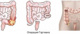
When assessing the colonoscopic picture, we focused on 3 types of tuberculous intestinal lesions: miliary, infiltrative and infiltrative-ulcerative form (see figure).
Miliary (a), infiltrative (b) and infiltrative-ulcerative (c) forms of intestinal tuberculosis.
During colonoscopy, erosions were most often found in the colon; tuberculous ulcers in all cases were localized in the right half of the colon, most often in the cecum, in the area of the bauhinine valve and the ileum. It was taken into account that the endoscopic picture of intestinal tuberculosis can be varied, although the changes are not specific. Oval or round ulcers, pseudopolyps, narrowing of the lumen, and rigidity of the intestinal walls may be observed. During intestinal endoscopy, swelling, hyperemia of the mucous membrane, the presence of ulcers with undermined edges, wall rigidity, and narrowing of the intestine are noted. Single, often multiple, ulcers are localized in the cecum, ileum, ascending, and colon. Tuberculous ulcers are often surrounded by a perifocal inflammatory shaft, often merge with each other, occupying a significant extent of the intestine, scarring, and produce ring-shaped narrowings of the intestine. Intestinal tuberculosis sometimes occurs in the form of an isolated hyperplastic pseudotumor process localized in the cecum, with a sharp thickening of the wall of the cecum, sometimes its ascending part, with an inflammatory process of specific tubercles, caseous decay, followed by scarring, narrowing of the intestinal lumen. To recognize intestinal tuberculosis and carry out differential diagnosis (with Crohn's disease, ulcerative colitis, intestinal tumors, amoebic dysentery, appendicitis), the results of histological examination are very important: with intestinal tuberculosis, along with sarcoid-like granulomas, foci of caseous necrosis appear. In some cases, the biopsy material reveals a picture of chronic nonspecific inflammation, but mycobacteria and epithelioid granulomas with Pirogov-Langhans cells, which form the basis of histological changes in tuberculosis, cannot be detected. This may be due to insufficient biopsy depth or predominantly submucosal localization of the pathological process. In such cases, the diagnosis is established based on a combination of clinical, laboratory and instrumental data.
Causes
Koch's bacillus has a shell that is resistant to acidic environments. It is able to survive freezing, drying, exposure to alkalis, and much more. etc. Characterized by the ability to form so-called L-forms with increased adaptability. The most pathogenic microbacteria for humans are:
- Mycobacterium tuberculosis humans;
- Mycobacterium bovis.
Microbacteria enter the body through contact, air, mixed and other routes. This is how the first inflammatory focus is formed. A child can also be infected during the mother's pregnancy - through the placenta or during childbirth, if it swallows amniotic fluid. Source: https://www.ncbi.nlm.nih.gov/pubmed/24548085 Marais BJ Tuberculosis in children J Paediatr Child Health . 2014 Oct;50(10):759-67. doi: 10.1111/jpc.12503. Epub 2014 Feb 19
Children at risk include:
- not vaccinated with BCG;
- long-term treatment with antibiotics, hormonal or cytostatic agents;
- having HIV status;
- living in unfavorable conditions (social and/or sanitary);
- having diabetes mellitus;
- with weakened immunity;
- under 2 years of age;
- and etc.
Results and discussion
Visual signs of intestinal tuberculosis were detected in 47 (74.6%) patients; they were characterized by the presence of erosions, infiltrates and ulcers in the colon. All patients underwent a biopsy of erosions or ulcers for histological examination, fluorescence microscopy and culture on solid nutrient media, and PCR. In 24 (38%) cases, microscopy revealed the presence of epithelioid giant cell granulomas, Ziehl-Neelsen staining revealed acid-fast mycobacteria, in 2 (3.2%) cases colon adenocarcinoma was diagnosed, in 4 (6.4%) - cytomegalovirus ulcerative colitis, which is typical for patients with HIV infection; in the remaining 37 (58.7%) cases, nonspecific colitis was detected. It was mandatory to take a biopsy from ulcers and erosions to test for MBT using fluorescent microscopy - in 17 (27%) cases MBT was found. When cultured on solid nutrient media, a positive result was obtained in 21 (33.3%) patients, and in 4 patients in this group, fluorescence microscopy gave a negative result. A PCR study to detect MBT fragments gave a positive result in 28 (44.4%) cases. However, research using the PCR method, which has high sensitivity, cannot be classified as a strictly specific diagnostic method, since the detection of MBT fragments in a biopsy from tuberculous ulcers is possible not only in intestinal tuberculosis, but also when ingesting sputum in patients with pulmonary tuberculosis.
Based on the results of colonoscopies with biopsies, the diagnosis of tuberculosis of the abdominal organs was established in 54 (85.7%) patients.
All patients received chemotherapy for tuberculosis in accordance with standard anti-tuberculosis chemotherapy regimens. In 46 (85.2%) patients, against the background of anti-tuberculosis therapy, clear positive dynamics were noted in the form of an improvement in general well-being, a decrease in the severity of symptoms of tuberculosis intoxication, the disappearance of abdominal pain, and normalization of stool. In 9 (16.6%) patients, despite anti-tuberculosis chemotherapy, no clear positive dynamics were noted: a slight improvement in general well-being, moderate abdominal pain and diarrhea persisted. These patients underwent control colonoscopy, 6 - once, 3 - twice. During the control colonoscopy, in comparison with the previous one, no clear positive dynamics were noted: tuberculous ulcers persisted or even new ones appeared, infiltration of the intestinal wall. Histological examination revealed an active tuberculosis process. These patients had their anti-tuberculosis therapy regimen changed, and all of them experienced clinical improvement.
The results obtained allowed us to conclude that colonoscopy is a fast, inexpensive and diagnostically valuable method for diagnosing tuberculosis of the abdominal organs (intestines), allowing us to reliably establish this diagnosis in 80.6% of patients. In some cases, performing a control colonoscopy against the background of anti-tuberculosis therapy allows one to assess the dynamics of treatment and, if necessary, change the anti-tuberculosis chemotherapy regimen.
T-SPOT.TB
T-SPOT.TB is a modern method of immunological diagnosis of latent tuberculosis infection using a blood test.
T-SPOT TEST FOR TUBERCULOSIS METHOD IS RECOMMENDED:
- If the patient refuses to undergo skin tests and fluorography;
- If extrapulmonary forms of TV are suspected;
- If the patient is found to have an allergy to tuberculin (according to the medical history), allergic diseases, skin diseases, epilepsy, acute and exacerbation of chronic diseases;
- Patients for whom subcutaneous tests are contraindicated or difficult due to exacerbation of the underlying disease.
- When a false positive Mantoux reaction is detected in children vaccinated with the BCG vaccine.
- In people who have visited countries with high incidence of tuberculosis (Africa, Asia)
- In pregnant women in case of contact with a patient with tuberculosis or presence of symptoms;
- Patients with HIV infection, especially those with a reduced level of CD4 protein cells;
- For tumor diseases, if a person is undergoing radiation or chemotherapy treatment
- For autoimmune and other pathologies leading to immune suppression;
Methods of transmission
- Airborne - through the sneezing, coughing of a patient with an open form of the disease, and even when drying out, the bacillus retains its pathogenicity.
- Alimentary – through the digestive tract. The infection enters the body due to poor hand hygiene or poorly washed and unprocessed food.
- Contact – the infection enters a person through the conjunctiva of the eyes, through kissing, sexual contact, through contact of contaminated objects with human blood, or the use of other people’s hygiene items.
How to treat tuberculosis?
Treatment of the pathology depends on its type, but most often a course of antibiotics is prescribed. Tuberculosis is a dangerous disease that requires immediate treatment. This allows the person to return to their normal lifestyle.
Antibacterial therapy is aimed at suppressing the proliferation of the tuberculosis pathogen.
Treatment takes place in 2 phases: in the first, several drugs are used at once to reduce the population of microbacteria, the second phase is maintenance therapy. Antibiotics stop the proliferation of bacteria and their release into the environment, the inflammatory process.
After taking such potent medications, a person needs additional supportive therapy that will strengthen the body and reduce the toxic effect. For this, immunostimulants (restore liver function), sorbents (remove toxic breakdown products of chemotherapy drugs) and vitamin complexes are prescribed.
After taking the drugs for two weeks, most people are no longer contagious and feel much better. However, it is very important to continue taking your medications as directed by your doctor and complete your entire course of antibiotics.
Diet for tuberculosis
Nutrition for tuberculosis should be aimed at strengthening the immune system.
- The patient should consume from 120 to 150 g of pure protein per day. It is needed for the production of antibodies. Sources of protein: fish, seafood, dairy products, lean poultry and fish, liver of cattle and fish.
- The amount of fat the patient needs is from 50 to 80 g per day. They are necessary to restore cell membranes that have been damaged by mycobacteria. To avoid a shortage of fats, you need to eat butter and vegetable oils, fish oil, lard, and animal fats in small quantities.
- Carbohydrates for tuberculosis should correspond to the age norm - about 400 g per day. They can be obtained from cereals and vegetables. Eating more than 80 g of confectionery products per day is not recommended.
- Mineral salts normalize metabolism and improve the functioning of the endocrine system, thereby increasing the body's defenses. Their sources can be: tomatoes, figs, cauliflower, herbs, cheeses, cottage cheese.
The first symptoms of tuberculosis
In the early stages, the disease is practically asymptomatic. As it develops, the patient's condition worsens, but no specific symptoms are observed. Increased fatigue, weakness, sudden weight loss for no apparent reason, a temperature of 37-38 ° C that does not subside for a long time, and night sweats appear. In children, the disease progresses faster than in adults.
The pulmonary form of tuberculosis is accompanied by a cough. Mild at first, it begins to progress over time. If your cough continues for more than three weeks, you should seek medical help immediately. The cough is initially dry, paroxysmal, especially at night and in the morning. Later, yellow-green sputum begins to be released, and at the cavity stage, hemoptysis is observed.
In the form of tuberculosis that affects the brain and its membranes, in addition to symptoms of general intoxication, sleep disturbances and headaches are observed, the intensity of which gradually increases.
Patient's appearance
Patients with tuberculosis appear emaciated in appearance, as they lose a lot of weight. They also have pale skin and sharp facial features. Despite the pale face, a blush is always noticeable on the cheeks of such people.
Best materials of the month
- Coronaviruses: SARS-CoV-2 (COVID-19)
- Antibiotics for the prevention and treatment of COVID-19: how effective are they?
- The most common "office" diseases
- Does vodka kill coronavirus?
- How to stay alive on our roads?


