The Aspergillus mushroom belongs to the genus of higher mold aerobic fungi. Today there are about 200 species of these mushrooms. They are widespread on all continents, in all countries of the world.
Fungi of the genus Aspergillus cause severe diseases in humans (mycoses), but at the same time, many of them are of very significant practical importance and are successfully used in industry due to their ability to produce a number of substances and enzymes. Aspergillus has been carefully studied by physiologists, biochemists and geneticists for several centuries.
What to do in such a situation? To get started, we recommend reading this article. This article describes in detail methods of controlling parasites. We also recommend that you consult a specialist. Read the article >>>
Epidemiology of aspergillosis
Aspergillus is widespread in nature. Saudi Arabia and Sudan are considered to be the regions with the highest levels of spores in the environment. Infection with fungi occurs through inhalation of their spores; sometimes pathogens enter the human body with food, sometimes through damaged skin. Aspergillus are aerobes. For energy synthesis processes, they require access to free molecular oxygen. Fungi of the genus Aspergillus are saprophytes. They use exclusively organic substances to ensure their vital functions. They grow well in humid environments (swampy areas and upper humus) and indoors.
- Typically, Aspergillus grows as mold on the surface of many substrates: inside and on the surface of rotting trees, the upper tiers of manure, plants (grass, hay), on the surface of rotting vegetables, various feeds, grapes, peanuts, jam, nuts, ground black pepper, tea, etc. packages. There are especially many of them on products containing starchy substances (potatoes, grains, flour, bread). Some Aspergillus species produce aflatoxin, which causes severe poisoning.
- Fungi can grow in nutrient-depleted environments. Thus, A. niger (black aspergillus) grows well on the walls of damp rooms.
- Fungi are found in the dust of rooms where hides and wool, hemp and hemp are processed. They attack cotton bolls and fibers and skin.
- Sources of fungi include showers and ventilation systems, air conditioners, humidifiers, books, shoes, pillows, soil of indoor plants, building materials, and textiles.
- Birds and farm animals can be sources of Aspergillus. Cows, dogs, cats, sheep, horses, rabbits, bees, pigeons, chickens, geese and turkeys suffer from aspergillosis.
Aspergillosis most often occurs in flour millers, people caring for pigeons, agricultural workers, and weaving and paper spinning workers. Persons with weakened immune systems, patients with diabetes mellitus and those who have undergone transplantation, as well as persons with IgE-mediated atopy (type I hypersensitivity) to fungal spores are susceptible to the disease.
Morphology of Aspergillus
Taxonomy
The Aspergillus mushroom belongs to the genus of higher mold fungi Aspergillus, class Ascomycetes, family Aspergillaceae. The genus Aspergillus includes several hundred species that differ in the structure of their conidia.
Story
The fungus Aspergillus was first cataloged by Italian biologist and priest Pier Antonio Micheli in 1729. The genus of these mushrooms received the name Aspergillum because of their resemblance to the shape of holy water sprinkler - aspergillus. Mushrooms are constantly studied by physiologists, biochemists and geneticists, since they not only cause harm to humans and animals, but also have very significant practical significance. They are successfully used in industry due to their ability to produce a number of substances and enzymes useful to humans.
The structure of Aspergillus
- The vegetative body of the aspergillus fungus (mycelium) is represented by mycelium. It is strong, very branched, firmly attached to the substrate, 4 - 6 microns wide. In some cases, abundant aerial mycelium (sclerotium) develops. Sometimes it is colorless, sometimes it is brightly colored.
- The mycelium consists of branched hyphae with partitions. The hyphae (web) range in size from 7 to 10 microns. Their main function is to absorb nutrients.
- Conidiophores extend upward from the supporting cells of the mycelium. In different types of mushrooms they have different sizes, may have partitions, and sometimes branch. In most fungi of the genus Aspergillus, conidiophores are colorless. In A.nidulans and A.ochraceus they are brown or yellowish in color, smooth or spiny.
- At the top of the conidiophore, a bubble swells and has a round or elongated shape.
- Sterigmata and flag cells (phialides) are located radially from the bladder. From their narrow neck, single-celled conidia (exospores) emerge one after another, arranged in a chain. Each Aspergillus species has a conidial structure unique to it, on the basis of which interspecific differentiation of fungi is based. Staining of conidia occurs gradually, starting from the phialids. Mature conidia are more intensely colored. Conidia in their mass give mold colonies a certain color - black, green, yellow or grayish. Under a microscope, you can see that the top of the conidiophores with conidia looks like a watering can, from the holes of which streams of water flow. Hence the second Russian name for the mushroom - “leichny” mushroom or “shaggy head”.
- Aspergillus spores range in size from 2 to 3.5 microns. They enter the human body through the respiratory tract, with food, or affect damaged skin. Spore germination occurs at a temperature of 35 °C.
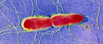
Rice. 5. Scheme of the structure of Aspergillus. 1 - conidia (exospores). 2 - sterigmata. 3 - bubble (swelling of the conidiophore).
Rice. 6. Under a microscope, the mycelium threads and fruiting organs - conidiophores, their characteristic swelling (bladder) and conidia (spores) are clearly visible.
Reproduction
As they mature, the conidia fall off and are transported to new places, where, provided favorable conditions exist, they germinate and form mycelium. This mode of reproduction is called asexual and is characteristic of most Aspergillus species. Some types of fungi of this species (A. fumigatus, A. flavus, A. lentulus and A. terreus) develop sexually (sporulation). In the colonies of such fungi, small balls (axospores) are visible to the naked eye, often yellow in color. These are cleistothecia (fruiting bodies).
Rice. 7. Fruiting bodies (photo on the left). Release of axospores (photo on the right).
Cultivation
Aspergillus grows well on various nutrient media. On Sabouraud's medium (Sabouraud agar) they form fluffy colonies, flat, initially white, and then different types of aspergillus acquire their own specific color, which is associated with sporulation and metabolites of the fungus.
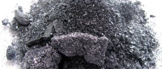
Rice. 8. Colonies of A. niger (Aspergillus nigra) are brown, chocolate or black in color.
Rice. 9. Fruiting organs of A. niger (Aspergillus nigra).
Rice. 10. Colonies of A. fumigatus are round, woolly, white aerial mycelium. Conidia give the colonies a soft bluish color.
Rice. 11. A. fumigatus. Chains of conidia on the conidiophore form a dense column.
Rice. 12. A.flavus-oryzae. Colonies are yellowish-green in color. The conidiophores on the swelling in some species bear only filiades or profilades.
Aspergillus resistance
Representatives of the genus Aspergillus grow wherever there is a high osmotic concentration of sugar, salt, etc. Formaldehyde and carbolic acid are used as disinfectants.
Habitats, distribution conditions, use in production
Representatives of this genus of fungi live mainly in soil, on plants, on rotting vegetable debris, in house dust, building materials, fabric, leather, textiles and on food products such as cheeses and canned meats.
Spores of fungi of this genus are present in the air almost constantly: every day each of us inhales about several hundred spores, which do not cause any diseases in a person with a normal immune system. And sometimes fungi of the genus Aspergillus can be found in the oropharynx of a healthy person.
As already described above, fungal spores can be present in indoor air, including in the air of hospitals, which can become a risk factor for nosocomial infection of an inpatient with a weakened immune system.
A number of representatives of fungi are used in industry to obtain organic acids, antibiotics, vitamins, enzymes, and for the industrial production of certain food products.
Why is Aspergillus dangerous?
Aspergillus causes the disease (mycosis) aspergillosis in humans, birds and animals. The main species pathogenic for humans are A.fumigatus and A.niger, other species - A.flavus, A.nidulans, A.terreus and A.clavatus are less common. These Aspergillus species grow at normal body temperature, which is not observed in all other species.
Aspergillus niger (black aspergillus)
Aspergillus nigra is also known as black mold. The main habitat of this species of fungi is damp places - soil, books, humidifiers, air conditioners, tile joints, washing machines, etc. Fungi cause diseases of cotton, peanut and sorghum seedlings in Sudan and India. Moldy feed containing aspergillus nigra is toxic to animals.
Black aspergillus most often affects the respiratory tract, less often the heart and central nervous system, it is the cause of otomycosis, aspergillus (infestation of fungal colonies in the lung cavity) and mycete (infestation of fungi in the sinuses).
Aspergillus fumigatus
Fungi grow in soil, manure, compost, forage, and attack grains, wool and cotton. Aspergillus fumigatus is the cause of severe mycoses in humans and animals. Fungi affect many internal organs, including the respiratory system, causing the development of bronchopulmonary allergic aspergillosis. Aspergillus fumigatus produces a toxin.
Aspergillus flavus
Fungi of the Aspergillus flavus species infect plants, insects, animals and humans. Cotton seedlings suffer from them. Fungi of this species cause paralysis of bees and diseases of silkworms. In humans, Aspergillus flavus often affects the lungs and various internal organs and is the cause of otomycosis. Mushrooms secrete aflatoxin, which accumulates in groundnuts, flax and cotton seeds, fish and liver, when consumed, severe poisoning develops in humans and animals. Researchers have established the fact that it (the toxin) has a carcinogenic effect.
Rice. 13. In the photo on the left is otomycosis (damage to the ear canal), on the right is a fungal infection of the skin of the foot.
Description of appearance
Externally, upon microscopic examination, fungi of the genus Aspergillus are mushrooms consisting of the same type of mycelium, 4–6 micrometers wide, on which “heads” with conidia are sometimes present.
A specific bacteriological nutrient medium for growing colonies of fungi of this genus is the so-called Sabouraud medium. On it, mushrooms form flat colonies, at first white, slightly fluffy, which subsequently take on bluish, yellowish, brown and other colors depending on the species. Their surface becomes powdery.
Clinical significance
A peculiarity of this genus of fungi is the ability to cause not only allergic diseases, but also infectious lesions.
In terms of the frequency of development of specific infectious diseases, fungi of the genus Aspergillus occupy second place after yeast-like fungi of the genus Candida.
Predisposing factors to the development of Aspergillus infections are immunodeficiencies, including secondary immunodeficiencies caused by taking high doses of systemic glucocorticosteroids, for which the cellular and molecular mechanisms of increased vulnerability of organs and tissues to fungal spores have been studied, as well as chronic pulmonary diseases.
Aspergillus can affect any organs and tissues.
Clinical manifestations include the following forms:
- bronchopulmonary aspergillosis and its varieties: infectious-allergic bronchopulmonary aspergillosis, purulent bronchitis, chronic aspergilloma, invasive pulmonary aspergillosis, chronic necrotizing pulmonary aspergillosis;
- generalized (septic) aspergillosis, which occurs in immunocompromised people (for example, HIV infection) and has a high incidence of deaths;
- aspergillosis of ENT organs: external and media otitis, rhinosinusitis, aspergillosis of the larynx;
- aspergillosis of the eye;
- skin aspergillosis in the form of erythematous scales and papules, in more severe cases – necrotic lesions of subcutaneous fatty tissue;
- bone aspergillosis;
- other forms of aspergillosis (damage to the mucous membranes of the mouth, genitals, mycotoxicosis).
The most common respiratory lesions occur against the background of chronic lung diseases:
- bronchial asthma, cystic fibrosis - for allergic bronchopulmonary aspergillosis;
- pre-existing cavities in the lungs (tuberculous cavities, cavities in patients with sarcoidosis or other diseases resulting from the formation of cavities) - for aspergilloma;
- chronic obstructive pulmonary disease during its treatment with glucocorticosteroids – for necrotizing pulmonary aspergillosis.
Risk factors for the occurrence of invasive pulmonary aspergillosis, in addition to those listed above, are secondary immunodeficiency states due to cancer, treatment with immunosuppressants, HIV infection, decompensated diabetes mellitus, massive treatment with antibiotics and other factors.
However, people with normal immune systems can also develop respiratory infections caused by Aspergillus fungi due to increased exposure to Aspergillus spores.
Massive inhalation of the spores of these fungi in healthy people can cause acute pneumonia, which usually resolves on its own.
Occupational risk factors for chronic diseases caused by spores of fungi of the genus Aspergillus are work in agriculture, weaving factories and paper spinning mills.
Allergic diseases associated with sensitization to mold allergens are widespread. These are primarily typical atopic diseases with fungal sensitization: year-round allergic rhinitis and bronchial asthma.
Much less often, exogenous allergic alveolitis can occur (see more in the corresponding section of the article “Mushroom Alternaria alternata: description and clinical significance”).
For fungi of the genus Aspergillus, this disease is called “malt workers’ lung” due to the high frequency of occupationally caused cases of diseases in these workers.
Also, some representatives of fungi of this genus can secrete toxic substances - aflatoxin, ochratoxin and sterigmatocystin, which, with chronic exposure, cause manifestations of mycotoxicosis - toxic hepatitis, kidney disease and even malignant tumors. However, the main feature of fungi of the genus Aspergillus, which distinguishes them from representatives of other genera of molds, is the ability to cause specific infectious diseases.
Mycoses of the lungs
Pulmonary mycoses are relatively rare and occur mainly in patients with sharply reduced body defense reactions (AIDS, cancer cachexia, long-term treatment with broad-spectrum antibiotics, glucocorticoids and immunosuppressive therapy). The causative agents of pulmonary mycoses are microscopic fungi (micromycetes). The manifestations of the disease and the nature of its course are significantly different from bacterial and viral infections. Identification of the causative agents of fungal pneumonia is the basis for successful treatment.
Aspergillosis
Invasive aspergillosis (pneumonia). The main causative agents of invasive aspergillosis are: A. fumigatus (60-90%), A. flavus (10-30%) and A. niger (2-15%). The frequency is 12-34 cases per 1 million population per year. Infection occurs through inhalation of Aspergillus conidia. Aspergillosis is not transmitted from person to person. It most often develops in patients with acute leukemia during cytostatic therapy, in patients receiving glucocorticoids and immunosuppressants for a long time. Clinical manifestations The duration of the incubation period is not determined. The most common signs of the disease are ineffectiveness of broad-spectrum antibiotics, fever over 38°C lasting more than 96 hours, nonproductive cough, chest pain, hemoptysis and shortness of breath. In 30% of cases, no increase in temperature may be observed. Sometimes the clinical picture of aspergillosis resembles signs of thrombosis of the branches of the pulmonary artery: sudden chest pain and severe shortness of breath. Often the only manifestation of the disease is changes on an x-ray or CT scan of the lungs.
Diagnostics
Main detection method:
- lesions - multislice computed tomography (MSCT);
- microbiological confirmation of the diagnosis - microscopy and culture of respiratory substrates;
- serological diagnosis - determination of Aspergillus antigen (galactomannan) in blood serum.
Diagnostic methods:
- radiography of the lungs and paranasal sinuses;
- Chest CT (MSCT);
- in the presence of neurological symptoms - MSCT or MRI of the brain;
- determination of Aspergillus antigen in blood serum;
- bronchoscopy, bronchoalveolar lavage (BAL), biopsy of lesions;
- microscopy and culture of BAL swabs, sputum, nasal discharge, and biopsy material.
Treatment includes antifungal (antifungal) therapy, elimination of risk factors, and surgical removal of affected tissue. Forecast. Without treatment, invasive aspergillosis almost always ends in death (within 1-4 weeks). During treatment, mortality is 30-50% and depends on the underlying disease (acute leukemia, etc.), as well as on the prevalence of aspergillosis or localization of the disease (dissemination, central nervous system damage)
Chronic necrotizing pulmonary aspergillosis is a relatively rare disease, accounting for approximately 5% of all cases of pulmonary aspergillosis. The causative agent is A. fumigatus, less commonly - A. flavus, A. terreus, etc. Risk factors are AIDS, diabetes mellitus, alcoholism, long-term treatment with steroids. Symptoms are characteristic, but not specific. Usually a chronic productive cough (with sputum) develops, often with moderate hemoptysis. Low-grade fever. General weakness and weight loss. The disease occurs “chronically” with periodic exacerbations and progressive impairment of lung function due to the development of fibrosis. Complications: damage to the pleura, ribs, vertebrae, pulmonary hemorrhage, invasive pulmonary aspergillosis (pneumonia) with hematogenous dissemination, damage to the brain and internal organs. Diagnosis - see invasive aspergillosis. Treatment. Long-term use of antifungal drugs. The indication for surgical treatment is a high risk of pulmonary hemorrhage.
Aspergilloma (non-invasive aspergillosis) , or “mushroom ball”, is the mycelium (mycelium) of the Aspergillus fungus that grows in the cavities of the lungs formed as a result of tuberculosis, tumors and other diseases. Aspergilloma can occur in the paranasal sinuses. The causative agent is Aspergillus fumigatus, less commonly A.flavus. Tuberculosis is the cause of cavity formation in aspergilloma in 40-70% of cases; destructive pneumonia - in 10-20%; bullous emphysema - in 10-20%; bronchiectasis - in 5-10%; tumors - in 3-7% of cases. The probability of developing aspergilloma in a cavity measuring 2 cm is 15-20%. Usually occurs between the ages of 40 and 70, more often in men. Initially, it is asymptomatic, but as it progresses, coughing begins to bother you, hemoptysis, and low-grade fever occur. With secondary bacterial infection of the cavity affected by fungi, signs of acute inflammation may develop. In most cases, aspergilloma occurs in the upper lobe of the right lung (50-75%), less often - in the upper lobe of the left lung (20-30%). In approximately 10% of patients, signs of aspergilloma resolve spontaneously, without treatment. During the course of the disease, most patients experience an episode of hemoptysis, and 20% have pulmonary hemorrhage. Complications of aspergilloma include pulmonary hemorrhage and invasive growth of Aspergillus with the development of chronic necrotizing pulmonary aspergillosis or specific pleurisy. Diagnosis - see invasive aspergillosis. Treatment is carried out in case of development or high risk of complications (repeated hemoptysis, pulmonary hemorrhage, etc.); observation is indicated for asymptomatic aspergilloma. The main treatment method is surgical removal of the affected area of the lung.
Allergic bronchopulmonary aspergillosis is characterized by the development of a type I hypersensitivity reaction when the respiratory tract is affected by Aspergillus. The frequency in patients with bronchial asthma is 1-5%, in patients with cystic fibrosis - 5-14%. Pathogens - Aspergillus fumigatus, A. clavatus, less often - other Aspergillus. Congenital predisposition contributes to its occurrence. No invasive damage to lung tissue is observed. The disease usually occurs chronically with periodic exacerbations of broncho-obstructive syndrome and/or the occurrence of eosinophilic infiltrates. The main signs of exacerbation are attacks of suffocation, increased body temperature, chest pain and cough with sputum containing brown inclusions and mucus plugs. Over a long period of time, bronchiectasis and pulmonary fibrosis develop, leading to respiratory failure.
- Invasive candidiasis
- Pulmonary cryptococcosis
- Zygomycosis of the lungs
- Hyalohyphomycosis
Invasive pulmonary candidiasis (candidal pneumonia) is relatively rare and accounts for approximately 5-15% of all cases of invasive candidiasis. It can be primary or secondary, resulting from hematogenous dissemination of Candida from another lesion. It develops mainly in patients against the background of severe pathology (surgical treatment of the gastrointestinal tract, infected pancreatic necrosis, long-term parenteral nutrition, the use of immunosuppressants, artificial ventilation, hemodialysis, repeated blood transfusions, diabetes mellitus). The main causative agents of candidal pneumonia are C. albicans, C. tropicalis, C. parapsilosis, C. glabrata, etc. Many Candida are inhabitants of the human body and are detected by culture from the mucous membrane of the oral cavity and gastrointestinal tract in 30-50% of healthy people . Clinical signs (fever, nonproductive cough, shortness of breath, and chest pain) are nonspecific and do not distinguish candidal pneumonia from bacterial or other pulmonary mycosis. The basis of diagnosis is the identification of Candida during histological examination and/or culture of a lung biopsy. Treatment. An important condition for successful treatment is identification of the pathogen. Candida species clearly correlates with sensitivity to antimycotics.
The incidence of cryptococcosis has increased significantly in recent years due to the HIV pandemic. In the vast majority of cases, the causative agent of cryptococcosis is Cryptococcus neoformans, which is distributed everywhere (in soil, on plants, in bird feces). The main risk factors are disorders of cellular immunity caused by AIDS, lymphoma, chronic lymphocytic leukemia, T-cell leukemia, as well as long-term use of glucocorticoids and immunosuppressants. The risk of developing cryptococcosis is determined by the severity of immunodeficiency. For example, the incidence of cryptococcosis in AIDS patients is up to 30%. Cryptococcosis rarely develops in immunocompetent patients. It occurs less frequently in women than in men, and less frequently in children than in adults. Infection occurs by inhalation. In patients with AIDS, the central nervous system, lungs, and skin are most often affected; disseminated variants of infection develop, involving the bones, kidneys, adrenal glands, etc. The main signs are fever (81%), cough (63%), shortness of breath (50%), weight loss (47%), rarely - chest pain and hemoptysis. Diagnosis: see invasive aspergillosis. For any localization of cryptococcal infection, a lumbar puncture is necessary (determination of intracerebral pressure, microscopy of cerebrospinal fluid and culture for flora). Treatment. The choice and duration of use of antimycotics are determined by the patient’s condition and the localization of the process.
Pathogens are lower fungi belonging to the class Zygomycetes. Zygomycetes are ubiquitous, live in the soil, and are often found on food products and in rotting plant waste. The pathogen enters the lungs by inhaling spores. The risk factors are the same as for other mycoses. Zygomycosis is characterized by an extremely aggressive course with very rapid destruction of all tissue barriers, damage to blood vessels, hematogenous dissemination with the subsequent development of thrombosis, infarction and tissue necrosis. With zygomycosis, any organs can be affected, but the paranasal sinuses (35-50% of all cases), lungs (20-30%), skin and subcutaneous tissue (10%), as well as the gastrointestinal tract (5-10%) are most often affected. %). Pulmonary zygomycosis is usually manifested by an increase in body temperature of more than 38 ° C, which does not decrease during treatment with broad-spectrum antibiotics, cough, chest pain, profuse hemoptysis or pulmonary hemorrhage.
Hyalohyphomycosis is a group of diseases caused by the fungi Fusarium spp., Acremonium spp., Paecilomyces spp., Scedosporium spp., Scopulariopsis brevicaulis and Trichoderma longibrachiatum. The pathogens are ubiquitous, often found in the soil and on various plants.
Fusarium is considered the second most common causative agent of invasive pulmonary mycoses after Aspergillus. In addition to pneumonia, hyalohifomycetes cause local lesions in immunocompetent patients, and fungemia (fungus in the blood) and disseminated infections, which are characterized by very high mortality, in immunocompromised patients. The unfavorable prognosis is associated both with the severity of immunosuppression in patients and with the low sensitivity of hyaloghiphomycetes to most used antimycotics.
Hyalohyphomycosis of the lungs most often develops in patients with hemoblastosis or recipients of bone marrow transplants, much less often in patients with widespread deep burns. Infection usually occurs by inhalation. One of the possible sources of the pathogen is the affected nails with onychomycosis. Pathogens can infect arteries with subsequent development of thrombosis, infarction and hematogenous dissemination. The disease usually begins as pneumonia or sinusitis, and as it progresses, hematogenous dissemination develops with damage to the skin, internal organs, bones and brain. The clinical picture of the disease is determined by the localization of the process; a common symptom is fever refractory to antibiotics. In 55-70% of patients, characteristic lesions of the skin and subcutaneous tissue develop: painful erythematous papules or subcutaneous nodules followed by the formation of a focus of necrosis in the center.
Diagnosis: see invasive aspergillosis.
Treatment. The causative agents of hyalohyphomycosis are characterized by low sensitivity and even resistance to antimycotics
Causes of aspergillosis
The causative agents of aspergillosis in humans can be the following types of mold fungi of the genus Aspergillus: A. flavus, A. Niger, A. Fumigatus, A. nidulans. A. terreus, A. clavatus. Aspergillus are aerobes and heterotrophs; are able to grow at temperatures up to 50°C and can be preserved for a long time when dried and frozen. In the environment, Aspergillus is ubiquitous - in soil, air, and water. Favorable conditions for the growth and reproduction of aspergillus are found in ventilation and shower systems, air conditioners and humidifiers, old clothes and books, damp walls and ceilings, long-term stored food products, agricultural and indoor plants, etc.
Infection with aspergillosis most often occurs through inhalation when inhaling dust particles containing the mycelium of the fungus. Agricultural workers, employees of paper spinning and weaving enterprises, flour millers, and pigeon breeders are at greatest risk of developing the disease, since pigeons are more likely than other birds to suffer from aspergillosis. The occurrence of a fungal infection is facilitated by infection during invasive procedures: bronchoscopy, puncture of the paranasal sinuses, endoscopic biopsy, etc. Contact transmission of aspergillosis through damaged skin and mucous membranes cannot be ruled out. Nutritional infection is also possible through consumption of food products contaminated with Aspergillus (for example, chicken meat).
In addition to exogenous infection with Aspergillus, cases of autoinfection (by activation of fungi that live on the skin, mucous membrane of the pharynx and respiratory tract) and transplacental infection are known. Risk factors for the incidence of aspergillosis include immunodeficiencies of any origin, chronic diseases of the respiratory system (COPD, tuberculosis, bronchiectasis, bronchial asthma, etc.), diabetes mellitus, dysbacteriosis, burn injuries; taking antibiotics, corticosteroids and cytostatics, radiotherapy. There are frequent cases of the development of mycoses of mixed etiology, caused by various types of fungi - aspergillus, candida, actinomycetes.
Causes of aspergillosis
The causative agents of the disease belong to the genus Aspergillus, and in human pathology, A. Flavus and A. Niger are of greatest importance, but other species may also occur, for example, A. Nidulans or A. Fumigatus. We can say that morphologically, these types of fungi consist of the same type of mycelium, having a width of 4-6 microns. Aspergilli, as a rule, have quite high biochemical activity, due to which they can form various enzymes.
The causative agents of pulmonary aspergillosis are widespread in nature. They are most often found in hay, flour, soil and grain, as well as dust. The pathogen usually enters the body with dust through the air. By aerogenous route it reaches the mucous membranes located on the upper respiratory tract. It is quite possible to become infected through the skin, which is often changed by another pathological process.
A decrease in the body's immune defense plays a leading role in the development of aspergillosis. This disease can be complicated by various pathological processes of the skin, internal organs and mucous membranes.
Classification of aspergillosis
Thus, depending on the routes of spread of fungal infection, endogenous (autoinfection), exogenous (with airborne and alimentary transmission) and transplacental aspergillosis (with vertical infection) are distinguished.
According to the localization of the pathological process, the following forms of aspergillosis are distinguished: bronchopulmonary (including pulmonary aspergillosis), ENT organs, skin, eye, bone, septic (generalized), etc. Primary damage to the respiratory tract and lungs accounts for about 90% of all cases aspergillosis; paranasal sinuses – 5%. Involvement of other organs is diagnosed in less than 5% of patients; dissemination of aspergillosis develops in approximately 30% of cases, mainly in weakened individuals with a burdened premorbid background.
Symptoms
The most studied form of pathology to date is pulmonary aspergillosis. The initial stages of bronchopulmonary aspergillosis are disguised as a clinical picture of tracheobronchitis or bronchitis. Patients are concerned about a cough with grayish sputum, hemoptysis, general weakness, and weight loss. When the process spreads to the lungs, a pulmonary form of mycosis develops - aspergillus pneumonia. In the acute phase, fever of the wrong type, chills, cough with copious mucopurulent sputum, shortness of breath, and chest pain are noted. There may be a musty odor coming from your mouth when you breathe. Microscopic examination of sputum reveals mycelial colonies and Aspergillus spores.
In patients with concomitant diseases of the respiratory system (pulmonary fibrosis, emphysema, cysts, lung abscess, sarcoidosis, tuberculosis, hypoplasia, histoplasmosis), pulmonary aspergilloma is often formed - an encapsulated lesion containing fungal hyphae, fibrin, mucus and cellular elements. Death of patients with aspergilloma can occur as a result of pulmonary hemorrhage or asphyxia.
Aspergillosis of the ENT organs can occur in the form of external or otitis media, rhinitis, sinusitis, tonsillitis, and pharyngitis. With aspergillus otitis media, hyperemia, peeling and itching of the skin of the external auditory canal initially occurs. Over time, the ear canal becomes filled with a loose grayish mass containing threads and fungal spores.
Aspergillosis may spread to the eardrum, accompanied by sharp stabbing pain in the ear. Lesions of the maxillary and sphenoid sinuses, the ethmoid bone, and the transition of fungal invasion to the orbits are described. Ocular aspergillosis can take the form of conjunctivitis, ulcerative blepharitis, nodular keratitis, dacryocystitis, blepharomeibomitis, panophthalmitis. Complications in the form of deep corneal ulcers, uveitis, glaucoma, and loss of vision are common.
Skin aspergillosis is characterized by the appearance of erythema, infiltration, brownish scales, and moderate itching. If onychomycosis develops, deformation of the nail plates, discoloration to dark yellow or brownish-greenish, and crumbling of the nails occur. Aspergillosis of the gastrointestinal tract occurs under the guise of erosive gastritis or enterocolitis: the smell of mold from the mouth, nausea, vomiting, and diarrhea are typical for it.
The generalized form of aspergillosis develops with hematogenous dissemination of aspergillus from the primary focus to various organs and tissues. With this form of the disease, aspergillus endocarditis, meningitis, and encephalitis occur; abscesses of the brain, kidneys, liver, myocardium; damage to bones, gastrointestinal tract, ENT organs; Aspergillus sepsis. Mortality from the septic form of aspergillosis is very high.
Symptoms of aspergillosis in humans
Since the respiratory system takes the first blow, the main symptoms of aspergillosis in humans begin to appear precisely from the respiratory system. In a third of cases, the fungus enters the body through the blood and lymph flow and spreads to all organs. This type of aspergillosis has a high mortality rate of about eighty percent. The rarest is cutaneous aspergillosis.
If the fungus has settled on the surface and has not penetrated the mucous membrane, as happens with tracheobronchitis or aspergilloma, then patients note the following symptoms: chronic cough with sputum, sometimes with blood during a strained cough. Most often in such cases there are pathologies of the lungs.
In response to the penetration of spores into the body, human tissues develop certain inflammatory reactions. The most common two types of inflammation are serous-desquamative and fibrous-purulent. With serous-desquamative inflammation, aspergillus causes exfoliation of the epithelium, membranes of the stomach, and lungs with the release of exudate (plasma with blood elements). In the second type - fibropurulent - aspergillus causes the release of exudate with fibrin (clotted blood protein) and a purulent component. The most severe reaction to aspergillosis is the formation of granulomas in the lungs.
Otherwise, aspergillosis gives an acute picture - a dense infiltrate forms in the lungs, which disintegrates. With the blood flow, infection of other organs occurs. At the onset of acute aspergillosis, the phenomenon of neutropenia is characteristic, which is expressed in sudden weakness, nosebleeds, fever, sudden chills, severe sweating, tachycardia, and a sharp decrease in pressure. In this case, a decrease in neutrophils is detected in the blood, which makes it difficult for the body to give an inflammatory response to the focus of aspergillosis. Therefore, with neutropenia, it is often not possible to diagnose aspergillosis - all indicators would seem to be normal. However, doctors know from experience that this may signal the onset of aspergillosis, so additional studies are prescribed. Most often, aspergillus settles in the sinuses. In this case, red lesions appear; after the tissue disintegrates, they lose their color and then turn black. This process is very rapid - it usually spreads to the eye sockets, facial tissues, and towards the brain. Typical symptoms in this condition are congestion, pain in the nasopharynx, sinuses, swelling of the mucous membrane. The sinuses are filled with pus, but they do not rupture.
Allergic aspergillosis is often associated with bronchial asthma. In this case, patients note asthmatic attacks, increased eosinophils in the blood, dark areas on x-ray, and the presence of antibodies in the serum (galactoman). To clarify the diagnosis, a sputum analysis is taken. In more than half of patients, aspergillus is detected during culture. In this case, a secondary culture is done to clarify the diagnosis (since conidia could have been accidentally introduced).
Diagnosis of aspergillosis
Depending on the form of mycosis, patients are referred for consultation to a specialist of the appropriate profile: pulmonologist, otolaryngologist, ophthalmologist, mycologist. In the process of diagnosing aspergillosis, much attention is paid to medical history, including professional history, the presence of chronic pulmonary pathology and immunodeficiency. If a bronchopulmonary form of aspergillosis is suspected, radiography and CT scan of the lungs, bronchoscopy with sputum sampling, and bronchoalveolar lavage are performed.
The basis for diagnosing aspergillosis is a complex of laboratory tests, the material for which can be sputum, washing water from the bronchi, scrapings from smooth skin and nails, discharge from the sinuses of the nose and external auditory canal, prints from the surface of the cornea, feces, etc. Aspergillus can be detected with using microscopy, cultural examination, PCR, serological reactions (ELISA, RSK, RIA). It is possible to carry out skin allergy tests with Aspergillus antigens.
Differential diagnosis of pulmonary aspergillosis is carried out with inflammatory diseases of the respiratory tract of viral or bacterial etiology, sarcoidosis, candidiasis, pulmonary tuberculosis, cystic fibrosis, lung tumors, etc. Aspergillosis of the skin and nails is similar to epidermophytosis, rubromycosis, syphilis, tuberculosis, actinomycosis.
Treatment of aspergillosis
Depending on the severity of the patient’s condition and the form of aspergillosis, treatment can be carried out on an outpatient basis or in a hospital of the appropriate profile. Antifungal therapy is carried out with the following drugs: amphotericin B, voriconazole, itraconazole, flucytosine, caspofungin. Antifungal drugs can be prescribed orally, intravenously, or inhaled. For aspergillosis of the skin, nails and mucous membranes, local treatment of the lesions with antifungal agents, antiseptics, and enzymes is carried out. Antifungal therapy lasts from 4 to 8 weeks, sometimes up to 3 months or longer.
For pulmonary aspergilloma, surgical tactics are indicated - economical lung resection or lobectomy. In the process of treating any form of aspergillosis, stimulating and immunocorrective therapy is necessary.
Forecast and prevention of aspergillosis
The most favorable course is observed with aspergillosis of the skin and mucous membranes. The mortality rate from pulmonary forms of mycosis is 20-35%, and in people with immunodeficiency - up to 50%. The septic form of aspergillosis has a poor prognosis. Measures to prevent infection with aspergillosis include measures to improve sanitary and hygienic conditions: combating dust in production, wearing personal protective equipment (respirators) by workers in mills, granaries, vegetable stores, weaving enterprises, improving ventilation of workshops and warehouses, regular mycological examination of persons from risk groups.
Pulmonary aspergillosis
Pulmonary aspergillosis is a very serious diagnosis. Since, due to the development of the disease caused by the mold fungi Aspergillus, aspergillomas begin to form in the human lungs, that is, tumor-like formations that consist of tightly woven fungi. There are also complications such as endocarditis, aspergillus pleurisy, otitis, meningoencephalitis and others.
However, at any time, aspergilloma can cause a serious complication - pulmonary hemorrhage, which can be massive and profuse. And in this case, there is no alternative to surgical treatment. Treatment of aspergillosis with conservative methods is possible when the mucous membranes or skin are damaged by the fungus.
It is possible to defeat parasites!
Antiparasitic Complex® - Reliable and safe removal of parasites in 21 days!
- The composition includes only natural ingredients;
- Does not cause side effects;
- Absolutely safe;
- Protects the liver, heart, lungs, stomach, skin from parasites;
- Removes waste products of parasites from the body.
- Effectively destroys most types of helminths in 21 days.
There is now a preferential program for free packaging. Read expert opinion.
Read further:
Fungi parasites of plants: representatives, life cycle of development
Pathogenic fungal spores: detection and destruction on nails
Allergic bronchopulmonary aspergillosis: symptoms and treatment methods
Fungi parasites of cereal plants: representatives, life cycle
Fungi parasites of animals: representatives, life cycle of development
Fungi parasites of humans: representatives, life cycle of development
Bibliography
- Centers for Disease Control and Prevention. Brucellosis. Parasites. Link
- Corbel MJ Parasitic diseases // World Health Organization. Link
- Young EJ Best matches for intestinal parasites // Clinical Infectious Diseases. — 1995. Vol. 21. - P. 283-290. Link
- Yushchuk N.D., Vengerov Yu.A. Infectious diseases: textbook. — 2nd edition. - M.: Medicine, 2003. - 544 p.
- Prevalence of parasitic diseases among the population, 2009 / Kokolova L. M., Reshetnikov A. D., Platonov T. A., Verkhovtseva L. A.
- Helminths of domestic carnivores of the Voronezh region, 2011 / Nikulin P. I., Romashov B. V.
An article for patients with a doctor-diagnosed disease. Does not replace a doctor's appointment and cannot be used for self-diagnosis.
The best stories from our readers
Topic: Parasites are to blame for all troubles!
From: Lyudmila S. ()
To: Administration Noparasites.ru
Not long ago my health condition worsened. I began to feel constant fatigue, headaches, laziness and some kind of endless apathy appeared. Problems also appeared with the gastrointestinal tract: bloating, diarrhea, pain and bad breath.
I thought it was because of the hard work and hoped that it would go away on its own. But every day I felt worse. The doctors couldn’t really say anything either. Everything seems to be normal, but I feel like my body is not healthy.
I decided to go to a private clinic. Here I was advised, in addition to general tests, to get tested for parasites. So in one of the tests they found parasites in me. According to doctors, these were worms, which 90% of people have and almost everyone is infected, to a greater or lesser extent.
I was prescribed a course of antiparasitic medications. But it didn’t give me any results. A week later, a friend sent me a link to an article where some parasitologist shared real tips on fighting parasites. This article literally saved my life. I followed all the advice that was there and after a couple of days I felt much better!
Digestion improved, headaches went away and the vital energy that I so lacked appeared. To be sure, I took the tests again and no parasites were found!
Anyone who wants to cleanse their body of parasites, no matter what types of these creatures live in you, read this article, I’m 100% sure it will help you! Go to article>>>
Still have questions? Ask them in our Anonymous group on VK
How to get rid of parasites in a week. The answer is here!
A reliable and effective remedy for combating worms. Removes all parasites in 21 days.
Go to website
Reviews
Read online
Symptoms that 100% indicate parasites! Take the Test.
How to rid your body of life-threatening parasites before it’s too late!
Read more
Website
To get a consultation
The doctor tells how to quickly get rid of parasites for adults and children!
A parasitologist explains what effective methods exist to combat helminths.
More details
Read completely
Comments
Search for cures for parasites
This service is a small help in finding cures for parasites. To start using it, select the type of parasite. If you don’t know what kind of parasite you are infected with, this parasite identification tool will help you by symptoms.
We recommend reading
Mosquito: description, types and characteristics, methods of feeding and reproduction
26.03.202126.03.2021red
How to cure Lyme disease
03/24/202103/22/2021ElenaKV
Centipede mosquito. Description, stages of development. What do big mosquitoes eat? Whether they bite or not. Damage from long-legged mosquitoes
24.03.202124.03.2021red
What do mosquitoes eat, besides blood, females and males, in the swamp and in nature?
23.03.202130.03.2021red