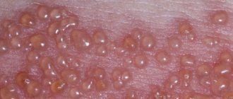
Lichen planus
Warts
6905 24 August
IMPORTANT!
The information in this section cannot be used for self-diagnosis and self-treatment.
In case of pain or other exacerbation of the disease, diagnostic tests should be prescribed only by the attending physician. To make a diagnosis and properly prescribe treatment, you should contact your doctor. Lichen planus: causes, symptoms, diagnosis and treatment methods.
Definition
Lichen planus is a chronic inflammatory disease of the skin and mucous membranes of unknown etiology. It is quite rare (according to WHO, 0.5% of all skin diseases) and, as a rule, occurs in people aged 45 to 60 years.
Causes of lichen planus
The causes of lichen planus still remain unclear. Experts classify it as a group of autoimmune diseases, when the body begins to produce antibodies that destroy its own tissues.
Provoking factors may include stressful situations, taking certain medications, possibly viral hepatitis C, or decreased immunity.
It has been noticed that patients with lichen planus are often characterized by increased excitability, suffer from insomnia and are prone to frequent mood swings. Some scientists believe that lichen planus is a delayed hypersensitivity reaction to various chemicals.
Proponents of the infectious nature of the disease talk about cases of the development of lichen planus after various injuries.
Classification of the disease
In patients with lichen planus, the following most common forms of skin lesions are distinguished:
- typical (the same type of rash in the form of papules),
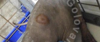
- hypertrophic (warty rashes with indentations),
- bullous (blisters turning into plaques),
- actinic (large plaques, light at the edges and dark in the center),
- ring-shaped, or anular (rashes in the form of rings with raised edges),
- atrophic (nodules that rise above the skin and leave behind scars),
- pigmented (brown rashes merging into lesions),
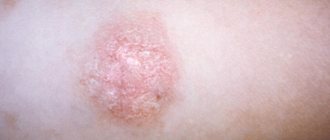
- erosive-ulcerative (in addition to papules there are erosions and, less commonly, ulcers),
- follicular (pointed papules covered with dense horny scales).
Symptoms of lichen planus
In lichen planus, rashes appear on the skin, localized symmetrically on the flexor surfaces of the limbs, torso, as well as on the mucous membranes of the oral cavity, sometimes the esophagus, perineal area and anus; less often, nails, hair, palms, soles and face are affected.
Patients are bothered by itching, the severity of which depends on the form of the disease. The presence of papules on the oral mucosa causes discomfort; in the erosive-ulcerative form, bleeding of the gums is possible; there are complaints of dry mouth and pain when eating hot food. Ulcerative skin lesions in the erosive-ulcerative form are characterized by pain that intensifies during movement if they are located on the lower extremities.
In rare cases, the disease affects the nails. You can observe thinning of the nail plate, longitudinal grooves, splitting of the nail, discoloration of the nail plate and even its rejection from the nail bed.
Diagnosis of lichen planus
In most cases, the diagnosis of lichen planus is made based on the clinical picture. However, if patients have hypertrophic, atrophic, pigmentary, cystic, erosive-ulcerative and follicular forms, the typical elements inherent in this disease may be absent. To clarify the diagnosis, a biopsy of skin lesions is performed, followed by histological examination.
Before prescribing drug therapy, it is necessary to conduct laboratory tests, including a general (clinical) blood test, a general urinalysis, a biochemical blood test: AST, ALT, total bilirubin, triglycerides, cholesterol, total protein.
Publications in the media
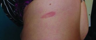
Lichen planus (Wilson's lichen, true lichen) is a chronic itchy dermatosis of unknown etiology, characterized by the appearance of flat red polygonal papules with a smooth shiny surface and a slight depression in the center. Frequency. 450:100,000. The predominant age is 30–60 years.
Risk factors • Dental diseases, unsatisfactory condition of dentures contribute to the development of lichen planus on the oral mucosa • Exposure to drugs (gold preparations, aminosalicylic acid, tetracyclines) and chemicals (paraphenylenediamine compounds - reagents for color photographic and film films) • SLE • Emotional stress .
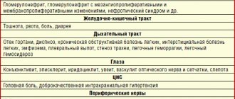
Clinical picture • Skin lesions •• Itching, often severe •• Flat, polygonal, brownish-bluish papules with a shiny surface, 1–10 mm in diameter. The rashes can merge, forming shagreen-like plaques covered with tightly packed scales. On the surface of some papules a peculiar mesh pattern is noticeable - the Wickham mesh, caused by uneven thickening of the granular layer of the epidermis (pathognomonic sign) •• Localization - the rashes are usually located on the flexor surface of the forearms, the anterior surface of the legs, the external genitalia; less often - on the dorsum of the feet, in the inguinal and sacral areas. There have been cases of generalization of dermatosis with the development of secondary erythroderma (erythematous form) •• Characterized by an isomorphic reaction at the sites of scratching (Köbner phenomenon) •• Depending on the clinical picture, the following forms are distinguished ••• Pigmented - flat papules are barely noticeable against the background of diffuse melasma ••• Erythematous - large itchy, swollen erythematous spots and papules, difficult to distinguish until the intensity of the erythema decreases; accompanied by general intoxication ••• Blistering (bullous) - blisters form against the background of papular rashes ••• Hypertrophic (warty) - large purple papules, often covered with horny layers, usually located on the anterior surface of the legs ••• Hyperkeratotic (horny) - on surfaces of papules - pronounced hyperkeratosis, localized on the legs ••• Coraloid - papules with a diameter of up to 1 cm in the form of beads of reddish-bluish color, alternating with areas of hyperpigmentation; usually located in the abdomen and neck ••• Atrophic - skin atrophy occurs in place of the papules ••• Dull (flattened) - large hemispherical, smooth, dense, low-itching papules are located on the legs, lower back and buttocks •• Depending on the location of the elements • •• Scattered single rashes ••• Linear rashes (lichen planus linear) - papules are located linearly, sometimes along the course of a nerve or along scratches ••• Ring-shaped rashes (lichen planus annulare) - papules are located in rings growing eccentrically, usually located on the trunk and mucous membranes. • Damage to the mucous membranes - in 40-60% of patients with skin lesions. In 20%, only the mucous membranes are affected. This condition is often considered as precancerous (the development of carcinoma is possible) •• The rash is painful, especially with ulceration •• Papular, often ring-shaped rashes of a whitish-pearly color in the form of a mesh, lace pattern, usually on the mucous membrane of the cheeks, less often - on the tongue, gums, the palate, on the red border of the lips - a continuous scaly stripe •• Depending on the clinical picture ••• The vesicular (bullous) form develops quite often ••• The erosive-ulcerative form with multiple erosions on the mucous membrane of the mouth and lips - the most severe form, may combined with diabetes and essential arterial hypertension (Grinshpan-Wilapol syndrome). • Damage to hair and nails •• Scalp ••• Lichen planus pilaris - rash of small papules around the mouths of follicles on the scalp ••• Skin atrophy and destruction of hair follicles, as a result of which persistent total alopecia may develop •• Nails - proximal formation - distal grooves and partial or complete destruction of the nail bed. The big toes are most often affected.
Research methods • Wickham's grid is better visible after local application of inorganic oils • Skin biopsy - inflammation with hyperkeratosis, thickening of the granular layer, uneven acanthosis, vacuolization of cells of the basal layer, hyaline bodies under the epidermis, heavy lymphocytic infiltration of the upper layers of the dermis • Serological tests to exclude syphilis.
Differential diagnosis • Secondary papular syphilides • Psoriasis • Atopic dermatitis • Leukoplakia (if localized on the mucous membrane) • Squamous cell and basal cell skin cancer • SLE • Erythema multiforme exudative • Exposure to chemicals • Rash due to drug allergies.
TREATMENT Management tactics • Clinical observation, especially of patients with the hypertrophic form of lichen planus, periodic examination of the skin • Timely detection and treatment of somatic diseases, functional changes in the nervous system, sanitation of the oral cavity, rational dental prosthetics (dental crowns must be made of the same metal) • PUVA therapy, heliocadmium laser therapy, inductothermy of the lumbar region (especially for generalized or intractable lesions). PUVA therapy is carried out periodically for 1–2 years • CBC, liver function tests, ANAT titer determination are carried out every six months • Anti-relapse therapy - repeated courses of vitamins, spa treatment • Due to the possibility of malignancy of ulcerative-erosive forms on the mucous membranes Regular monitoring by an oncologist is necessary.
General recommendations • A regimen with long sleep, rational employment with the exception of occupational factors that irritate the skin • Correction of the reaction to stress • For lichen planus of the oral mucosa, the exception of hot, hot, spicy and rough foods.
Drug therapy • For all forms of the disease •• Sedatives •• Penicillin preparations •• Vitamins (nicotinic acid, thiamine, pyridoxine, retinol) •• Histoglobulin 2 ml 2 times/week subcutaneously, per course - 8-10 injections • For skin lesions •• Locally - HA ointments (for example, triamcinolone 0.1%) •• Antihistamines (for example, diphenhydramine 25 mg every 6 hours) - to reduce itching • For lesions of the mucous membranes •• Retinol topically, tigazon 30 –75 mg/day orally (contraindicated during pregnancy) •• GC orally • For erosive-ulcerative and bullous forms - combined treatment •• GC (for example, prednisolone 20-25 mg every other day for 2-4 weeks) •• Chloroquine 0.25 g 1–2 times a day for 4–6 weeks •• Xanthinol nicotinate 150 mg 3 times a day •• Erosion of the oral mucosa is lubricated with a paste containing solcoseryl.
Complications • Malignant degeneration of long-existing hypertrophic, ulcerative-erosive lesions on the mucous membranes (1% of cases) • Alopecia • Nail destruction.
Prognosis • The disease has a benign, but long-term course, especially erosive-ulcerative forms • An individual therapeutic approach to the patient, taking into account pathogenetic factors, contributes to positive treatment results • Spontaneous resolution is possible within several weeks • A tendency to relapse is noted, especially against the background of emotional stress. A recurrent course is observed in 20% of cases, mainly in generalized forms. Prevention. Elimination of chemical factors and drugs that provoke lichenoid reactions, thorough sanitation of the oral cavity, reduction of stress.
ICD-10 • L43 Lichen planus
Proven methods for treating lichen ruber
Therapeutic methods are the main method of treatment and depend on the stage of the disease. At the same time, an integrated approach is being implemented aimed at blocking both the causes themselves and the unpleasant consequences of deprivation:
- impaired functioning of the immune system, which is the cause of the manifestation of lichen, is leveled with the help of immunosuppressants;
- the main means of treating lichen planus are hormonal drugs that reduce the supply of components to the foci of immune inflammation that cause the development of ulcers;
- an integral stage of recovery procedures will be the administration of anti-allergenic drugs and vitamin A, which promotes the effective healing of eroded layers of the skin or mucous membrane;
- in severe forms of the disease, antibiotics and synthetic interferons and interferonogens are prescribed in the treatment of lichen ruber.
In addition to taking medications, their analogues in the form of ointments are used, as well as physiotherapeutic procedures (UV and laser irradiation, magnetic therapy). As part of an integrated approach, folk remedies based on natural substances are effective.
Our center’s specialists will be able to determine the optimal and effective treatment. They have the appropriate equipment and sufficient experience to make a detailed diagnosis of the factors causing lichen planus.
Causes of the disease
Lichen planus occurs under the influence of a number of factors, among which the most common is immuno-allergic. When the body's immunity decreases under the influence of external and internal factors, T-lymphocytes cannot cope with their protective function.
Factors that provoke the development of oral lichen planus include:
- Chronic stress;
- Strong shocks;
- Diseases of the gastrointestinal tract;
- Diabetes;
- Hypertension;
- Grynszpan syndrome;
- Injuries to the oral mucosa;
- Incorrectly installed dentures;
- Fillings that injure the oral mucosa;
- Prostheses made of dissimilar materials;
- Long-term use of tetracycline;
The risk group also includes patients whose work involves developing camera film or who come into contact with paraphenyldiamine.
Factors causing the disease
This disease is chronic and can be transmitted at the genetic level. However, among the reasons for its appearance or exacerbation, doctors name several factors:
- weakening of the immune system and increased infections;
- exposure to third-party chemicals (medicines, household and industrial preparations);
- the body’s own synthesis of dangerous components due to stressful situations;
- existing diseases (allergies, gastrointestinal disorders, damage to the mucous membrane, diabetes).
Accurate identification of the most likely causes of the disease subsequently determines the most effective method of treating lichen ruber.
General notes on therapy
- The choice of treatment method depends on the severity and localization of clinical manifestations, the form and duration of the disease, information about the effectiveness of previously administered therapy.
- Treatment is not required for damage to the oral mucosa that is limited to reticular rashes of a typical form, not accompanied by subjective sensations. In other cases, patients require therapy.
- During the period of exacerbation of the disease, patients are recommended to take a gentle regimen with limited physical and psycho-emotional stress. In the food regime, salted, smoked, fried foods should be limited. In patients with damage to the oral mucosa, it is necessary to exclude irritating and rough foods.
treatment goals
- regression of rashes;
- improving the quality of life of patients.
indications for hospitalization
- ineffectiveness of outpatient treatment;
- widespread and severe lesions of the skin and mucous membranes, including hyperkeratotic, bullous, erosive and ulcerative.
External therapy
In the presence of limited rashes, treatment begins with the prescription of topical glucocorticosteroid drugs of medium and high activity (possibly alternating them):
- betamethasone, cream, ointment 2 times a day externally to the lesions for 12 weeks, or
- clobetasol, cream, ointment 2 times a day externally to the lesions for 4-8 weeks, or
- fluocinolone acetonide, cream, gel, ointment 2 times a day externally to the lesions for 4-8 weeks, or
- hydrocortisone-17 butyrate, cream, ointment 2 times a day externally to the lesions for 4-8 weeks, or
- triamcinolone ointment 2 times a day externally to the lesions for 4-8 weeks, or
- mometasone, cream, ointment, lotion 1-2 times a day externally to the lesions for 4-8 weeks, or
- betamethasone + salicylic acid, ointment 2 times a day externally to the affected areas for 4-8 weeks, or
- salicylic acid + flumethasone, ointment 2 times a day externally on the affected areas for 4-8 weeks.
Alternative Treatments
- tacrolimus ointment 2 times a day for 2 weeks
- calcipotriol ointment 2 times a day for 2 weeks
Systemic therapy
1. Systemic glucocorticosteroid drugs:
- prednisolone 20-30 mg per day orally for 1-2 months followed by gradual withdrawal or
- betamethasone 1 ml once every 2-3 weeks intramuscularly or intralesional, for a course of 3-4 injections.
2. In the treatment of patients with lichen planus, antimalarial drugs can be used, which are used as systemic therapy and can be prescribed with glucocorticosteroid drugs:
- Hydroxychloroquine 200 mg orally 2 times a day for 5 days, then a break of 2 days, treatment courses are repeated for 1-2 months or
- chloroquine 250 mg orally 2 times a day for 5 days, then break for 2 days, courses of treatment are repeated for 1-2 months.
3. To relieve itching, one of the antihistamines is prescribed
1st generation, which is used both orally and in injection forms:
- mebhydrolin 100 mg orally 2-3 times a day for 7-10 days or
- clemastine 1 mg orally or intramuscularly 2-3 times a day for 7-10 days.
Also, in order to reduce itching, an antipsychotic with non-blocking activity can be prescribed: hydroxyzine 25-100 mg per day orally for 28 days.
Alternative Treatments
- Metronidazole 500 mg 2 times a day for 10 days
Non-drug treatment
1. For minor infiltration of the lesions, narrow-band medium-wave phototherapy with a wavelength of 311 nm is prescribed 3-4 times a week for 6-12 weeks.
2. For patients with more pronounced infiltration in the lesions, PUVA therapy with oral or external use of a photosensitizer is indicated:
- PUVA therapy using oral photosensitizers: methoxsalen 0.6 mg per kg body weight or
- PUVA therapy with external use of photosensitizers: methoxalen 0.5-1 mg/l for a course of 8 to 23 procedures.
Treatment of lichen planus of the oral mucosa
1. First-line drugs for the treatment of patients with LP of the oral mucosa are topical glucocorticosteroid drugs:
- betamethasone, cream, ointment 2 times a day externally to the lesions for 8 weeks, or
- triamcinolone ointment 3 times a day externally to the lesions for 12 weeks, or
- fluocinolone acetonide, cream, gel, ointment 2 times a day externally to the lesions for 4-12 weeks, or
- clobetasol, cream, ointment 2 times a day externally to the lesions for 4-8 weeks.
2. In case of ineffectiveness of topical corticosteroid drugs, retinoids are prescribed for external use:
- isotretinoin gel 2 times a day externally to the lesions for 8 weeks.
3. Additionally, painkillers and wound healing agents are used:
- aloe vera leaves, liniment 2 times a day externally on the lesions;
- lidocaine + chamomile flower extract, gel: a strip 0.5 cm long is applied to painful or inflamed areas of the oral mucosa and rubbed in with light massaging movements 3 times a day;
- choline salicylate + cetalkonium chloride, dental gel 1 cm for adults and 0.5 cm for children, squeezed onto a clean finger and rubbed with light massaging movements into the affected area of the oral mucosa 2-3 times a day before or after meals and before bed.
4. In the case of severe lichen planus of the oral mucosa, resistant to therapy, systemic glucocorticosteroid drugs are used: prednisolone 0.5-1 mg per kg of body weight for 3 weeks.
For the treatment of children
topical glucocorticosteroid drugs are used.
Tactics in the absence of treatment effect
If the therapy is ineffective, patients may be prescribed acitretin or cyclosporine:
- acitretin 30 mg per day orally for 3-8 weeks or
- isotretinoin 0.5 mg/kg/day for 8 weeks
- cyclosporine 5 mg per kg body weight per day orally for 3-8 weeks.
Due to the possibility of developing undesirable effects during retinoid therapy (changes in the level of transaminases, hepatitis, hypertriglyceridemia, hypercholesterolemia, hyperglycemia, etc.), it is necessary to monitor the level of lipids, blood glucose, and liver function. Due to the teratogenic properties of retinoids, women of reproductive age should use reliable contraception 4 weeks before, during and for 2 years after the end of acitretin therapy. If pregnancy occurs, it should be terminated for medical reasons.
During treatment with cyclosporine, regular monitoring of plasma creatinine concentration is necessary - an increase may indicate a nephrotoxic effect of the drug and requires a dose reduction: by 25% if creatinine increases by more than 30% from the original and by 50% if its level doubles; When dose reduction within 4 weeks does not lead to a decrease in creatinine, cyclosporine is discontinued. It is recommended to monitor blood pressure, blood levels of potassium, uric acid, bilirubin, transaminases, and lipid profile. During the treatment period, immunization with live attenuated vaccines is contraindicated.
Treatment
For different forms of the disease, a different course of medications is prescribed. If the disease is asymptomatic, drug treatment may not be necessary. If erosions and ulcers have already formed, it is necessary to begin treatment as quickly as possible to avoid worsening the condition.
Inflammatory processes are controlled by the use of corticosteroids. Local treatment is carried out to relieve pain and accelerate tissue regeneration. To avoid the development of candidiasis, antifungal drugs are prescribed.
Electrophoresis and phonophoresis are also effective. In rare cases, when erosions do not heal for a long time, surgical intervention is indicated.
In all cases, without exception, the oral cavity is sanitized and the source of inflammation is removed (for example, periodontitis, caries or pulpitis).
Genital lesions in LP
Therapy for genital lesions with LP is fundamentally similar to treatment tactics for lesions of the oral mucosa. The main goal of therapy is to prevent or minimize the formation of scarring, synechiae, vaginal stenosis in women and phimosis in men. TGCS are used as the first line of therapy, while standard antipruritic drugs are used to relieve itching. Initially, an intensive regimen is used, usually clobetasol 2 times a day for 1-2 months to limit the inflammatory process, followed by a transition to a maintenance regimen - using the drug 2-3 times a week. The above treatment tactics are appropriate in the treatment of lichen sclerosus, and their effectiveness has been confirmed in a number of clinical case reports. To relieve friction and pain, it is possible to use intravaginal applications of emollients. If the anal area is affected, topical corticosteroids in the form of foam or suppositories can be used. In case of disease refractory to therapy, as well as severe damage to the genitals, systemic oral corticosteroids can be used in a course with a gradually decreasing dosage and transition to topical maintenance therapy. Topical retinoids have a pronounced irritant effect and are usually poorly tolerated in the erosive form of the disease. There are a number of retrospective studies and case reports with successful use of topical calcineurin inhibitors. Despite a burning sensation when applied to erosive lesions, topical calcineurin inhibitors are better tolerated than topical retinoids and are widely used in the treatment of LP affecting the genitals.
Follicular form of LP
The goal of therapy for the follicular form of LP (lichen planopilaris) and its variants is to stop the inflammatory process as early as possible to minimize the death of epithelial stem cells of the hair follicle through inflammation-induced apoptosis, as well as control of concomitant symptoms of the disease until the spontaneous occurrence of clinical remission. Strong and very strong topical corticosteroids are used as the first line of therapy in clinical practice. Intralesional administration of corticosteroids is practiced by many specialists, despite the lack of sufficient data in favor of this technique, as well as the risk of developing scalp atrophy with prolonged use of drugs. Many doctors prefer twice daily use of strong and very strong topical corticosteroids for 6-8 weeks, with a gradual transition to use 3 times a week or as required. With an aggressive, rapidly progressive course of the disease, systemic corticosteroids (30-80 mg/day in terms of prednisolone) are often prescribed, and in some cases, systemic cyclosporine (3-10 mg/kg/day), although there is insufficient data on the effectiveness of drugs in these groups. In the presence of pronounced inflammatory changes, in addition to topical therapy, a course of prednisolone with a gradual dose reduction (starting from 40 mg/day) may be prescribed, and hydroxychloroquine may be prescribed simultaneously. It is believed that the latter helps to reduce the immune response to self-antigens and also modulates T-cell-mediated gene expression. Its immunomodulatory effects as well as its good safety profile may be useful in the treatment of follicular LP, making hydroxychloroquine a preferred treatment option for lichen planopilaris. In case of intolerance to hydroxychloroquine or if there are contraindications to its use, tetracycline antibiotics, which are second-line drugs, can be used. Also, second-line drugs include mycophenolate mofetil and cyclosporine.
Nail damage
Despite significant functional and cosmetic impairments associated with nail lesions in LP, evidence-based data are currently insufficient. The goal of therapy is to prevent or minimize permanent scarring (as with the follicular form of LP), and it is also important to stop the inflammatory process at the earliest possible stage to achieve optimal results. Topical and systemic corticosteroids are used as the first line of therapy, as well as their intralesional administration; systemic use of drugs is indicated when several nail plates are affected. According to the literature, the use of alitretinoin appears to be effective. For many dermatologists, including the authors of the article, the use of systemic corticosteroids (in the form of pulse therapy or a short course with a gradual dose reduction) is preferable to intralesional administration of corticosteroids, due to the convenience and effectiveness of this technique. Also, there is evidence of an increase in the effectiveness of topical therapy with strong and very strong topical corticosteroids when applied under occlusion.
Traditional Treatments
Key Points
- Traditional medications used for LP include topical and systemic corticosteroids, phototherapy, retinoids, topical calcineurin inhibitors, and systemic immunosuppressive drugs.
- Lesions of the mucous membrane of the oral cavity, genitals, and the follicular form of LP do not tend to spontaneously resolve and often require systemic therapy.
- To date, an insufficient number of clinical studies have been conducted on the treatment of LP. Lack of data does not always indicate the lack of effectiveness of a treatment method.
LP is an important and understudied clinical problem. Thus, the evidence base for the large number of treatment options widely used in clinical practice remains inadequate. Next, the basics of pharmacology and the evidence base for each drug used in routine practice will be discussed, separately for the most common variants of the disease in clinical practice. A brief overview of traditionally used drugs, their pharmacodynamics and level of evidence is given in Tables 1-5.
Cutaneous form of LP
Topical and systemic corticosteroids
The goal of therapy for the classic self-limited cutaneous form of LP is to accelerate the process of resolution of the lesion, as well as symptomatic relief of itching. The standard first-line therapy is topical corticosteroids, and the choice of drug in most cases is based on the clinical experience of the physician rather than on clinical trial data. Commonly used drugs include clobetasol propionate, fluocinolone acetonide, betamethasone dipropionate, triamcinolone acetonide (can also be used as intralesional injections in cases of hypertrophic LP that are refractory to treatment). The authors recommend the use of ointment bases, as they have more pronounced moisturizing properties, as well as a less complex composition compared to creams. TGCS applications are used for lesions up to 15% of the body area; for more extensive lesions, in some cases it is preferable to use systemic drugs. Systemic corticosteroids, both for oral and intramuscular use, can be used for forms of LP that are resistant to topical corticosteroids. The most commonly used is prednisolone at a dose of 30-60 mg/day; the duration of therapy depends on the severity and refractoriness of the disease. A placebo-controlled randomized clinical trial showed that patients refractory to topical corticosteroids experienced significant improvement with a 10-day course of systemic prednisolone at a dose of 30 mg/day. For severe cases of LP, the course duration is up to 4-6 weeks. It is necessary to remember the side effects of systemic corticosteroids, especially when prescribing drugs of this group for a long period. Long-term monitoring of blood pressure and urine glucosuria levels is necessary. Repeated courses of systemic corticosteroids should be avoided whenever possible. A thorough examination of the patient is mandatory to detect symptoms of long-term use and toxicity of systemic corticosteroids using appropriate diagnostic methods. The mechanism of action of systemic GCS is rather nonspecific, based on the induction of immunosuppression and an anti-inflammatory effect, realized through the regulation of the action of proinflammatory mediators at the genetic, cytokine and cellular level. The lack of specificity of pharmacological action leads to a number of side effects, so further research is needed to find a targeted drug.
Phototherapy, photochemotherapy
Phototherapy and photochemotherapy have been used for many years in the treatment of inflammatory dermatoses, including LP, due to their complex immunosuppressive effects. Recent randomized controlled trials show the superiority of phototherapy over treatment with systemic corticosteroids; In a study of patients with TGCS-refractory disease, a 6-week course of narrowband UV-B phototherapy was associated with a complete response in 52% of patients, while a response was observed in 13% of patients with systemic prednisolone therapy for the same duration (however, prednisolone was used in a dose less than the standard one). According to a retrospective cohort study, narrowband UVB phototherapy and PUVA (psoralen+UVA phototherapy) have comparable clinical efficacy in the classic cutaneous form of LP, however, it is preferable to use narrowband UVB phototherapy, since the method is simpler and has a more acceptable safety profile.
Retinoids
Retinoids have been used long-term to successfully treat cutaneous LP, with acitretin being the most commonly prescribed drug. It is believed that the pharmacological effects of acitretin are realized through the activation of retinoic acid receptors, which control the maturation of the epidermis and the inflammatory response in the skin. A double-blind, placebo-controlled study of patients with cutaneous LP showed that an 8-week course of acitretin at a dose of 30 mg/day is associated with long-term remission of the disease. However, a higher incidence of side effects has been reported in patients treated with acitretin, which explains the lack of widespread use of acitretin and other systemic retinoids in patients with cutaneous LP. Other studies have shown the clinical effectiveness of other systemic retinoids - etretinate, isotretionin, tretinoin, however, the indications for the use of drugs in routine practice are limited.
Damage to the oral mucosa in LLP
Topical and systemic corticosteroids
Topical corticosteroids are first-line drugs for the treatment of localized forms of LP of the oral mucosa. Strong and very strong drugs - clobetasol, triamcinolone, betamethasone, fluocytonide, fluticasone - have proven effectiveness and safety. These drugs are usually applied 2 times a day, either undiluted (eg clobetasol propionate 0.05%) or mixed with Orabase (eg triamcinolone) for 1-2 months, followed by maintenance use. It is also possible to use GCS in the form of intralesional injections, but there is insufficient data to support the advantage of this method over others (there are only isolated clinical cases). Systemic therapy using oral corticosteroids may be indicated in case of significant severity of the disease, in case of refractory to external therapy, however, the evidence base for this approach is insufficient; adequate placebo-controlled studies have not been conducted. Taking into account the wide range of side effects associated with the use of systemic corticosteroids, this group of drugs is used in the form of a short course with subsequent transition to topical therapy.
Topical calcineurin inhibitors
Interest in the possibility of using topical calcineurin inhibitors was attracted primarily by their anti-inflammatory effect, which is different from tGCs. The mechanism of action of the drugs is realized by suppressing the activity of T cells at the level of transcription factors. Published studies examining the effectiveness of this group of drugs are very contradictory - a meta-analysis performed in 2012, evaluating 3 randomized controlled trials, showed that the effectiveness of pimecrolimus was no greater than placebo. Subsequent studies were more encouraging - according to the data obtained, the effectiveness of topical calcineurin inhibitors was comparable to the effect of tGCs. A 2014 study found that pimecrolimus was slightly more effective than tacrolimus, while more recent studies found that tacrolimus and pimecrolimus were equivalent. These data were confirmed in an open randomized controlled trial (2017), a pilot randomized controlled trial, and a subsequent meta-analysis. Many physicians have found tacrolimus to be more effective in their clinical practice. Side effects of topical calcineurin inhibitors include local irritation and burning, especially on eroded lesions (which limits their use in the case of the erosive form of LP), but in general their safety profile is not inferior to long-term use of corticosteroids. The absence of side effects of TGCS determines increasing interest in the use of these drugs in clinical practice.
Topical and systemic retinoids
Retinoids have certain immunomodulatory effects due to their direct effects on T cells through nuclear retinoic acid receptors. Topical retinoids are an alternative to corticosteroids for non-erosive LP and are recommended as a second-line drug according to World Workshop in Oral Medicine IV. Despite the proven effectiveness of topical tretinoin and isotretinoin 0.1% (in placebo-controlled studies), one study showed lower effectiveness compared to tGCs. Side effects include local irritation, photosensitivity, and teratogenicity. The possibility of disease relapse within 2-5 weeks after stopping use of the drug significantly limits the widespread use of topical retinoids in everyday practice. The use of systemic retinoids (etretinate, isotretinoin) is limited due to an insufficient risk-benefit ratio, however, the use of alitretinoin may be more successful.


![Rice. 2. Relationship between dose of diclofenac (D), naproxen (Nap) and etoricoxib (Eto) and effect size in osteoarthritis [3]](https://irknotary.ru/wp-content/uploads/ris-2-zavisimost-mezhdu-dozoj-diklofenaka-d-naproksena-nap-i-330x140.jpg)