It branches like a tree, first into large branches (trunks), then into smaller branches and twigs, and is conventionally divided into several parts or sections:
- 1. The ascending aorta is the area from the aortic valve to the brachiocephalic trunk.
- 2. The aortic arch is a short section from which all the vessels supplying the arms and head (brachiocephalic arteries) depart. They anatomically form an arch connecting the ascending and descending aorta.
- 3. The descending (thoracic) aorta begins from the mouth of the left subclavian artery and continues to the diaphragm.
- 4. Below the diaphragm and before the bifurcation of the aorta (bifurcation) is the abdominal aorta.
Dividing the aorta into sections is very important for assessing risk and choosing optimal treatment tactics in patients with aortic aneurysms.
An aortic aneurysm is an area of local expansion.
What is the aorta
The aorta is the largest vessel in the human body, which carries blood from the heart to the organs and limbs. The upper section of the aorta runs inside the chest, this section is called the thoracic aorta . The lower part is in the abdominal cavity and is called the abdominal aorta . It delivers blood to the lower part of the body. In the lower abdomen, the abdominal aorta divides into two large vessels - the iliac arteries , which carry blood to the lower extremities.
The aortic wall consists of three layers: internal (intima), middle (media), external (adventitia).
Abdominal aortic aneurysm
Abdominal aortic aneurysm is a chronic degenerative disease with life-threatening complications. An abdominal aortic aneurysm is defined as an increase in its diameter by more than 50% compared to the norm or a local bulging of its wall. Under the pressure of blood flowing through this vessel, the dilatation or bulging of the aorta may progress. The diameter of a normal aorta in the abdominal region is approximately 2 cm. However, at the site of an aneurysm, the aorta can be dilated to 7 cm or more.
Why is an aortic aneurysm dangerous?
An aortic aneurysm poses a major health risk as it can rupture. A ruptured aneurysm can cause massive internal bleeding, which in turn leads to shock or death.
An abdominal aortic aneurysm can cause other serious health problems. Blood clots (thrombi) often form in the aneurysm sac or parts of the aneurysm tear off, which move with the blood flow along the branches of the aorta to the internal organs and limbs. If one of the blood vessels becomes blocked, it can cause severe pain and lead to organ death or loss of a lower limb. Fortunately, if an aortic aneurysm is diagnosed early, treatment can be timely, safe and effective.
The human arterial system is characterized by pronounced individual structural features. Often there are atypical variants of the origin of arteries from the main trunk, different forms of branching and topography of vessels, an unequal number of vascular routes and sources of blood supply to organs [1]. In connection with the development of cardiac surgery, angiosurgery, including high-tech interventions, the problem of variant anatomy of the vascular system is of particular relevance [2].
Along with the typical structure of the branches of the aortic arch, various options for their origin, number and topography are described in the literature [2-6]. Thus, C. Bhattarai et al. [5], P. Durai et al. [7], B. Junagade et al. [8] describe the origin of two branches from the aortic arch (the left subclavian artery and the common arterial trunk, which divides into the brachiocephalic trunk and the left common carotid artery), as well as four branches (the brachiocephalic trunk, the left common carotid artery, the left vertebral artery and the left subclavian artery ). P. Rekha et al. [6] established the origin of the right and left brachiocephalic trunks from the aortic arch. Abnormally branching arteries can be compressed by neighboring organs and become deformed, which leads to disruption of the blood supply to organs and tissues. The possibility of atypical origin of the branches of the aortic arch is important to consider when performing surgical interventions, since in this case the probability of their damage is quite high [3, 4, 6, 8].
The purpose of the study is to study the variant anatomy of the branches of the aortic arch.
The study was conducted on anatomical preparations of the heart and aorta of adults using the dissection method.
In typical cases, 3 arteries depart from the aortic arch from right to left: the brachiocephalic trunk, the left common carotid artery and the left subclavian artery (Fig. 1).
Rice. 1. Typical structure of the branches of the aortic arch. 1 - aortic arch; 2 - brachiocephalic trunk; 3 - left common carotid artery; 4 - left subclavian artery.
Along with the typical origin of the branches of the aortic arch, we found 2 atypical variants of the structure of the branches of the aortic arch. In our report we provide a description of these cases.
In the first case, 4 branches departed from the convex surface of the aortic arch from right to left in the following order: brachiocephalic trunk, left external carotid artery, left internal carotid artery and left subclavian artery (Fig. 2).
Rice. 2. Variant of the structure of the branches of the aortic arch. 1 - aortic arch; 2 - brachiocephalic trunk; 3 - left external carotid artery; 4 - left internal carotid artery; 5 - left subclavian artery.
In the second case, two branches departed from the convex surface of the aortic arch from right to left in the following order: the brachiocephalic trunk and the left subclavian artery. The left common carotid artery began atypically from the brachiocephalic trunk (Fig. 3).
Rice. 3. Variant of the structure of the branches of the aortic arch. 1 - aortic arch; 2 - brachiocephalic trunk; 3 - left common carotid artery; 4 - left subclavian artery.
Along with the typical variant of the origin of the branches of the aortic arch, there are various atypical variants. We found cases of origin of four branches (brachiocephalic trunk, left external carotid artery, left internal carotid artery, left subclavian artery) and two branches (brachiocephalic trunk and left subclavian artery). The discovered variants of the origin of the branches of the aortic arch are of great clinical importance in cardiac surgery and angiosurgical practice.
The authors declare no conflict
of interest
.
Information about authors
Plotnikova Nadezhda Alekseevna
- Doctor of Medical Sciences, Prof., Head. department normal and pathological anatomy with a course of forensic medicine FSBEI HE "Mordovia State University named after. N.P. Ogareva"; 430032, Saransk, st. Ulyanova, 26;
Rybakov Alexey Gennadievich
- Candidate of Medical Sciences, Associate Professor of the department. normal and pathological anatomy with a course of forensic medicine FSBEI HE "Mordovia State University named after. N.P. Ogareva"; e-mail
Loshkarev Igor Alexandrovich
- Candidate of Medical Sciences, Associate Professor of the department. normal and pathological anatomy with a course of forensic medicine FSBEI HE "Mordovia State University named after. N.P. Ogareva"; e-mail
Machinsky Petr Alexandrovich
- Candidate of Medical Sciences, Associate Professor of the department. normal and pathological anatomy with a course of forensic medicine FSBEI HE "Mordovia State University named after. N.P. Ogareva"; e-mail
Types of aortic aneurysms
There are “true” and “false” aortic aneurysms. A true aneurysm develops due to the gradual weakening of all layers of the aortic wall. A false aneurysm is usually the result of trauma. It is formed from the connective tissue surrounding the aorta. The cavity of the false aneurysm is filled with blood through a crack in the aortic wall. The aortic walls themselves do not participate in the formation of an aneurysm.
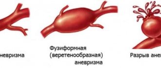
Depending on the form there are:
- saccular aneurysm - expansion of the aortic cavity on only one side;
- spindle-shaped (fusiform) aneurysm - expansion of the aneurysm cavity on all sides;
- mixed aneurysm - a combination of saccular and fusiform forms.
Causes and risk factors for the development of abdominal aortic aneurysm
The causes of the development of abdominal aortic aneurysms are very diverse. The most common cause of aneurysm development is atherosclerosis. Atherosclerotic aneurysms account for 96% of the total number of all aneurysms. In addition, the disease can be either congenital (fibromuscular dysplasia, Erdheim's cystic medianecrosis, Marfan syndrome, etc.) or acquired (inflammatory and non-inflammatory). Inflammation of the aorta occurs when various microorganisms invade (syphilis, tuberculosis, salmonellosis, etc.) or as a result of an allergic-inflammatory process (nonspecific aortoarteritis). Non-inflammatory aneurysms most often develop with atherosclerotic lesions of the aorta. Less commonly, they are the result of injury to its wall.
Risk factors for developing an aneurysm
- Arterial hypertension;
- Smoking;
- The presence of aneurysms in other family members. Which indicates the role of hereditary factors in the development of this disease;
- Gender: men over 60 years of age (abdominal aortic aneurysms occur less frequently in women).
What it is
We should start by defining the aorta. The aorta is the largest vessel in our body, and accordingly its importance is high. An aneurysm is an enlargement of a blood vessel. An aortic aneurysm can form in the thoracic or abdominal regions. Moreover, aneurysm of the abdominal aorta is much more common. This condition of the vessel may not cause any harm for a long time, however, an aneurysm is very dangerous due to the unpredictable course of expansion and the threat of rupture of the vessel walls when they become thinner - and this condition already poses a threat to life due to severe internal bleeding. In addition, blood clots can form at the site of vessel expansion due to changes in blood flow - the danger of this condition is the risk of the blood clot breaking off and clogging a smaller vessel.
Symptoms and signs of abdominal aortic aneurysm
In most patients, abdominal aortic aneurysms occur without any symptoms and are an incidental finding during examinations and operations for other reasons.
When signs of an aneurysm develop, the patient experiences one or more of the following symptoms:
- A feeling of pulsation in the abdomen, similar to a heartbeat, an unpleasant feeling of heaviness or fullness.
- Dull, aching pain in the abdomen, in the navel area, usually on the left.
Indirect signs of abdominal aortic aneurysm are important :
- Abdominal syndrome. Manifested by the appearance of belching, vomiting, unstable stool or constipation, lack of appetite and weight loss;
- Ischioradic syndrome. Manifested by lower back pain, sensory disturbances and movement disorders in the lower extremities;
- Syndrome of chronic ischemia of the lower extremities. Manifests itself in the appearance of pain in the muscles of the lower extremities when walking, sometimes at rest, coldness of the skin of the lower extremities;
- Urological syndrome. It manifests itself as pain and heaviness in the lower back, difficulty urinating, and the appearance of blood in the urine.
Harbingers of rupture may be increased abdominal pain.
When an aneurysm ruptures, the patient suddenly feels an increase or appearance of abdominal pain, sometimes “radiating” to the lower back, groin area and perineum, as well as severe weakness and dizziness. These are symptoms of massive internal bleeding. The development of such a situation is life-threatening! The patient needs emergency medical care!
Symptoms of the disease
As a rule, the disease occurs without symptoms. In most cases, an aneurysm is discovered accidentally - during a routine examination. If symptoms appear, they are expressed in the area of the aortic arch. Some of the most striking signs of the development of the disease include:
- heavy snoring;
- shortness of breath, cough;
- sharp chest pain;
- discomfort during swallowing.
In some cases, individual signs of a cardiac aortic aneurysm may appear.
Diagnosis of the disease
Diagnosis of the disease begins with the patient visiting a therapist. In this case, the specialist collects anamnesis, analyzes the existing symptoms, and then refers the patient to a highly specialized specialist. To confirm the diagnosis, additional laboratory and hardware tests are prescribed:
- clinical, general urine and blood tests: allow you to determine the presence of pathologies that affect the further development of the disease;
- echocardiography: helps determine the type, shape, size of the aneurysm;
- X-ray: shows enlarged heart, pulmonary edema.
In some cases, MRI of the heart vessels is prescribed to obtain important data.
Make an appointment Make an appointment and get a high-quality examination of the heart and blood vessels in our center
Prevention
When diagnosing this disease, it is important to resort to effective preventive measures. These include the following activities:
- maintaining a healthy lifestyle;
- refusal to drink alcohol, smoke;
- timely examination by a doctor.
In addition, the patient must form the correct diet. Meals for aortic aneurysm should be nutritious. At the same time, the menu should not include fatty foods. Sufficient physical activity is another component of prevention. If a patient has acute chest pain for more than 6 minutes, the patient should seek immediate medical attention.
Diagnosis of abdominal aortic aneurysms
Most often, abdominal aortic aneurysms are detected by ultrasound examination of the abdominal organs. Typically, the discovery of an aneurysm is an incidental finding. If a doctor suspects a patient has an aortic aneurysm, modern diagnostic methods are used to clarify the diagnosis.
Methods for diagnosing abdominal aortic aneurysm
- Computed tomography in angio mode;
- Magnetic resonance imaging in angio mode;
- X-ray contrast aorto- and angiography;
- Ultrasound duplex or triplex angioscanning of the abdominal aorta.
If necessary, the abdominal and thoracic aorta is examined.
Treatment methods for aortic aneurysm
There are several methods for treating aortic aneurysm. It is important to know the advantages and disadvantages of each of these techniques. Approaches to the treatment of abdominal aortic aneurysms:
Monitoring the patient over time
If the aneurysm is less than 4.5 cm in diameter, the patient is recommended to be monitored by a vascular surgeon, since the risk of surgery exceeds the risk of rupture of the aortic aneurysm. Such patients should undergo repeated ultrasound examinations and/or computed tomography at least once every 6 months.
When the aneurysm diameter is more than 5 cm, surgical intervention becomes preferable, since as the size of the aneurysm increases, the risk of aneurysm rupture increases.
If the size of the aneurysm increases by more than 1 cm per year, the risk of rupture increases and surgical treatment also becomes preferable.
Open surgery: aneurysm resection and aortic replacement
Surgical treatment is aimed at preventing life-threatening complications. The risk of surgery is associated with possible complications that include heart attack, stroke, limb loss, acute intestinal ischemia, male sexual dysfunction, embolization, prosthetic infection, and renal failure.
The operation is performed under general anesthesia. The essence of the operation is to remove the aneurysmal expansion and replace it with a synthetic prosthesis. The average mortality rate for open procedures is 3-5%. However, it may be higher if the renal and/or iliac arteries are involved in the aneurysm, as well as due to the patient’s concomitant pathology. Observation in the postoperative period is carried out once a year. Long-term treatment results are good.
Endovascular repair of aortic aneurysm: installation of a stent graft
Endoprosthetics of aortic aneurysm is a modern alternative to open surgery.
The operation is performed under spinal or local anesthesia through small incisions/punctures in the groin areas. Through the above approaches, catheters are inserted into the femoral artery under X-ray control. According to which, in the future, the endoprosthesis will be brought to the aneurysmal extension. An endoprosthesis or stent-graft of the abdominal aorta is a mesh frame made of a special alloy and wrapped in synthetic material. The last stage of the operation is the installation of a stent graft at the site of the aneurysmal expansion of the aorta. Eventually, the aneurysm is “switched off” from the bloodstream and the risk of rupture becomes unlikely. After aortic replacement, the patient is observed in the hospital for 2-4 days and discharged.
This technique allows to reduce the incidence of early complications, shorten the length of patient stay in the hospital and reduce the mortality rate to 1-2%. Observation in the postoperative period is carried out every 4-6 months using ultrasound techniques, CT angiography, X-ray contrast angiography. The endovascular treatment method is certainly less traumatic. About 40,000 such operations are performed annually in the United States alone.
Thus, the choice of treatment method for abdominal aortic aneurysm is based on the individual characteristics of the patient.
Treatment
As already noted, the most dangerous complication of an aortic aneurysm is the threat of its rupture and internal bleeding. If the patient did not consult a doctor and the aneurysm was not detected in a timely manner, then in the event of a rupture, the only option would be open surgery: the surgeon will remove the affected area of the aorta and install a prosthesis in its place. This method of emergency treatment of an aneurysm has contraindications; in addition, the operation is a serious intervention with associated risks and a long rehabilitation period (up to three months). Therefore, it is so important to contact a specialist if you are at risk and feel periodic pain and/or throbbing in the abdominal area.
Once an aneurysm is diagnosed, its further expansion is difficult to predict; regular monitoring by an experienced specialist, lifestyle adjustments, and control of risk factors are required. When the attending physician realizes that there is a risk of rupture, an alternative to emergency open surgery will be the planned installation of a stent graft. The installation is minimally invasive, i.e. the stent is inserted through the vessel, advanced to the site of the aneurysm and secured there. The structure of the stent resembles a vessel; it is fixed with one edge above the expansion of the aorta, and with the other below. After the operation, blood will flow through the stent, and the resulting cavity between it and the aortic wall will decrease over time. The stent graft is inserted under local anesthesia and requires only a couple of days of recovery.
Of course, any surgical intervention carries its own risks, which is why it is so important to choose a good medical center with experienced cardiovascular surgeons. Understanding the danger of aortic aneurysm as a disease, not only doctors in the cardiology department, but also in other departments of our center, when conducting routine examinations or examinations due to other diseases, monitor the patient’s condition and analyze the entire volume of data received: if a problem with the aorta is suspected, the patient within one center will be transferred for consultation with a cardiologist. For our patients, we try to organize the most convenient logic for staying in our center and consulting with specialists in order to avoid double examinations and unnecessary manipulations. Comprehensive patient management is one of the main principles of our work. And the quality of surgical and conservative care is maintained thanks to a team of experienced specialists, provided with the necessary diagnostic and treatment facilities.
You can sign up for a consultation using a special form on the website or by phone.