The main purpose of ultrasound of the abdominal aorta is to identify an aneurysm or atherosclerotic lesion of the aorta and damage to its large branches: splenic, hepatic, renal and mesenteric arteries. Ultrasound of the abdominal aorta is a non-invasive diagnostic method that has high value in identifying many vascular diseases. The results of ultrasound are very dependent on the examination technology, the quality of the equipment and the qualifications of the specialist. Today, the low price of ultrasound of the aorta and abdominal organs, the simplicity and safety of the study make it indispensable for the primary detection of abdominal aortic aneurysm.
The innovative vascular center specializes in vascular surgery, so we strive to diagnose vascular pathology at the highest level. Our center has excellent stationary and mobile ultrasound machines that allow us to study in detail the structure of the aortic wall and its branches, as well as identify aneurysms. The specialists of our medical center are dedicated to working specifically with vascular pathology, therefore they identify it much better than general ultrasound doctors.
Preparing for the study
To successfully perform an ultrasound of the abdominal cavity, preparation is necessary aimed at reducing the amount of gases in the intestines that interfere with seeing the retroperitoneal organs, including the aorta. The preparation rules for examining the abdominal aorta and its branches using ultrasound are simple:
- 2 days before the examination, exclude legumes, potatoes, cabbage, melon, dairy products, soda and all foods high in carbohydrates from the diet. These products may cause increased gas formation.
- On the eve of the ultrasound, start taking medications to improve bowel function. For these purposes, taking espumisan or regular activated carbon is suitable.
- It is advisable to skip dinner the evening before the procedure, and breakfast in the morning.
Unpaired visceral branches of the abdominal aorta
.
1
.
The celiac trunk
is a short artery (~1.5 ÷ 2.0
cm
).
It starts from the anterior semicircle of the aorta at the level of the XII thoracic vertebra. Above the superior edge of the body of the pancreas, the celiac trunk is divided into three arteries: the left gastric artery, the common hepatic artery and the splenic artery. eleven
.
The left gastric artery
goes up and to the left, towards the cardiac part of the stomach.
Next, the artery passes along the lesser curvature of the stomach (between the leaves of the lesser omentum). The esophageal branches extend from the left gastric artery to the abdominal part of the esophagus. The branches extending from the left gastric artery on the lesser curvature of the stomach run along the anterior and posterior surfaces of the organ and anastomose with the branches of the arteries following along the greater curvature. 12
.
The common hepatic artery
runs from the celiac trunk to the right and is divided into two arteries: the hepatic artery itself and the gastroduodenal artery.
The proper hepatic artery passes through the hepatoduodenal ligament to the liver. At the porta hepatis, the artery divides into right and left branches. The gallbladder artery departs from the right branch and goes to the gallbladder. The thin right gastric artery arises from the proper hepatic artery. The right gastric artery on the lesser curvature of the stomach anastomoses with the left gastric artery. The gastroduodenal artery passes behind the pylorus and divides into the right gastroepiploic and superior gastroduodenal arteries. The right gastroepiploic artery runs to the left along the greater curvature of the stomach. It anastomoses with the left artery of the same name and gives off numerous branches to the stomach and greater omentum (omental branches). The superior posterior and anterior pancreatic-duodenal arteries give branches to the duodenum (duodenal branches) and to the pancreas (pancreatic branches). 13
.
The splenic artery
is the largest of the branches of the celiac trunk.
Along the upper edge of the body of the pancreas, it goes to the spleen, giving short gastric arteries and branches to the pancreas to the bottom of the stomach - pancreatic branches. Having entered the hilum of the spleen, the splenic artery branches into vessels of smaller diameter. At the hilum of the spleen, the left gastroepiploic artery departs from the splenic artery. It goes along the greater curvature of the stomach to the right. On its way, the left gastroepiploic artery gives branches to the stomach - gastric branches and to the omentum - omental branches. The terminal branches of the left gastroepiploic artery at the greater curvature of the stomach anastomose with the right gastroepiploic artery. 2
.
The superior mesenteric artery
arises from the abdominal aorta behind the body of the pancreas at the level of the XII thoracic - I lumbar vertebra.
This artery follows in a caudal direction between the head of the pancreas and the lower part of the duodenum and enters the root of the mesentery of the small intestine. Here it is divided into a number of branches. Let's look at these branches. 2.1
.
from the superior mesenteric artery (2 cm
below its beginning).
It goes to the head of the pancreas and to the duodenum. Here, the inferior pancreatic-duodenal artery anastomoses with the superior pancreatic-duodenal arteries (branches of the gastroduodenal artery). 2.2
.
jejunal arteries
and
ileal arteries
arise from the left semicircle of the superior mesenteric .
They are directed to the loops of the mesenteric part of the small intestine. On the way to the intestinal wall, they form arcades in the mesentery - arcuate anastomoses convex towards the intestine. Anastomoses provide continuous blood flow to the intestine even when individual arteries are compressed during intestinal peristalsis. 2
.3.
The ileocolic artery courses caudally and to the right to the terminal ileum, the cecum, and the appendix. On its way, it gives off ileal branches, the anterior and posterior cecal arteries, as well as the artery of the appendix. In addition, it gives off colonic branches to the ascending colon. 2.4
.
The right colonic artery
begins somewhat proximal to the previous one, and sometimes branches off from it.
The right colic artery runs to the right towards the ascending colon. In the wall of this intestine, the right colic artery anastomoses with the colic branch of the ileocolic artery and with the branches of the middle colic artery. 2.5
.
The middle colic artery
arises from the superior mesenteric artery distal to the beginning of the right colon, follows upward and supplies blood to the transverse colon and the upper part of the ascending colon.
The middle colonic artery has two branches. The right branch of the artery anastomoses with the right colon artery. The left branch of the artery forms an anastomosis along the colon with the branches of the left colon artery (from the inferior mesenteric artery). The anastomosis is called a Riolan arc. Jean Riolan the Younger (1580-1657), French anatomist and physiologist. 3
.
The inferior mesenteric artery
arises from the left semicircle of the abdominal aorta at the level of the third lumbar vertebra.
It passes behind the peritoneum distally and to the left and gives off a number of branches to the sigmoid colon, to the descending colon and to the left part of the transverse colon. A number of branches arise from the inferior mesenteric artery. Let's look at these branches. 3.1
.
The left colic artery
passes anterior to the left testicular (ovarian) artery and the left ureter.
The artery divides into descending and ascending branches. They supply blood to the descending colon and the left transverse colon. The left colic artery anastomoses with a branch of the middle colic artery. As a result, the above-mentioned long anastomosis (riolan arch) is formed along the edge of the colon. 3.2
.
Two or three sigmoid arteries
go to the sigmoid colon.
They pass first retroperitoneally, and then in the thickness of the mesentery of this intestine. 3
.3.
The superior rectal artery
is the terminal branch of the inferior mesenteric artery. It runs distally and divides into two branches. Its first branch supplies blood to the lower parts of the sigmoid colon. This branch of the superior rectal artery forms an anastomosis with a branch of the sigmoid artery. The second branch of the superior rectal artery passes in front of the left common iliac artery and descends into the pelvic cavity. Here, in the walls of the rectal ampulla, it branches and forms anastomoses with the branches of the middle rectal arteries and with the branches of the internal iliac arteries.
Paired visceral branches of the abdominal aorta
.
4
.
The middle adrenal artery
arises from the aorta at the level of the first lumbar vertebra, just below the beginning of the superior mesenteric artery, and goes to the adrenal hilum.
On its way, this artery anastomoses with the superior adrenal arteries (from the inferior phrenic artery) and with the inferior adrenal artery (from the renal artery). 5
.
The renal artery
arises from the aorta at the level of the I-II lumbar vertebrae, ~1 ÷ 2
cm
below the origin of the superior mesenteric artery, and goes laterally to the renal hilum.
The right renal artery is slightly longer than the left artery. It passes behind the inferior vena cava. The inferior adrenal artery (to the adrenal gland) and the ureteral arterial branches (to the ureter) depart from the renal artery. In the renal parenchyma, the renal artery branches according to the internal structure of the kidney. 6
.
The testicular (ovarian) artery
is a thin, long blood vessel that arises from the aorta at an acute angle below the renal artery. Sometimes the right and left arteries arise from the aorta through a common trunk. The testicular artery passes through the inguinal canal as part of the spermatic cord to the testicle. The ovarian artery passes through the suspensory ligament and reaches the ovary. The testicular artery gives off ureteric branches and branches of the epididymis. The testicular artery anastomoses with the cremasteric artery (a branch of the epigastric artery) and with the artery of the vas deferens (a branch of the umbilical artery). The ovarian artery also gives off ureteric branches and tubal branches and anastomoses with the ovarian branch of the uterine artery. At the level of the middle of the IV lumbar vertebra, the abdominal part of the aorta divides into two common iliac arteries, forming the aortic bifurcation and the distal thin vessel - the median sacral artery. The median sacral artery passes caudally along the pelvic surface of the sacrum into the small pelvis. The branches of the abdominal aorta are connected by numerous anastomoses both among themselves and with the branches of the thoracic aorta and the branches of the iliac arteries.
Parietal (parietal) branches of the abdominal aorta
.
7
.
The inferior phrenic artery
is the first branch from the beginning of the abdominal aorta.
This paired branch departs from the aorta in the aortic opening of the diaphragm at the level of the celiac trunk from the aorta or slightly higher. On its way to the diaphragm, the artery gives off ~1 ÷ 24 superior adrenal arteries, which run caudally to the adrenal glands. 8
.
The lumbar arteries
(4 pairs) arise from the posterior semicircle of the aorta and go to the abdominal muscles. In this case, the lumbar arteries are located between the transverse and internal oblique abdominal muscles. Each lumbar artery gives off a dorsal branch to the muscles and skin of the back in the lumbar region. A spinal branch departs from the dorsal branch, penetrating through the intervertebral foramen to the spinal cord and its membranes.
Abdominal veins
Venous blood from the gastrointestinal tract and from the auxiliary organs of the digestive system flows through the largest visceral vein of the human body - the portal vein, which flows into the inferior vena cava. Inferior vena cava
- the largest vein of the circulatory system.
Unlike many smaller veins, the inferior vena cava does not have valves. The inferior vena cava is formed by the confluence of the left and right common iliac veins and is located retroperitoneally. The confluence of the common iliac veins is located slightly below the division of the aorta into arteries of the same name at the level of the intervertebral disc between the IV and V lumbar vertebrae. Initially, the inferior vena cava follows cranially along the anterior surface of the right psoas major muscle to the right of the abdominal aorta. Next, the inferior vena cava passes behind the horizontal part of the duodenum, behind the head of the pancreas and the root of the mesentery. Then the inferior vena cava passes along the hepatic groove of the same name. Here the hepatic veins flow into it. After emerging from the groove, the inferior vena cava passes through its own opening of the tendinous center of the diaphragm into the posterior mediastinum of the thoracic cavity. Here the inferior vena cava passes into the pericardial cavity, becomes covered with the epicardium and flows into the right atrium. In the abdominal cavity behind the inferior vena cava there are the right sympathetic trunk, the initial sections of the right lumbar arteries and the right renal artery. The inferior vena cava has parietal and visceral tributaries. Parietal tributaries carry blood from the walls of the abdominal cavity and from the pelvic organs. Visceral tributaries carry blood from the internal organs of the abdominal cavity. Consider the parietal tributaries of the inferior vena cava. 1
.
Lumbar veins
(~3 ÷ 4
pieces
) arise from the walls of the abdominal cavity.
The hierarchical distribution of their branches corresponds to the distribution of the lumbar arteries of the same name. Often the first and second lumbar veins flow not into the inferior vena cava, but into the azygos vein. The lumbar veins of each side anastomose with their branches - the right and left ascending lumbar veins. Blood flows from the vertebral venous plexuses into the lumbar veins through the spinal veins. 2
.
The inferior phrenic veins
(two each on the right and left) are adjacent to the arteries of the same name.
These veins flow into the inferior vena cava after its exit from the hepatic groove of the same name. Let's consider the visceral tributaries of the inferior vena cava. 3
.
The testicular (ovarian) vein
is a paired (right, left) vein that is formed at the posterior edge of the testicle (at the ovarian hilum) at the confluence of numerous veins entwining the artery of the same name.
These numerous veins form the pampiniform plexus. In men, the pampiniform plexus encircles the spermatic cord. The small veins of the plexus merge and form one venous trunk on each side. The right testicular (ovarian) vein drains at an acute angle into the inferior vena cava, slightly below the right renal vein. The left testicular (ovarian) vein flows at right angles into the left renal vein. 4
.
The renal vein
is a paired (right, left) vein that runs from the hilum of the kidney in a horizontal direction (in front of the renal artery).
At the level of the intervertebral disc between the I and II lumbar vertebrae, the renal vein flows into the inferior vena cava. The left renal vein (longer than the right) passes in front of the aorta. Both veins anastomose with the lumbar veins, as well as with the right and left ascending lumbar veins. 5
.
The adrenal vein
is a short, valveless, paired (right, left) vein that emerges from the adrenal hilum.
The left adrenal vein drains into the left renal vein, and the right adrenal vein drains into the inferior vena cava. Part of the superficial adrenal veins flows into tributaries of the inferior vena cava (into the inferior phrenic, lumbar, renal veins), and the other part into tributaries of the portal vein (pancreatic, splenic, gastric veins). 6
.
Hepatic veins
(~3 ÷ 4
pieces
) are short veins located in the liver parenchyma (they do not always have valves).
The hepatic veins drain into the inferior vena cava in the region of the hepatic sulcus. One of the hepatic veins (usually the right one), before flowing into the inferior vena cava, has a connection with the venous ligament of the liver (an overgrown venous duct that functions in the fetus). 7
.
The portal vein of the liver
is the largest vein of all the visceral veins, collecting blood from the internal organs.
Its length is ~5 ÷ 6 cm
, external diameter is ~11 ÷ 18
mm
.
The hepatic portal vein is the main afferent blood vessel of the so-called hepatic portal system
.
| Scheme. Portal vein of the liver . Modified by : Gray H., (1821–1865), Drake R., Vogl W., Mitchell A., Eds. Gray's Anatomy for Students. Churchill Livingstone, 2007, 1150 p., see: Human Anatomy: Literature. Illustrations. | |
The portal vein of the liver is located in the portal of the liver in the thickness of the hepatoduodenal ligament. The hepatic artery (in front of the portal vein), the common bile duct, nerves, lymph nodes and lymphatic vessels are also located here. The hepatic portal vein is formed by the fusion of the veins of unpaired abdominal organs. These are the veins of the stomach, pancreas, small intestine, colon, and spleen. From these organs, venous blood enters the liver through the portal vein, and from the liver through the hepatic veins into the inferior vena cava. The main tributaries of the portal vein are the superior mesenteric vein, the splenic vein, and the inferior mesenteric vein. They all merge with each other and empty into the portal vein behind the head of the pancreas. After entering the porta hepatis, the portal vein divides into two branches: the right branch (larger) and the left branch. Each of the branches of the portal vein, in turn, breaks up first into segmental branches, and then into branches of smaller and smaller diameter up to the interlobular veins. These veins pass into wide vessels - sinusoidal capillaries, located radially inside the hepatic lobules. Sinusoidal capillaries flow into the central veins of the hepatic lobules. See three diagrams in a separate window: Structure of the hepatic lobule. Sublobular veins emerge from each hepatic lobule. They sequentially merge into three or four hepatic veins. Thus, blood flowing from the liver into the inferior vena cava through the hepatic veins passes through two microhemacirculatory beds on its way. One microhemacirculatory bed is located in the walls of the digestive tract. The tributaries of the portal vein originate from it. Another microhemacirculatory bed is located in the liver parenchyma. Sublobular veins originate from it, merging into the hepatic veins. Before entering the portal of the liver (in the thickness of the hepatoduodenal ligament), the gall bladder vein from the gallbladder, the right and left gastric veins and the prepyloric vein from the corresponding parts of the stomach flow into the portal vein. The left gastric vein anastomoses with the esophageal veins - tributaries of the azygos vein flowing into the superior vena cava. In the thickness of the round ligament of the liver, the peri-umbilical veins follow to the liver. They begin in the anterior abdominal wall, in the navel area. Here they anastomose with the superior epigastric veins (tributaries of the internal thoracic veins, draining into the superior vena cava) and with the superficial and inferior epigastric veins (tributaries of the femoral and external iliac veins, draining into the inferior vena cava). In addition, three more large veins flow into the portal vein.
| Scheme. Typical arterial blood supply to the wall of the colon . Modified by : Gray H., (1821–1865), Standring S., Ed. Gray's Anatomy: The Anatomical Basis of Clinical Practice. 39th ed., Churchill Livingstone, 2008, 1600 p., see: Human Anatomy: Literature. Illustrations. | |
7
.1.
The superior mesenteric vein
is located at the root of the mesentery of the small intestine to the right of the artery of the same name.
Its tributaries are the veins of the jejunum and ileum, pancreatic veins, pancreatic-duodenal veins, ileocolic vein, right omental vein, right and middle colic veins, vein of the appendix. All these veins bring blood to the superior mesenteric vein from the walls of the jejunum and ileum and the appendix, the ascending colon and transverse colon, from the stomach, duodenum and pancreas, and the greater omentum. 7.2
.
The splenic vein
is located along the superior edge of the pancreas caudal to the splenic artery.
This vein runs from left to right, crossing the aorta in front. Posterior to the head of the pancreas, it merges with the superior mesenteric vein. The tributaries of the splenic vein are the pancreatic veins, short gastric veins and the left gastroepiploic vein. The latter anastomoses along the greater curvature of the stomach with the right vein of the same name. The splenic vein collects blood from the spleen, part of the stomach, pancreas and greater omentum. 7.3
.
The inferior mesenteric vein
is formed by the confluence of the superior rectal vein, the left colic vein, and the sigmoid veins. It is located next to the left colon artery. The inferior mesenteric vein runs upward, passes behind the pancreas and drains into the splenic vein (sometimes into the superior mesenteric vein). The inferior mesenteric vein collects blood from the walls of the upper rectum, sigmoid colon, and descending colon.
| Scheme. Arteries of the colon. . | |
| Scheme. Veins of the colon . | |
Literature. Illustrations. References. Illustrations
Click here to access the site's library! Click here and receive access to the reference library!
- Koch TR Colonic Diseases = Diseases of the colon. Humana Press, 2003, 572 p. Illustrated textbook.
Physiology, pathophysiology, medicine . . Access to this source = Access to the reference. URL: https://www.tryfonov.ru/tryfonov/serv_r.htm#0 quotation - Webber E. Basic In-Home Colon Cleansing: An Illustrated Guide = Colon Cleansing at Home. Illustrated guide. Health Management Research Institute, 2003, 134 p. Excellently illustrated study guide
. . Access to this source = Access to the reference. URL: https://www.tryfonov.ru/tryfonov/serv_r.htm#0 quotation - Messmann H., Ed. Atlas of Colonoscopy: Techniques, Diagnosis, Interventional Procedures = Atlas of Colonoscopy: technique, diagnosis, treatment procedures, Thieme Medical Publishers, 2005, 236 p. A beautifully illustrated guide
. . Access to this source = Access to the reference. URL: https://www.tryfonov.ru/tryfonov/serv_r.htm#0 quotation - Wexner SD, Stollman N., Eds. Diseases of the Colon = Diseases of the colon. Informa Healthcare, 2007, 809 p. A collection of beautifully illustrated articles.
First chapter: Embryology, structure and functions of the colon. Access to this source = Access to the reference. URL: https://www.tryfonov.ru/tryfonov/serv_r.htm#0 quotation - Wolff BG, Fleshman JW, Beck DE, Pemberton JH The ASCRS Textbook of Colon and Rectal Surgery. Guide, Springer, 2007, 816 p. Contains illustrated reviews: Anatomy and embryology of the colon, rectum, anus.
Physiology of the colon. Physiology of the anus . . Access to this source = Access to the reference. URL: https://www.tryfonov.ru/tryfonov/serv_r.htm#0 quotation
See: Gastroenterology: dictionary, Gastroenterology: Literature. Illustrations,
| LIBRARY = LIBRARY Human Physiology = Human Physiology, Human Anatomy = Human Anatomy, Human Biochemistry = Human Biochemistry, Human Psychology = Human Psychology, Medicine = Medicine, Mathematics = Mathematics, Chemistry = Chemistry, Physics = Physics, General Scientific Literature = General Science Lexis. Click here to access any source in the site's library! Click here and receive access to the any reference of the library! “I AM LEARNED. . . N E D O U C H A ?” T E S T V A S H E G O I N T E L L E C T A Premise : The effectiveness of the development of any branch of knowledge is determined by the degree of compliance with the methodology of knowledge - the knowable entity. |
Error? Click here and fix it! Search on the site E-mail of the author (author)
How is an ultrasound of the abdominal aorta and its branches performed?
The principle of ultrasound scanning of the abdominal cavity is to process the ultrasound signal reflected from the organs. This allows you to evaluate the structure and size of this organ. Ultrasound examination of the abdominal aorta is not dangerous and does not cause any discomfort. The diagnostic process itself includes only a few steps:
- In the ultrasound diagnostic room, the patient sits on the couch to the right of the doctor and opens his stomach.
- The doctor lubricates a special sensor and the patient’s abdomen with a transparent gel, which reduces tissue resistance and helps the ultrasound wave penetrate inside.
- The specialist then slowly moves the sensor along the abdominal wall and records the results of the observations.
The capabilities of ultrasound devices allow not only to assess the size, shape and structure of the vessel wall, but also to study the characteristics of blood flow. This is achieved using Doppler ultrasound and color Doppler mapping. Dopplerography is based on recording the ultrasonic signal from moving red blood cells and changing the speed of this signal depending on the approach or distance of these particles to the sensor. The speed of blood flow, determined by Doppler ultrasound, is an indicator of the patency of blood vessels below the study site or in areas of narrowing of the vessel. In narrowed places it is significantly strengthened; if there is an obstacle to the outflow of blood below the test site, then the speed decreases.
Interpretation of ultrasound data of the abdominal aorta
During an ultrasound of the aorta and abdominal vessels, the doctor examines the size of the aorta and its lumen, how evenly it is colored during Doppler mapping. Dilatation of the abdominal aorta of more than 4 cm speaks in favor of an aneurysm. Ultrasound diagnostics allows you to assess the condition of the arteries of the kidneys, intestines, liver and stomach. It is possible to detect dilation of the arteries of internal organs. With complicated aneurysms, it is possible to detect a retroperitoneal hematoma due to rupture, fluid in the abdominal cavity, thrombosis of the aorta or visceral arteries. Often, a good ultrasound scanner can detect dissection of the vessel wall, its tears and atherosclerotic plaques. Ultrasound of the abdominal aorta and visceral branches lasts about 25-40 minutes. After the examination, you can immediately return to normal activities.
Diagnostics of abdominal ultrasound:
Doppler ultrasound OR ultrasound of the abdominal cavity is done if there is a feeling of heaviness and pulsation, bloating and pain. If the cause of persistent pain is unknown, this examination will help identify the cause. To identify pathology, waves of different frequencies are used. The result is shown on the screen in the form of graphic images.
Doppler ultrasound of the abdominal region and its branches helps to identify blood flow disturbances and pathological lesions.
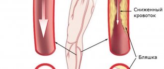
For example, you can determine atherosclerosis, which develops due to damage to the arteries. Cholesterol plaques form in them and clog blood vessels. Aneurysms when the vessels are dilated by more than two mm. Narrowing of the celiac trunk. It occurs because the arteries in the abdominal cavity narrow and the blood supply to the liver and kidneys is blocked. Atherosclerotic damage to blood vessels and the resulting blood obstruction. Blood clots in the abdominal aorta. Congenital bending, elongation, tortuosity of the aorta. An aorta that is dissecting due to an aneurysm. Ultrasound examination of the celiac trunk is done as soon as there is a suspicion of the disease. Because the pressure increases and the aorta can rupture. Severe bleeding will occur and death is possible. If the symptoms are bothering you for the first time and are not very pronounced, then you can do a duplex scan. This examination contains ultrasound and Doppler scanning. It is possible to do color Doppler ultrasound. View the lumen of the vessel, the structure of the walls, the degree and extent of the disturbance in blood flow. Spectral Doppler sonography is done to make a final diagnosis.