Arterial disease, known as atherosclerosis obliterans, is the irreversible growth of low-density lipoprotein plaques under the inner lining of the arteries.
The term “obliteration” means the gradual complete closure of the lumen of a vessel with the development of ischemia of organs and tissues where blood stops flowing. It reflects the most severe variant of the course of the atherosclerotic process with damage to large and medium-sized arteries. Such vessels include: the aorta, arteries of the kidneys, abdominal cavity, upper and lower extremities.
In medical practice, the diagnosis is more common when describing circulatory disorders in the legs, although it is possible to develop atherosclerosis in an obliterating form in the arms, in the renal and mesenteric arteries.
What does the ICD say?
The International Classification of Diseases (ICD-10) does not have a characteristic name for the disease. It is taken into account in the class “Diseases of the heart and blood vessels”, in the subclass “Diseases of the arteries”, in the group “Atherosclerosis” with code I70.
For atherosclerosis of the vessels of the extremities, the general code I70.2 is established. Separate codes take into account lesions of the renal, mesenteric and other peripheral arteries.
Obliterating atherosclerosis of the arteries of the legs is most clearly and often manifested. Let's consider the reasons for its development, possible approaches to diagnosis and treatment.
Causes
The main cause of atherosclerosis is smoking. The nicotine contained in tobacco causes the arteries to spasm, thereby preventing blood from moving through the vessels and increasing the risk of blood clots in them.
Additional factors that provoke atherosclerosis of the arteries of the lower extremities and lead to an earlier onset and severe course of the disease:
- increased cholesterol levels due to frequent consumption of foods rich in animal fats;
- high blood pressure;
- excess weight;
- hereditary predisposition;
- diabetes;
- lack of sufficient physical activity;
- frequent stress.
Frostbite or prolonged cooling of the legs suffered at a young age of frostbite can also be a risk factor.
Why does this form of atherosclerosis develop?
The reasons for the development of obliterating atherosclerosis of the legs include general factors that accelerate the atherosclerotic process, negatively affect the level of cholesterol in the blood, and typical conditions for damage specifically to the vessels of the lower extremities.
General factors:
- age-related characteristics - it has been established that in people over 40 years of age, immunity is significantly reduced due to the cessation of the activity of the thymus gland, this allows infectious agents to easily destroy the arterial wall, the help of cholesterol in “patching holes” becomes excessive and causes deposits in the form of plaques;
- the greatest changes in men are associated with the protection of the female body by sex hormones and changes during pregnancy;
- nicotine intoxication in smokers promotes spastic contractions of the vascular bed and disrupts the regulation of tone by the nervous system;
- stress, uncontrollable situations, stressful working conditions, nervous breakdowns lead to an increased role of arterial spasm;
- poor nutrition causes an excess intake of fats and light carbohydrates, which are not processed into energy, but are stored as excess weight or increase the level of cholesterol derivatives in the blood, from which plaques are built;
- passive sedentary lifestyle;
- presence of concomitant diseases (hypertension, diabetes mellitus);
- hereditary predisposition.
Local factors contributing to damage to the blood vessels of the legs include:
- frequent hypothermia, frostbite of the legs;
- wearing tight shoes that impede blood supply;
- previous injuries (bruises, fractures).
What it is?
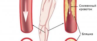
Obliterating atherosclerosis is one of the forms of atherosclerosis. With this disease, cholesterol plaques form on the walls of the arteries, they disrupt normal blood flow, causing vasoconstriction (stenosis) or its complete blockage, called occlusion or obliteration, therefore they speak of occlusive-stenotic damage to the arteries of the legs.
According to statistics, the prerogative of having pathology belongs to men over 40 years of age. Obliterating atherosclerosis of the lower extremities occurs in 10% of the total population of the Earth, and this number is constantly growing.
Mechanism of arterial damage
All of these reasons disrupt fat metabolism, namely the accumulation of low-density lipoproteins and triglycerides. These substances are normally carriers of molecules, but during illness they change, binding to antibodies, becoming accumulations of unnecessary reserves in cells.
The process intensifies with diabetes, hypertension, decreased thyroid function, gout, during menopause, and under the influence of stressful situations.
The other side is impaired utilization of lipoproteins by the liver. This depends on the loss of sensitivity of hepatocyte cells to the changed complexes. Nerve endings (receptors) do not recognize them, so they are not accepted for processing.
Heredity is explained by mutation of genes that control the fat metabolism of cholesterol compounds.
Development mechanism
Most often, atherosclerosis of the vessels of the lower extremities manifests itself in old age and is caused by disorders of lipoprotein metabolism in the body. The development mechanism goes through the following stages.
- Cholesterol and triglycerides that enter the body (which are absorbed into the intestinal walls) are captured by special transport proteins - chylomicrons and transported into the bloodstream.
- The liver processes the resulting substances and synthesizes special fatty complexes - VLDL (very low density cholesterol).
- In the blood, VLDL molecules are affected by the enzyme lipoprotein lipase. In the first stage of the chemical reaction, VLDL is transformed into intermediate-density lipoproteins (or IDL), and then in the second stage of the reaction, IDL is transformed into LDL (low-density cholesterol). LDL is the so-called “bad” cholesterol and it is the one that is more atherogenic (i.e., capable of provoking atherosclerosis).
- Fatty fractions enter the liver for further processing. Here, high-density cholesterol (HDL) is formed from lipoproteins (LDL and LPPP), which has the opposite effect and is able to cleanse the walls of blood vessels from cholesterol deposits. This is the so-called “good” cholesterol. Part of the fatty alcohol is processed into digestive bile acids, which are necessary for normal food processing and are sent to the intestines.
- At this stage, the liver cells may fail (genetically or due to old age), as a result of which, instead of HDL at the output, low-density fat fractions remain unchanged and enter the bloodstream.
Mutated or otherwise altered lipoproteins are no less, and perhaps more atherogenic. For example, oxidized under the influence of H2O2 (hydrogen peroxide).
- Low-density fat fractions (LDL) settle on the walls of the arteries of the lower extremities. Prolonged presence of foreign substances in the lumen of blood vessels promotes inflammation. However, neither macrophages nor leukocytes can cope with cholesterol fractions. If the process is delayed, layers of fatty alcohol - plaques - form. These deposits are very dense and interfere with normal blood flow.
- Deposits of “bad” cholesterol are encapsulated, and when the capsule is ruptured or damaged, blood clots form. Blood clots have an additional occlusive effect and further clog the arteries.
- Gradually, the cholesterol fractions, together with blood clots, take on a rigid structure due to the deposition of calcium-containing salts. The walls of the arteries lose their normal extensibility and become fragile, resulting in possible ruptures. In addition to this, persistent ischemia and necrosis of nearby tissues is formed due to hypoxia and lack of nutrients.
What do diseased leg arteries “look like” from the inside?
Plaques form more often in places of greatest blood flow pressure, in the area of vascular branches. From the inside, the artery wall is yellowish in color, dense, elasticity is lost, deformation and deposition of calcium salts are possible.
The femoral and popliteal arteries have five types of lesions according to location and extent:
- type 1 - limited areas of narrowing (occlusion) in individual segments;
- type 2 - widespread changes in the entire surface of only the external femoral vessel;
- type 3 - widespread occlusion of the external femoral and popliteal arteries, but preservation of patency at the site of the fork of the popliteal branch;
- type 4 - obliteration of the superficial femoral and popliteal arteries in combination with the level of the fork of the popliteal branch, but with preserved patency of the deep femoral artery;
- type 5 is the most severe lesion, since there is narrowing at the level of the superficial, deep femoral and popliteal arteries.
In combined lesions of the arterial vessels of the leg with the popliteal, 3 types are distinguished:
- type 1 - with complete obliteration of the popliteal segment and the initial parts of the tibial arteries, patency in the middle and lower part of the leg is preserved;
- type 2 - narrowing at the level of one or two arteries of the leg, but there is blood flow in the lower part of the popliteal and tibial arteries;
- type 3 - patency is preserved only at the level of small branches on the legs and feet.
Progressive atherosclerosis causes ulceration of the plaque with tissue breakdown. The detached masses migrate to more distant areas of the legs, cause thrombosis, and impair blood circulation.
Another outcome is the formation of an aneurysmal sac, thinning of the wall and internal bleeding from the damaged vessel.
Features of the flow
All symptoms of the disease develop gradually, but in rare cases, obliterating atherosclerosis of the vessels of the lower extremities manifests itself in the form of arterial thrombosis. Then, at the site of arterial stenosis, a blood clot appears, which instantly and tightly blocks the lumen of the artery. Such a pathology develops unexpectedly for the patient; he feels a sharp deterioration in health, the skin of the leg turns pale and becomes cold. In this case, a quick visit to a vascular surgeon (counting the time to irreversible events by the clock) allows the person to save his leg.
With a concomitant disease—diabetes mellitus—the course of obliterating atherosclerosis has its own characteristics. The history of such pathologies is not uncommon, and the disease develops so rapidly (from several hours to several days) that in a short time it leads to necrosis or gangrene in the lower extremities. Unfortunately, doctors often resort to leg amputation in such situations - this is the only thing that can save a person’s life.
Clinical manifestations, classification of the disease
Symptoms of obliterating atherosclerosis depend on the degree of vasoconstriction and the severity of the disease. In accordance with these signs, clinical stages are distinguished.
Initial (asymptomatic) - from the name it is clear that the patient does not present any complaints, considers himself a healthy person, but a blood test reveals an increase in lipid levels.
Stage of primary signs. The following symptoms are of concern:
- numbness (patients say “as if my leg was sitting”);
- constant chilliness of the feet;
- rarely – muscle cramps;
- unexpressed pain in the legs.
Stage of clinical manifestations. Complaints:
- severe pain in the legs, worsening when walking and causing limping;
- the skin on the feet and legs is pale and cold;
- ulcers or non-healing wounds on the toes may appear.
Unlike endarteritis (thrombangitis), there is no such pronounced intermittent claudication, the pain is constant.
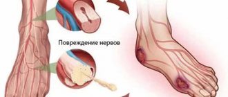
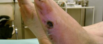
Stage of trophic disturbances. Manifestations:
- severe pain;
- atrophy of the muscles of the leg and thigh;
- trophic ulcers on the feet;
- the onset of gangrene.
The symmetry of the lesion is more typical of atherosclerosis
Symptoms
Symptoms of OASNK in the initial stages are usually quite vague or absent altogether. Therefore, the disease is considered insidious and unpredictable. It is this damage to the arteries that tends to develop gradually, and the severity of clinical signs will directly depend on the stage of development of the disease.
The first signs of obliterating atherosclerosis of the lower extremities (second stage of the disease):
- feet begin to constantly freeze;
- legs often go numb;
- swelling of the legs occurs;
- if the disease affects one leg, then it is always colder than the healthy one;
- Pain in the legs appears after a long walk.
These manifestations appear in the second stage. At this stage of development of atherosclerosis, a person can walk 1000–1500 meters without pain.
People often do not attach importance to symptoms such as freezing, periodic numbness, pain when walking long distances. But in vain! After all, by starting treatment at the second stage of the pathology, you can 100% prevent complications.
Symptoms that appear at stage 3:
- nails grow slower than before;
- hair begins to fall out on the legs;
- pain can occur spontaneously both day and night;
- pain appears after walking short distances (250–900 m).
When a person has stage 4 of obliterating atherosclerosis of the legs, he cannot walk 50 meters without pain. For such patients, even going shopping, and sometimes just going out into the yard, becomes an impossible task, as going up and down the steps turns into torture. Often patients with stage 4 disease can only move around the house. And as complications develop, they stop getting up at all.
At this stage, treatment of the disease, obliterating atherosclerosis of the lower extremities, often becomes powerless; it can only briefly alleviate the symptoms and prevent further increases in complications, such as:
- darkening of the skin on the legs;
- ulcers;
- gangrene (this complication requires amputation of the limb).
Diagnostics
Diagnosis of different stages of the disease usually does not cause difficulties for the doctor. Cold sweaty foot, decreased pulsation in the dorsal artery, muscle atrophy and trophic changes are beyond doubt.
In the clinic, therapists together with surgeons deal with the problems of atherosclerotic lesions of the blood vessels of the legs. For consultation, referral to a vascular surgery center or department is possible.
In addition to blood tests for cholesterol and lipoproteins, as well as glucose levels, the doctor needs to assess the degree of patency of the arteries.
For this use:
- angiography with the introduction of a contrast agent into the femoral artery - the technique is performed in a hospital setting;
- Vascular ultrasound, Doppler ultrasound in the clinic.
It is necessary to differentiate the diagnosis with obliterating endarteritis, Raynaud's disease, Monkeberg's disease, and sciatic nerve neuritis.
In the picture a) the radiograph shows the branches of the arteries below the knee on the right leg, color Doppler sonography b), taken from the front and back, confirms the obstruction of the vessels from the level of the middle of the shin on the left leg (S)
Raynaud's disease is more characterized by a paroxysmal nature and damage to the hands. Monckeberg's sclerosis is a rare genetic disease in which calcification of not only the peripheral but also the coronary arteries rapidly develops.
With neuritis of the sciatic nerve, the pain is shooting in nature, radiating along the outer surface of the thigh through the lower leg to the big toe. Positive symptoms of tension (Lassegue), pathological knee reflexes, increased pain when moving the spine, bending forward, and palpation at the nerve exit points are detected. There are no pulsation disorders in the arteries of the foot.
Characteristics of the disease and features of its development
The development of atherosclerosis of the lower extremities is associated with the presence of pathologies and bad habits in a person, which negatively affect blood counts and the possibility of its movement through the vessels.
This disease can be characterized as damage to the large arteries of the lower extremities, associated with the formation of atherosclerotic plaques on the internal surfaces of their walls. Such deposits slow down blood flow in the legs, as they partially or completely block the lumen of the blood vessels.
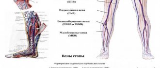
The most commonly affected arteries are:
- femoral;
- anterior tibial;
- posterior tibial;
- popliteal;
- common iliacs.
The formed plaque does not remain stable: if provoking factors are not excluded, its size increases to the point that the formation completely clogs the blood vessel.
The development of pathology occurs gradually. The following stages of the pathological process are distinguished:
- Formation of lipid stains. The walls of arteries locally absorb lipid compounds. Externally, the areas that were saturated with them look like light yellow stripes. At this stage, there are no visible disturbances in blood flow, so the disease does not manifest itself in any symptoms.
- Formation of fibrous plaque (atheroma). Areas of lipid stains become inflamed due to the immune system's reaction to the changes occurring. As a result, the fats accumulated in the vessel wall disintegrate, and connective tissue begins to grow in their place. This process is called sclerosis. The forming plaque protrudes into the lumen, preventing the full passage of blood in this area.
- Formation of complicated atheroma. This is the most severe stage, during which pathological changes occur: the cholesterol plaque gradually disintegrates, and its contents (fat molecules and cells surrounded by connective tissue) penetrate into the bloodstream. Erosion appears at the site of the destroyed plaque. Platelets and other elements responsible for blood clotting are concentrated here. This starts the process of accelerated thrombus formation. The affected area in the structure of the vessel grows and thickens, which significantly impedes blood circulation.
The development of this disease is a long process: 10-15 years pass from the appearance of the first changes to the appearance of the initial symptoms. This makes it difficult to start treatment in a timely manner.
Treatment
Treatment of obliterating atherosclerosis is carried out taking into account the stage of circulatory disorders. Measures to change lifestyle and diet will be required.
- The patient is required to categorically quit smoking.
- In terms of nutrition, you will have to introduce a low-cholesterol diet with limited consumption of fatty foods, meat, and light carbohydrates.
- It is necessary to observe hygienic foot care (wash your feet twice a day, wipe with a disinfectant solution, change socks more often).
- You should wear comfortable loose shoes, use woolen socks for insulation, and avoid hypothermia. Even minor injuries (cuts, calluses, rubbing) can be dangerous.
You can read more about nutrition principles in this article.
The need for warmth is not a whim, but a real need for the patient
Medicines are conservative methods of therapy and are prescribed only by a doctor. They have to:
- cause dilatation of arterial vessels;
- promote the development of collateral (auxiliary) blood circulation;
- prevent further development of atherosclerosis;
- improve microcirculation at the tissue level.
Prevention
Lost health due to atherosclerosis is the result of your attitude towards yourself at random, therefore, already having such a disease, you must at least now be more attentive to yourself and be sure to carry out prevention. In case of OASNK, it is necessary to choose spacious, comfortable shoes to avoid calluses, bruises, avoid any injuries to the legs, and when sitting, do not cross your legs over each other, because At the same time, the vessels are pinched and the blood supply to the affected leg is disrupted. It is necessary to take walks every day; it is very good for your legs. This also includes proper nutrition with the exception of animal fats, salt, smoked meats, fried, red meat, full-fat milk, and cream.
It is necessary to normalize weight and control blood pressure - the numbers should not exceed 140/85. Reducing blood lipids will protect you from myocardial infarction; eliminating physical inactivity from your daily routine and introducing moderate physical activity will also be useful. Quitting smoking is mandatory (this alone reduces the mortality rate from 54% to 18%). It is better to avoid alcohol in any dose.
It is necessary to promptly treat any chronic diseases, control blood sugar levels, avoid stress, regularly visit a doctor for examinations, and systematically conduct courses of conservative treatment. The prognosis is determined by the presence of other forms of atherosclerosis in the vicinity: cerebral, coronary - which, of course, do not improve health.
When is the use of conservative methods justified?
Vascular surgeons warn about the danger of delaying conservative methods in the presence of severe obliteration of blood vessels and the threat of trophic changes and gangrene.
A course of drug therapy is allowed for:
- stages of chronic arterial insufficiency;
- accompanying severe diseases (myocardial and cerebral ischemia, diabetes mellitus, chronic kidney and lung diseases);
- multilevel vascular lesions with occlusion of the main and terminal arteries.
Diagnostic methods for OASNK
If atherosclerosis of the arteries of the lower extremities is suspected, further examination is necessary to confirm (or exclude) the diagnosis of OASNK, establish the stage of the disease and determine treatment tactics. For this purpose, the following series of diagnostic measures are carried out:
- Examination by a vascular surgeon to determine the pulsation of the main arteries of the extremities, neck and abdominal cavity, as well as listening to noises in the projection of large vessels;
- Ultrasound duplex scanning
- X-ray contrast angiography;
- Computed tomography in angio mode;
- Magnetic resonance imaging in angio mode.
Through the above studies, local damage or multifocal (multi-level, multi-story) atherosclerosis of the main arteries, atherosclerosis of the aorta and arteries of the lower extremities is revealed. Areas of complete closure of the lumen (occlusion) of the artery and thrombosis may be detected.
Medicines and their action
To relieve pain, use:
- painkillers;
- novocaine blockades (intraarterial, paravertebral at the level of 2–3 lumbar vertebrae).
Vasospasm is relieved with No-shpa, Halidor, Nicotinic acid, Hexonium.
To prevent thrombosis, Trental, Pentoxifyline, Agapurin, and the aspirin group are used.
In hospital settings, Trental is administered intravenously by drip, then tablets are prescribed in a maintenance dosage
Indirect anticoagulants (reduced blood clotting) are prescribed.
You can stimulate metabolism in tissues with vitamins, Solcoseryl, Parmidine, Prodectin.
The antioxidant effects of vitamins A, C, E, and Probuctol are used.
To restore protective forces, immunomodulatory drugs, ultraviolet irradiation of blood, and hemosorption are used.
Taking into account the autoimmune component in the pathology, desensitizing drugs are prescribed (Pipolfen, Diphenhydramine, Loratadine).
If the diet continues to maintain high levels of lipids in the blood, medications are prescribed to eliminate excess cholesterol.
This is what a removed blood clot looks like along with cholesterol plaques during atherectomy surgery
Stages of development of atherosclerosis of the lower extremities
The most detailed is the modified classification of chronic arterial insufficiency of the lower extremities (CANF), which takes into account in detail the phenomena of critical limb ischemia, which is necessary when determining treatment tactics.
| Stage 1 | Muscle pain only during heavy physical activity (when walking over a distance of more than 1 km). Initial signs of stenosis appear - the skin turns pale, there is a feeling of goosebumps, it seems that the legs are always cold, fatigue quickly sets in when walking, excessive sweating is observed |
| Stage 2A | Feeling of fatigue and stiffness in the calf muscles, intermittent claudication after 200-1000 m |
| Stage 2B | Intermittent claudication in less than 200 m |
| Stage 3A | Intermittent claudication after a few steps or pain at rest when it is possible to keep the lower limb in a horizontal position for more than 2 hours |
| Stage 3B | Pain at rest, ischemic edema, inability to keep the lower limb in a horizontal position for 2 hours |
| Stage 4A | Gangrene of the fingers or part of the foot with the prospect of maintaining the supporting function of the limb |
| Stage 4B | Extensive necrotic changes in the limb without the possibility of maintaining its supporting function |
Surgical methods of treatment
If success with conservative therapy has not been achieved, the level of cholesterol in the blood exceeds 7.5 mmol/l, then angiosurgeons routinely offer:
- performing bypass surgery (creating a bypass for blood flow);
- vascular plastic surgery with removal and replacement with a segment of one’s own vein or alloplast;
- installation of a stent in an artery.
These techniques help restore blood circulation in the affected limb.
In case of severe changes, tissue necrosis, gangrene of the fingers, emergency amputation of the limb is necessary. The level of amputation is determined by the pattern of preserved vessels on the angiogram.
The severe consequences of the disease require patients to take a responsible attitude towards their health. Any pain symptoms in the legs should be consulted with a specialist. Treatment cannot be delayed.
Treatment of obliterating atherosclerosis
Conservative treatment of patients with obliterating atherosclerosis of the arteries of the lower extremities is carried out in the following cases:
- at the stage of chronic arterial circulatory failure in the extremities according to the classification of A. V. Pokrovsky - Fontane;
- with severe concomitant pathology: coronary disease, cerebral vascular damage, chronic diseases of the lungs, liver, kidneys, diabetes mellitus;
- multiple (multi-story) occlusions and stenoses of the main arteries;
- lesions of the distal vascular bed.
It assumes:
- sedative therapy (seduxen, elenium);
- desensitizing therapy (diphenhydramine, pipolfen);
- pain relief (analgesics, intra-arterial agents, blockades of 1% novocaine solutions, paravertebral blockades at the L2 - L3 level, epigastric blockades);
- exclusion of vascular risk factors (smoking, alcohol, excessive cooling, nervous stress, physical inactivity, diabetes mellitus);
- improving the rheological properties of blood, i.e. reducing its viscosity (plasma substitutes - dextrans, defibrinogenic enzymes - acrod, pentoxifylline, trental, vasonite, agapuria);
- elimination of vascular spasm (antispasmodics - no-shpa, halidor, xanthinol nicotinate; gangioblockers - hexonium, dicaine);
- normalization of the blood coagulation system (anticoagulants);
- inhibition of platelet adhesive-aggregation activity (acetylsalicylic acid, ticlide);
- restoration of oxidant-antioxidant balance - protection of cell membranes (antioxidants - vitamins A, E, C, probucol);
- activation of metabolic processes in tissues (vitamins, nicotinic acid, complamin, solcoseryl, bradykinin inhibitors - prodectin, parmidine);
- elimination of immune disorders (immunomodulation, immunosorption, ultraviolet radiation of blood);
- normalization of lipid metabolism. It includes diet therapy, the prescription of lipid-lowering drugs, the use of extracorporeal methods for correcting the composition and properties of circulating blood, partial jejunoileobypass surgery, and gene therapy.
Diet therapy for obliterating atherosclerosis is based on limiting the energy value of food intake to 2000 kcal per day with a decrease in the proportion of fat (up to 30% or less) and cholesterol (less than 300 mg). It is justified to prescribe antiatherogenic nutritional supplements to patients, such as polyunsaturated fatty acids, fish oil, eikonol (a nutritional supplement obtained from certain types of fish).
If there is no normalization of lipid metabolism parameters during diet therapy, without stopping it, drug treatment is carried out. Currently, five groups of lipid-lowering drugs are used for the treatment and prevention of atherosclerosis:
- enterosorbents - cholestyramine, which are sequestrants of bile acids;
- statins - lovastatin (Mevacor), simvastatin (Zocor), privastatin (Lipostat), fluvastatin (Leskol)
- fibrates - mofibrate, otofibrate;
The effectiveness of conservative therapy is assessed by factors affecting lipid metabolism, primarily by the level of total cholesterol and LDL cholesterol.
Normal triglyceride levels are 150 mg/dL. Extracorporeal methods for correcting the composition and properties of circulating blood: plasmapheresis; selective immunosorption, including on sorbents with monoclonal antibodies to LDL (especially effective in the treatment of patients with severe hetero- and homozygous hypercholesterolemia); hemosorption. These methods make it possible to obtain a persistent hypolipidemic effect, which consists in reducing the level of LDL in the blood and increasing the content of HDL, reducing the atherogenic coefficient. This slows down the progression of atherosclerotic arterial occlusion. However, if conservative correction of hyperlipidemia is unsuccessful, there is a tendency for the process to progress, especially in early atherosclerosis, significant clinical manifestations of atherosclerosis in patients with its generalized form, which is usually observed in people with familial hypercholesterolemia, when the cholesterol level exceeds 7.5 mmol/l, In cases of severe xanthomatosis, a partial jejunoileal shunt operation (Buchwald operation) can be performed.
The essence of this surgical intervention is to exclude the distal third of the small intestine from digestion and anastomose the proximal 2/3 of the small intestine with the dome of the cecum. The colon has the ability to synthesize and secrete several types of lipids and their apoproteins, influence hepatic synthesis and secretion of lipids through absorption and enterohepatic circulation of bile acids (BAs) and cholesterol. A decrease in the length of the functioning part of the small intestine leads to impaired absorption of FAs and acceleration of their excretion, an increase in the synthesis of fatty acids in the liver, increasing the oxidation of cholesterol, a decrease in the intestinal synthesis of cholesterol, chylomicrons, VLDL, a decrease in lipid absorption and subsequent inhibition of the synthesis of atherogenic lipoproteins in the liver. A side effect of Buchwald's operation is the frequent development of diarrhea, impaired absorption of vitamin B12 and folic acid.
Two main methods of gene therapy for obliterating atherosclerosis have been developed. The essence of the first of them is to introduce a gene encoding a normal protein - a receptor for LDL, with the help of a retrovirus, into a culture of hepatocyte cells of the patient, and then, through a catheter installed in the portal vein, to deliver a suspension of such cells to the patient's liver. After their engraftment, the donor's normal receptors begin to function. The disadvantage of the method is the need for the patient to take significant doses of statins and a gradual decrease in the function of the introduced genes.
The second (direct) method is performed on a patient without prior manipulation of target cells, while the gene is complexed with a carrier (vector) and directly introduced into the patient, but locally - into the cardiovascular system to avoid dissemination of the gene in the body. Direct administration is carried out using a viral infection, chemical or physical method,
In the complex of conservative treatment of patients with atherosclerosis, especially with stages III - IV of chronic arterial insufficiency of the extremities, it is advisable to include drugs with a complex mechanism of action; 1) tanakan - stimulates the production of relaxation factor by the vascular endothelium. The drug has a vasodilating effect on small arterioles, reduces capillary permeability, reduces platelet and red blood cell aggregation, protects cell membranes by suppressing lipid peroxidation reactions, improves the absorption of glucose and oxygen by tissues; 2) prostaglandins and their synthetic derivatives (vasoprostan). They influence all stages of the development of ischemic syndrome in the limb, have a vasodilating effect, suppress platelet aggregation, improve microcirculation, and normalize metabolic processes in ischemic tissues.
Patients with obliterating atherosclerosis of the vessels of the lower extremities are prescribed physiotherapeutic, balneological and sanatorium-resort treatment (magnetic therapy with pulsed and direct currents with effects on the lumbar sympathetic ganglia and lower extremities, interference currents on the lower extremities and lumbar spine, massage of the lower extremities, reflex-segmental massage of the spine , radon, hydrogen sulfide baths, acupuncture, hyperbarotherapy).
One of the most modern methods of physiotherapeutic treatment of patients with obliterating atherosclerosis of the vessels of the lower extremities is electrical stimulation of the spinal cord. It is performed if it is impossible to perform reconstructive operations on the arteries due to the prevalence of occlusive lesions with systolic pressure at the ankle level less than 50 mm Hg. Art. The essence of the method is the percutaneous introduction of a quadripolar electrode into the epidural space of the lumbar spine, extending its apex to the T12 level and positioning it in the midline. During the first week, electrical stimulation of the spinal cord is carried out with a pulse frequency of 70 - 120 Hz from an external source. If a positive clinical result is obtained, the generator is implanted into the subcutaneous tissue of the anterior abdominal wall and programmed for continuous or intermittent operation. Electrical stimulation is carried out for a long time (months).
For obliterating atherosclerosis of the vessels of the lower extremities, training walking is also used (kinesitherapy, muscle training, walking through a walking throuth). Kinesitherapy aims to increase the distance of pain-free walking. The essence of the method is as follows: if hypoxic pain appears in the calf muscles when the patient overcomes a certain distance, he temporarily slows down his step. A few minutes after this, the patient is again able to perform movements without pain. The mechanism of the beneficial effect of training walking in occlusive-stenotic lesions of the arteries of the extremities is explained by an improvement in the utilization of oxygen by myocytes, an increase in the activity of their mitochondrial enzymes and anaerobic energy production, the transformation of white muscle fibers into red ones, stimulation of collateral circulation, and an increase in the ischemic pain threshold.
For the surgical treatment of patients with obliterating atherosclerosis of the main arteries of the lower extremities, arterial reconstructive and palliative operations are used. Reconstructive methods for restoring arterial blood flow include: endarterectomy, bypass surgery, prosthetics, X-ray endovascular reconstruction (see “Treatment of Leriche syndrome”). An indispensable condition for their implementation is good patency of the distal vascular bed.
Endarterectomy (thrombendarterectomy), as a rule, is used in patients with short (segmental) single occlusions of the main arteries 7-10 cm long. The essence of the operation is to remove the atheromatically changed intima along with the blood clots located next to it. Endarterectomy can be open, semi-closed, closed, eversion, as well as using mechanical and physical methods.
In open endarterectomy, the isolated artery is dissected longitudinally above the location of the plaque. Then, under visual control, the altered intima is peeled off from the underlying layers of the wall to the level of transition to visually unaffected areas and is cut off. The edges of the intima adjacent to the manipulation zone are fixed to the artery wall with atraumatic sutures, which is a reliable way to prevent it from wrapping and blocking the lumen of the arteries. To prevent narrowing of the endarterectomized artery, an autovenous patch is sewn into the incision.
The semi-closed endarterectomy method involves: 1) exposure of the affected segment of the arteries along its entire length; 2) dissection of arteries (longitudinal, transverse) in the projection of the distal end of the occlusion; 3) circular separation in this place of the atheromatically changed intima from the muscular membrane; 4) transverse intersection of the selected segment and passing along it in the proximal direction a special instrument - a disobliterator, mainly a ring (ring stripper), exfoliating the altered ingima; 5) opening the lumen of the artery above the area of the proximal end of the occlusion and removing the detached cylinder of the affected intima through it; 6) suturing the artery wall, if necessary with an autovenous patch.
Endarterectomy with the closed method is carried out in the same way as the semi-open one, but without isolating the artery along its entire length.
When using the eversion endarterectomy method, the artery is transversely dissected below the location of the plaque. Next, the layer of its wall, consisting of the muscular layer and adventitia, peels off from the affected intima and contracts (turns out) in the proximal direction along the upper border of the plaque. At this level, the resulting cylinder of altered intima is cut off. The inverted muscularis propria and adventitia return to their original position. The patency of the vessel is restored by applying a circular suture. It is also possible to perform reverse eversion thromboendarterectomy.
Bypass operations for obliterating atherosclerosis are performed for extended and multi-storey occlusive-stenotic lesions of the main arteries of the lower extremities. A segment of the great saphenous vein isolated from its bed, reversed and anastomosed with the artery above and below the obstacle is most often used as bypasses. Less commonly used are the human umbilical cord vein, homoarterial grafts, synthetic prostheses made of polytetrafluoroethylene, and the great saphenous vein without isolating it from the bed. The essence of the latter method is that the vein is not isolated from the subcutaneous tissue and is not reversed, but is intersected above and below the site of occlusion. Before forming an arteriovenous anastomosis, the venous valves are destroyed using valvulotomes of various designs. The presence of tributary veins capable of acting as arteriovenous fistulas after arterial blood flow is started through it is established on the basis of data from angiography, Dopplerography, palpation, etc., followed by their ligation.
The success of the shunt operation is determined, in addition to the condition of the peripheral bed, and the diameter of the shunt used, which must exceed 4-5 mm.
In case of severe damage to the arteries of the leg, obstruction of the plantar arch, in addition to the usual femoral-popliteal (tibial) autovenous shunting, additionally c. After leaf anastomosis, an arteriovenous fistula is formed, which leads to the discharge of part of the blood directly into the vein, increases the speed of blood flow through the vein and thereby reduces the likelihood of thrombosis. During surgery, a side-to-side anastomosis is first performed with the receiving artery, then a fistula is created by anastomosing the distal end of the shunt with the adjacent popliteal or tibial vein. The diameter should be 2-4 mm, i.e. 40 - 60% of the shunt diameter.
Prosthetics of the main arteries of the lower extremities for atherosclerosis is used extremely rarely.
If it is not possible to restore blood flow through the main arteries, primarily due to occlusion of the distal vascular bed, plastic surgery of the deep femoral artery is performed. At the same time, quite frequent damage to both the deep femoral artery and the popliteal and leg arteries, poor development of collaterals between them lead to unsatisfactory results of the operation.
In case of occlusion of the distal vascular bed or poor condition of the deep femoral artery, palliative surgical interventions are performed aimed at enhancing collateral circulation in the limb. These include lumbar sympathectomy, revascularizing osteotrephination, methods of P. F. Bytka, G. A. Ilizarov, microsurgical transplantation of the greater omentum to ischemic tissue of the extremities.
Lumbar sympathectomy for obliterating atherosclerosis involves extra-, transperitoneal removal of the II - III lumbar sympathetic ganglia on the affected side (Diez operation). The main mechanism of action of the operation is to eliminate the influence of the sympathetic nervous system.
When using revascularizing osteotrephination for obliterating atherosclerosis, 6-9 burr holes with a diameter of 4-6 mm are made on the medial surface of the tibia at biologically active points (as in acupuncture) in the area of a well-developed subcutaneous network of collaterals without damaging the bone marrow. In the postoperative period, subthreshold irritation at biologically active points caused by trepanation stimulates the opening of reserve collaterals. At the same time, through burr holes, unconventional intervascular connections are formed between the arteries of muscle tissue and the bone marrow. In addition, the content of bone marrow mediators—myelopeptides, which have analgesic, trophic and angioprotective properties—increases in the general bloodstream (G. A. Ilizarov, F. N. Zusmanovich, 1983).
The essence of P.F. Bytok’s method is the introduction of autologous blood through certain points on the foot and lower leg into their soft tissues (Fig. 42). Treatment is carried out for 30 days. The tissues are infiltrated twice - on the lower leg on the 1st and 14th days, on the foot on the 7th and 21st days. One session consumes 60 - 80 ml of blood for the foot, 150 - 180 ml for the lower leg. The clinical effect of the operation becomes noticeable after 2-3 months. after completion of the course of treatment and is associated with the formation of well-vascularized connective tissue in the extravasation area.
G. A. Ilizarov’s method (longitudinal compactectomy according to G. A. Illizarov) involves the formation of a longitudinal bone flake 10-16 cm long from the anterior internal surface of the tibia. 2-3 distraction wires are passed through it and attached to the Ilizarov apparatus applied to the bone. From the 8th to 9th postoperative day, the bone flake is removed daily from the tibia by 0.5 mm. The procedure is performed for 31-36 days, until the gap between the tibia and its fragments is 15-20 mm. After this, fixation of the flake continues for 45–60 days, which depends on the degree of maturity of the connective tissue. According to G. A. Illizarov, when the flake is distracted, regional stimulation of the vascular network occurs under the influence of tensile stress. At the same time, the main vessels expand, the number and caliber of small vessels of muscles, fascia and bones increase; at the site of hematoma formation, well-supplied connective tissue develops; due to increased blood supply, regenerative processes in the limb are activated.
During microsurgical transplantation of the greater omentum onto ischemic tissues of the extremities, the greater omentum is placed subfascially on the thigh, moving to the popliteal region and lower leg. The feeding vessel of the graft, usually the right gastroepiploic artery, is implanted into the common femoral artery, and the vein into the femoral vein.
The disadvantage of these methods of surgical treatment of obliterating atherosclerosis, which occurs with occlusion of the entire distal vascular bed of the lower extremities, is the long period of time required for the development of collateral circulation - from 1 to 3 months. This limits the use of such operations in the treatment of patients with critical limb ischemia of stages III - IV, who need a rapid increase in blood circulation in the limb. In such cases, arterilization of the venous system of the foot is carried out: arterilization of the superficial venous network with preliminary destruction of its valves - arterilization into the sources of the great saphenous vein, and in case of occlusion of the superficial veins - into the deep venous system. Arterilization into the origins of the great saphenous vein on the foot involves performing a bypass (reversed autovein, vein in situ, prosthesis) between the patent segment of the popliteal artery or the distal segment of the superficial femoral artery and the origins of the great saphenous vein on the foot. The arterilization of the deep venous network is based on the inclusion of the posterior tibial vein into the bloodstream using a similar technique.
If it is impossible to perform reconstructive surgery in patients with thrombotic occlusions of the arteries of the lower extremities or abdominal aorta due to atherosclerosis, systemic or local thrombolysis with well-known thrombolytic drugs (streptokinase, decase) can be used.
The greatest effect from its use is achieved: 1) with a period of occlusion not exceeding 12 months. in patients with damage to the abdominal aorta and iliac arteries, 6 months. - with the appearance of the femoral and popliteal arteries, 1 month. - renal arteries; 2) with the extent of occlusion up to 13 cm, 3) with a satisfactory condition of the distal vascular bed (the arteries of the lower leg are patent).
Systemic lysis is carried out according to the traditional scheme, local involves the introduction of a thrombolytic in a lower dosage through a catheter directly into the body of the thrombus, antegrade or retrograde, which is accompanied by activation, in contrast to systemic lysis, only of plasminogen, which is part of the structure of the thrombus.
There are several methods of local thrombolysis: 1) continuous infusion with an initial administration of a large dose, and then a maintenance dose; 2) administration of a thrombolytic drug through a catheter with multiple holes along the entire length of the occluding thrombus (pulsatile spray technique); 3) administration of a thrombolytic in a large dose while pulling the catheter along the length of the thrombus. The maximum duration of thrombolytic therapy does not exceed 48 hours. Its effectiveness is monitored angiographically or using ultrasonography.
In the postoperative period, patients continue complex conservative treatment aimed at preventing purulent and thrombotic complications of the operation. Subsequently, they must annually undergo 1-2 courses of inpatient therapy for the disease, and while on outpatient treatment, constantly take antiplatelet agents, indirect anticoagulants and other pathogenetically based drugs.
Treating atherosclerosis
- Shishkin A.A.
- Volkov A.M.
- Kabirov A.V.
- Baranov V.S.
Shishkin Andrey Andreevich
Candidate of Medical Sciences. Surgeon, proctologist, phlebologist at SM-Clinic. Proficient in all modern methods of conservative and surgical treatment of diseases of the veins of the lower extremities (including sclerotherapy, EVLT - endovasal laser coagulation, traditional phlebectomy)
Read moreVolkov Anton Maksimovich
Phlebologist, surgeon at SM-Clinic. Performs operations with a modern proprietary method of treating varicose veins using a laser (modified endovenous laser coagulation. M-EVLC).
Surgical treatment of varicose veins of any complexity (phlebectomy, miniphlebectomy) More detailsKabirov Alexander Vitalievich
Cardiovascular surgeon at SM-Clinic. Candidate of Medical Sciences Proficient in all modern methods of conservative and surgical treatment of diseases of the veins of the lower extremities (including sclerotherapy, EVLT - endovasal laser coagulation, traditional phlebectomy)
More detailsBaranov Vladimir Sergeevich
Cardiovascular surgeon at SM-Clinic. Candidate of Medical Sciences Treats lower varicose veins using non-surgical and surgical methods (aesthetic sclerotherapy, ECHO sclerotherapy, stem sclerotherapy, phlebectomy, miniphlebectomy, EVLT).
More details
Prevention of atherosclerosis
For patients with a family history and age-related predisposition, a screening examination is recommended to determine the risk of developing cardiovascular pathology.
It aims to detect laboratory and genetic markers of the disease. Prevention of atherosclerosis includes weight control, proper nutrition, sufficient physical activity, giving up bad habits, and timely treatment of chronic diseases.
Author:
Pugonina Tatyana Alekseevna, Therapist
Prevention methods
For prevention purposes, you need to start controlling the levels of lipids, sugar and cholesterol in the blood as early as possible. Even with a slight increase in numbers relative to the norm, you should consult a doctor. It is also recommended to regularly do ultrasound of the lower extremities - once every 1-2 years.
It is important to watch your diet, because the reason for the increase in “bad” cholesterol in the blood is the saturated fats that we consume in food. These are products of animal origin - fatty meat, lard, butter. There is a lot of cholesterol in eggs, or rather in the yolks. All these products should be consumed in the smallest possible quantities.
| Name of service (price list incomplete) | Price, rub.) | In installments* |
| Appointment (examination, consultation) with a cardiovascular surgeon, primary, therapeutic and diagnostic, outpatient | 1 750 | — |
| Program “Risk of atherosclerosis and ischemic heart disease, predisposition to dyslipidemia” | 19 000 | — |
* You can read more about the conditions here - Treatment on credit or in installments.
Causes
The main cause of the disease is systemic atherosclerosis. The formation of cholesterol plaques on the walls of blood vessels of the lower extremities is associated with the same pathological mechanisms that cause vascular damage in any other place.
Factors in the development of atherosclerosis:
- dyslipidemia – an imbalance in the content of fats (triglycerides and lipoproteins) in the blood;
- weakness and loss of firmness and elasticity of the vessel wall;
- disruption of cellular metabolism;
- genetic predisposition.
Risk factors for developing pathology:
- gender – men suffer from atherosclerosis several times more often than women;
- age – the disease usually begins to progress after 40 years;
- smoking - nicotine has a vasoconstrictor property and negatively affects the condition of the walls of blood vessels;
- overweight;
- unhealthy diet – a large amount of fat in the diet worsens lipid metabolism and contributes to the formation of cholesterol deposits;
- sedentary lifestyle;
- hormonal disorders;
- unfavorable environmental conditions.
Source: freepic.diller / ru.freepik.com
People with chronic diseases are predisposed to obliterating atherosclerosis: hypertension, endocrine disorders, ischemia and a number of others.