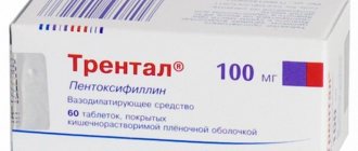
Pentoxifylline
The dimethylxanthine derivative pentoxifylline belongs to the group of angioprotectors and agents that facilitate the movement of blood through the web of capillaries. Pentoxifylline improves the rheological properties of blood, promotes the “unsticking” of sticky platelets, makes red blood cells more elastic, improves microcirculation and saturates tissues with oxygen. If we look at its action through a biochemical prism, the drug increases the concentration of the energy substrates cAMP and ATP, respectively, in platelets and erythrocytes with a logical increase in energy potential, resulting in a vasodilating effect, a decrease in the total peripheral vascular resistance, an increase in systolic (shock) and minute blood volume against the background of the absence or very slight change in heart rate. Pentoxifylline also expands the arteries of the heart, which facilitates the delivery of oxygen to the heart muscle, and the blood itself, due to the expansion of the blood vessels of the lungs, becomes more oxygenated. In addition to this, the drug tones the muscles involved in the breathing process - the diaphragmatic and intercostal muscles. When administered intravenously, pentoxifylline stimulates collateral (in other words, “roundabout”) blood circulation.
The concentration of ATP under the influence of the drug increases not only in red blood cells, but also in brain tissue. There, in the brain, neurologists have the right to expect their patients to improve microcirculation in ischemic areas. Another “bonus” of pentoxifylline for the central nervous system is stimulation of bioelectrical activity.
Pentoxifylline is available in the form of tablets and solution for intravenous and intra-arterial administration. The treatment regimen and dose of the drug are determined individually. During treatment with pentoxifylline, it is recommended to monitor blood pressure indicators, and when combining the drug with antithrombotic agents - also blood clotting indicators. Injection of pentoxifylline requires special care: in this situation, it is advisable to place the patient in a supine position. Low and intermittent blood pressure is a reason to reduce the recommended dose of the drug. This condition is also true for older people. But for smokers the dose should be higher, because... this human defect reduces the therapeutic effectiveness of the drug. Although it would be much better for the body to quit smoking altogether.
L.I. Bogdanets, V.M. Koshkin, A.I. Kiriyenko RGMU, City Clinical Hospital No. 1 named after. N.I. Pirogov, Moscow
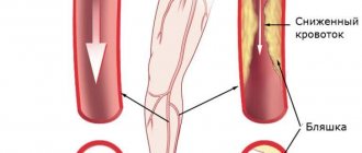
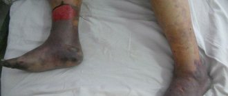
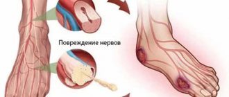
Chronic venous insufficiency (CVI) and chronic occlusive diseases of the arteries of the lower extremities (COLD) are the most common vascular diseases, which in the stage of decompensation are complicated by the formation of trophic ulcers. Chronic arterial insufficiency of the lower extremities affects 2-3% of the population, among whom obliterating atherosclerosis of the arteries accounts for 80-90% [8]. Various forms of chronic venous outflow insufficiency affect about 14% of men and 20% of women [6]. In the general structure of ulcers of the lower extremities of various origins, venous ulcers predominate (about 70%), which occur in 1-2% of the adult working population and 4-5% of elderly patients [2, 7], while arterial ulcers account for about 7 % [9]. However, the vast majority of patients in the departments of purulent surgery are patients with obliterating atherosclerosis in the stage of critical ischemia with the presence of trophic disorders in the form of ulcers, necrosis or gangrene of the extremities. Every year, up to 22 amputations per 100 thousand population are performed for this reason [3]. The development of venous ulcers (stage 5-6 according to the CEAP classification) is based on venous hypertension against the background of long-standing varicose or postthrombotic disease of the lower extremities. As a rule, their formation is preceded by a number of pathological processes at the tissue (hypoxia), microcirculatory (microthrombosis and slugging of formed elements) and cellular (accumulation and fixation of leukocytes in the microvasculature with their subsequent penetration into paravascular tissues and activation with the release of lysosomal enzymes) levels, which is caused by venous hypertension. In addition, in severe forms of CVI, systemic hemostasiological disorders develop (the level of thrombin-antithrombin III complexes, plasmin-antiplasmin, fibrinogen and prothrombin increases), which cause blood hyperviscosity syndrome and enhance microcirculatory stasis [11]. The formation of a venous ulcer (in the classic version, in places where direct perforating veins are localized - the inner surface of the lower third of the leg, less often - its outer or posterior surface) is preceded by the appearance of an area of hyperpigmentation, in the center of which, after some time, a thickening of the skin appears, which over time acquires a whitish, varnished appearance , reminiscent of paraffin deposits (so-called white skin atrophy). In the future, any minimal trauma in this place (bruise, insect bite, etc.) leads to the development of an ulcerative defect, cellulite, dermatitis. The leading pathogenetic mechanism underlying the formation of arterial ulcers (stage 4a according to the Fontaine-Pokrovsky classification) is the progression of the atherosclerotic and stenotic process in the main arteries, causing a hemodynamically significant (75% or more) narrowing of the lumen of the arteries or their complete obliteration [17 ] as a result of steadily progressing morphofunctional changes in the vascular wall [5, 10]. This process, together with disturbances occurring in the system of hemorheology, lipid peroxidation, platelet hemostasis (platelet activation occurs with increased thrombin formation and slow platelet movement in the vessel), leads, as a result of the formation of an atherosclerotic plaque, to a decrease in perfusion pressure, the threshold value of which is considered to be , equal to 20-30 mm Hg. Art. As a result of this, adequate exchange between blood and tissues ceases, accumulation of metabolic products in tissues (acidosis), damage to the microvasculature and the appearance of pathophysiological phenomena characteristic of critical ischemia: • arteriovenous shunting of the blood; • ischemic edema; • imbalance of humoral regulatory systems; • hyperproduction of biologically active substances (cytokines) by platelets and neutrophilic leukocytes [8]. Microcirculation disorders lead to pain and trophic disorders with the development of necrotic lesions of soft tissues. Common sites for the occurrence of atherosclerotic ulcers are the terminal phalanges of the fingers and nails, the nail bed, as well as the heads of the 1st and 2nd metatarsal bones, and with even more severe disorders of the arterial blood supply, trophic disorders are usually localized on the lateral edge of the foot, the heel. One of the reasons for their occurrence is the pressure of shoes on noticeable bone protrusions or abrasions, damage to soft tissues when treating nails, which at first looks like hemorrhage. The color of these lesions ranges from dark blue to black. Despite the various pathogenetic mechanisms of ulcer formation and the clinical picture in patients with diseases of the arteries and veins of the lower extremities, one of the main causes of trophic changes seems to be microcirculation disorders, the state of which largely determines the nature of the course of reparative processes. There is no doubt that the therapy of venous trophic ulcers should include a complex of measures aimed at correcting pathological phlebohemodynamics, the most important among which are elastic compression, therapeutic and protective regimen, systemic drugs and, in the absence of contraindications, surgical methods with the primary use of special endovideosurgical equipment, as well as the use of various topical medications (adequate to the stage of the wound process). Treatment of patients with severe stages of ischemia should also be comprehensive, including primarily reconstructive surgical operations on the arteries, as well as X-ray endovascular interventions (balloon angioplasty, stenting, prosthetics) aimed at eliminating the main cause - arterial blood flow disorders. Such interventions should be carried out against the background and in combination with various methods of conservative treatment, both local and systemic. In this case, it is fundamentally important to carry out conservative treatment for life and continuously [8], taking into account the etiopathogenesis: exclusion of risk factors (smoking, alcohol consumption), treatment of concomitant diseases (hypertension, weight loss, correction of dyslipoproteinemia, blood sugar). Improvement of arterial circulation is achieved through drug therapy, physical therapy, and physiotherapy. Carrying out local treatment involves surgical treatment of the wound (it must be borne in mind that even minimal damage can quickly spread over a large area in just a few days): necrosis, infected tissue and plaque must be removed, purulent leaks should be wide open. Subsequent treatment with various dressings is aimed at cleansing the wound of necrosis, pus, stimulating granulation and epithelization. Thus, treatment of patients with trophic ulcers of vascular origin is a complex and lengthy process, since elimination of disturbances in venous outflow and arterial inflow by surgery is not always fully clinically and technically possible, and interruption of the cascade of pathological reactions leading ultimately to the formation of ulcerative defect, requires the simultaneous administration of several drugs with different mechanisms of action that affect certain parts of the pathogenesis: phlebotropic drugs, low-molecular dextrans, non-steroidal anti-inflammatory drugs, platelet disaggregants, systemic enzyme therapy. To date, it has been proven that in patients with venous trophic ulcers, in addition to local therapy, it is most effective and cost-effective to prescribe drugs that have a pronounced venotonic effect, a high lymphatic drainage effect, and also prevent migration, adhesion and activation of leukocytes - an important link in the pathogenesis of trophic disorders in CVI [12]. Among them, pentoxifylline should occupy an important place, the effectiveness of which is confirmed by numerous clinical studies that compared the effectiveness of its use (as monotherapy or in combination with compression treatment), placebo or other methods of treating venous ulcers lasting from 8 weeks to 6 months. Healing of ulcers was noted in 88% of cases when using pentoxifylline, while in the control group (placebo) only 44% of patients, the reduction in the area of the ulcer defect in the main group averaged 93%, in the placebo group - 56%. The results of treatment of venous ulcers using pentoxifylline are more significant with the simultaneous use of elastic compression [4, 14]. Pentoxifylline (the original drug Trental® from Sanofi-Aventis) is a methylxanthine derivative with high activity in influencing blood rheology. Trental® was first registered as a treatment for intermittent claudication and has been used for a long time in patients with chronic occlusive diseases of the arteries of the lower extremities. It is believed that the effect of this drug is associated with improved deformability of red blood cell membranes, improving oxygen delivery to tissues and suppressing sludge. Later it was found that it suppresses cytokine-mediated activation of neutrophils and adhesion of leukocytes to the endothelium, and also reduces the release of free oxygen radicals [18], takes an active part in reducing the aggregation and adhesion of platelets, erythrocytes, increasing the level of activity of plasminogen and plasmin, antithrombin III , a decrease in fibrinogen in the blood plasma, the level of antiplasmin, antitrypsin and macroglobulin [16]. Thus, Trental promotes faster healing of trophic ulcers, although the results obtained in some cases turned out to be statistically unreliable. The most important area of pharmacotherapy for chronic obliterating diseases of the arteries of the lower extremities, along with the correction of lipid metabolism, is the use of platelet disaggregants, i.e. inhibitors of increased platelet activity that occurs when the arterial wall is damaged. A series of studies have confirmed the effectiveness of using synthetic analogues of prostaglandin E1 and pentoxifylline for this purpose [15]. The effectiveness of Trental in patients with chronic obliterating diseases of the arteries of the lower extremities is expressed in an increase in the distance of pain-free walking, the presence and severity of “rest pain,” and the severity of reparative processes in the area of trophic disorders. Clinical placebo-controlled trials have overwhelmingly confirmed the positive effect of pentoxifylline - from 59 to 74%, while when using placebo it was observed from 5 to 29% of cases [14]. Our many years of experience in using Trental in patients with venous and arterial trophic ulcers also indicates its positive effect on reparative processes. Clinically, this was expressed in a decrease in swelling and hyperemia of paraulcerous tissues, a decrease in exudation, and faster relief of pain, which ultimately led to the appearance of active granulation tissue and epithelization of the ulcerative defect. The drug was usually prescribed at a dose of 1200 mg per day for two months, for persistent, long-term non-healing venous ulcers - in combination with phlebotonics for a course of two to three months. We also consider it appropriate and pathogenetically justified to use Trental in patients with severe trophic changes in the skin (pre-ulcerative condition), indurative cellulite against the background of CVI, and to prepare patients for surgical treatment. It should be noted that the use of pentoxifylline, as well as other vasoactive drugs, in patients with chronic obliterating diseases of the arteries of the lower extremities, regardless of any circumstances (inpatient treatment, surgical interventions with restoration of the main blood flow), should be carried out in longer courses - from 6 months or more (only alternation of different antiplatelet drugs is possible), since after their withdrawal the clinical manifestations of arterial insufficiency increase again. In everyday practice, pentoxifylline is still widely used in a dose of 100 mg three times a day, which does not give a noticeable clinical result. It has been proven that its greatest effectiveness is manifested in a dose of 1200 mg per day [13], for this purpose it is advisable to prescribe Trental-400. It should be noted that this drug is well tolerated, easy to use, and has a small number of side effects (usually associated with violation of the dosage regimen or individual intolerance). An important direction in the treatment of trophic ulcers is the prevention of relapse or its occurrence, which in patients with CVI should include mandatory elastic compression (it is preferable to use medical compression hosiery of compression class 3), the use of pentoxifylline as monotherapy or in combination with compression treatment and phlebotonics (it should be a course student, at least two months two to three times a year), rational organization of work and rest, limitation of physical and static exercise, adherence to a diet with a predominance of protein foods, vitamins, microelements and limitation of spicy, fatty, salty foods, unconditional abstinence from alcohol and smoking. Patients with chronic obliterating diseases of the arteries of the lower extremities, in order to prevent relapse or the formation of ulcers, should be recommended to eliminate risk factors (smoking, alcohol, physical inactivity, excess weight), lifelong intake of platelet disaggregants, antioxidants, as well as a set of measures to improve lipid metabolism. All this should be carried out under conditions of clinical observation with a frequency of three to four weeks to six months, careful care and daily examination of the feet for any changes (cornea, cracks, the presence of a fungal infection). Feet must be treated promptly; the use of sharp cutting instruments for foot care is unacceptable, as is walking barefoot, the use of external heat sources (heat pads, hot foot baths); It is mandatory to wear orthopedic shoes. Thus, Trental-400 is currently one of the most frequently and successfully used drugs in phlebological and angiological practice for the treatment and prevention of trophic ulcers of vascular origin. This is due to its effectiveness, proven not only by numerous clinical studies, but also by the results of practical use, good tolerability, versatility of action, the ability to combine with other drugs, and economic accessibility (taking into account the need for long-term use).
Literature 1. Vasyutkov V.Ya. Trophic ulcers of the foot and leg. M: Medicine, 1993. 2. Vin F. Trophic ulcers of the lower extremities // Phlebolymphology. 1998. No. 7. pp. 10-12. 3. Goryunov S.V., Romashov D.V., Butivshchenko I.A. Purulent surgery. Atlas. M.: BINOM. Laboratory of Knowledge, 2004. pp. 297-327. 4. De Santis M.T., Belcaro D., Cesarone M.R. The use of pentoxifylline for the treatment of venous ulcers: a 12-month double-blind, placebo-controlled study, microcirculation and healing // Angiology. 2002. January-February. pp. 49-51. 5. Klimov A.N., Nikulcheva N.G. Lipids, lipoproteins and atherosclerosis. M.: Medicine, 1984. 166 p. 6. Kuleshov S.E. Diabetes mellitus and surgical diseases. M.: Resurrection, 1996. 7. Savelyev V.S. Modern trends in the surgical treatment of chronic venous insufficiency // Phlebolymphology. 1996. No. 1. P. 5-7. 8. Savelyev V.S., Koshkin V.M. Critical ischemia of the lower extremities. M.: Medicine, 1997. 9. Savelyev V.S., Kirienko A.I., Bogachev V.Yu. Trophic ulcers // Phlebology. M.: Medicine, 2001. P. 510-551. 10. Chazov E.I. History of the study of atherosclerosis: truths, hypotheses, speculations // Therapeutic archive. 1998. pp. 9-16. 11. Coleridge-Smith Ph. D. Microcirculation in Venous Disease (second edition) // Landes Bioscience. 1998. 234 rub. 12. Colerige-Smith Ph. D. From Skin Disoders to Venous Leg Ulcers: Pathophysiology and Efficacy of Daflon 500 mg in ulcer Healing // Angiology. 2003. No. 54. R. 45-50. 13. Colgan MP, Dormandy JA, Jones PW, Schraibman IG, Shanik DG, Young RAL. Oxpentifylline treatment of venous ulcers of the leg. Br Med J 1990; 300:972-5. 14. Dale JJ, Ruckley CV, Harper DR, Gibson B, Nelson EA, Prescott RJ. A randomized double-blind placebo controlled trial of oxpentifylline in the treatment of venous leg ulcers. Phlebology 1995; Suppl 1:917-18. 15. Hamer J., Ramelet A., Schmeller W., Brunner U. Management of Leg Ulcers. KARGER. 199; 294. 16. Pipinos II, Boska MD et al. Pentoxifylline reverses oxidative mitochondrial defect in claudicating skeletal muscle // J Surg Res. 01-Feb-2002; 102(2):126-32. 17. Rodino W., Panetta TF, Burack JH, Bryan DH, Williams RF Combined carotid endarterectomy, innominate artery reconstruction, and coronary artery bypass grafting: Case report // J. Vasc. Surg. 1996, no. 24: 1017-21. 18. Sullivan GW, Carper HT, Novick WJ, Mandell GL. Inhibition of the inflammatory action of interleukin-1 and tumor necrosis factor (alpha) on neutrophil function by pentoxifylline // Infect Immunol. 1988; 56:1722-9.