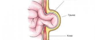
The internal organs are usually located in the hernial sac - part of the peritoneum (the inner membrane lining the abdominal cavity from the inside), which has fallen out along with the contents. The hernial sac can consist of several sections - chambers and contain any internal organs of the abdominal cavity inside. Symptoms of an umbilical hernia depend on its volume, the size of the hernial orifice, the presence of adhesions and obesity in the patient. Umbilical hernias can be reducible or unreducible; in the latter case, the walls of the hernial sac are fused to the surrounding tissues.
Small umbilical hernias with wide hernial orifices and the absence of adhesions usually do not bother patients too much - they quickly learn to repair it themselves. Large hernias with a narrow orifice (especially unreducible ones and in patients suffering from concomitant obesity) are an obstacle to the movement of feces through the intestines, which causes habitual constipation, pain, nausea and vomiting.
Often, umbilical hernias begin in women after childbirth. In this case, loops of the small and large intestines, the stomach and any other abdominal organs enter the hernial sac. With large hernias, the hernial sac is usually divided by septa into several sections (chambers).
A symptom of an umbilical hernia is swelling in the area of the anterior abdominal wall, which occurs when straining or when the patient is standing. In this case, in the lying position the swelling disappears. If there are no adhesions, then the patient can repair such a hernia himself, and it does not cause him much concern (including his ability to work), but sometimes pain occurs. This often asymptomatic course leads to the fact that patients consult a doctor when the hernia becomes unreducible or strangulated.
With a large hernia with a narrow hernial orifice, patients are often bothered by constipation and other discomfort from the gastrointestinal tract. In this regard, large hernias are an obstacle to physical labor. Such hernias are often complicated by strangulation and intestinal obstruction.
Symptoms of a strangulated umbilical hernia in adults
An umbilical hernia is often almost asymptomatic, but can also be strangulated.
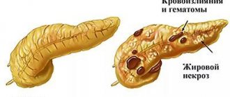
When the contents of the hernial sac are compressed in the hernial orifice, signs of strangulation of the umbilical hernia appear. Strangulation can begin with a hernia of any size after severe physical stress, for example, after lifting something heavy. The patient's excess weight contributes to the injury. Usually the loops of the small intestine are strangulated, and symptoms of acute intestinal obstruction appear. Rapid compression of internal organs causes poor circulation and necrosis (death) of their tissues. In the case when the compression of tissues does not occur too sharply, only the veins are compressed and venous stagnation occurs, and then the arteries are gradually compressed. In this case, tissue necrosis occurs more slowly and the patient’s condition is not so severe.
Symptoms of strangulated umbilical hernia are sharp, sudden pain in the hernia area, its non-reducibility (even if it was previously reducible freely), nausea and vomiting. After some time, an area of redness with a bluish tint appears near the navel.
TREATMENT
For complete recovery, only surgical removal of the umbilical hernia is required. Doctors decide individually what method of intervention will be carried out, taking into account:
- localization and size of the hernial formation;
- are there any complications (inflammation, strangulation);
- general health condition and age of the patient.
Doctors at our Center prefer minimally invasive methods. Most operations (even for large postoperative hernias) are performed endoscopically. Such an intervention is less traumatic for the patient and less likely to cause postoperative complications.
UMBILICAL HERNIOPLASTY
Surgery is the only method that can completely eliminate a hernia. Taking into account the indications, laparoscopic intervention or open abdominal surgery is chosen. Each method has its own characteristics:
- Laparoscopy. Several punctures are made in the abdominal wall and a mesh is installed to close the hernial opening. The entire course of the operation is visible to the surgeon on the monitor, which ensures maximum precision of the intervention. Tissue damage is minimal, and the patient is discharged from the hospital a day later.
- Open hernioplasty. A tissue incision is made in the peri-umbilical area and a mesh is installed. It is used if the hernia in the navel area reaches a very large size.
The surgeon decides which method to choose. In our medical center, preference is given to laparoscopic hernioplasty, if indications permit.
FEATURES OF LAPAROSCOPY AND OPEN INTERVENTION
The cost of removing an umbilical hernia using the closed (laparoscopic) method is usually more expensive than open repair. But it has a number of advantages:
- short postoperative rehabilitation (5-7 days);
- minimum hospital stay (1 day);
- pain is insignificant;
- low risk of complications;
- minimal cosmetic defect.
The price of surgery for an umbilical hernia using the open method seems more attractive to many. But then, in addition to a noticeable postoperative suture on the abdomen, the person receives a number of inconveniences. First of all, this means a longer hospital stay (minimum 2 days) and an increase in the recovery period to 2 weeks. And the cut hurts much more than a small puncture.
The mesh used for hernia repair is perceived by the body as a foreign body, and in the early postoperative period lymphatic exudate almost always accumulates near it. This is a natural reaction of the body. But if during laparoscopy the amount of fluid is insignificant and does not cause concern, then with open intervention the accumulation of lymphatic exudate can be large, and it is removed using a puncture.
Treatment of umbilical hernia
An umbilical hernia is treated surgically.
An operation is performed in which the hernial orifice is expanded, the internal organs located in the hernial sac are returned to their place in the abdominal cavity, after which the hernial orifice is closed and the abdominal wall is strengthened. This operation should be done as planned. Contraindications for elective surgery for an umbilical hernia are any acute diseases, exacerbations of chronic diseases, severe conditions associated with various diseases of internal organs. In this case, conservative treatment is carried out: wearing a bandage and limiting physical activity are prescribed. Obese patients are also advised to lose weight.
A strangulated hernia requires emergency surgery. In this case, the same operation is performed as during planned treatment, and if tissue necrosis occurs, they are removed.
If an umbilical hernia appears, you need to consult a doctor as soon as possible and undergo a planned operation, without waiting for the formation of adhesions and strangulation of the hernia.
Surgery for umbilical hernia according to compulsory medical insurance at the Central Clinical Hospital of the Russian Academy of Sciences
The clinic of the Central Clinical Hospital of the Russian Academy of Sciences provides the full range of medical services for umbilical hernia: from examination and diagnosis to high-quality rehabilitation after surgery.
- An experienced doctor performs an examination, assesses the patient’s condition and, if the diagnosis is confirmed, prescribes a planned herniotomy operation.
- Surgical treatment is carried out in a modern operating room using high-quality instruments and drugs;
- The operation is performed by experienced, highly qualified surgeons.
Hernia treatment under compulsory medical insurance is a service that every citizen of the Russian Federation can receive at the Central Clinical Hospital of the Russian Academy of Sciences. In this case, the treatment will be carried out at the highest level with a caring and attentive attitude of all medical staff towards the patient.
The clinic has a rehabilitation treatment department, where you can undergo high-quality rehabilitation, which will allow you to return to your normal lifestyle as soon as possible.
The advantages of contacting the Central Clinical Hospital of the Russian Academy of Sciences for the treatment of umbilical hernia:
- consultations with experienced specialists;
- carrying out an operation under compulsory medical insurance;
- careful and attentive attitude towards the patient;
- use of modern surgical techniques, high-quality equipment, safe drugs;
- opportunity to receive rehabilitation treatment.
To make an appointment for examination and treatment at the Central Clinical Hospital of the Russian Academy of Sciences and clarify the cost of services, you can call or use a special form on the website.
Four main symptoms, diagnosis
The most typical symptoms of the disease:
- Puffiness, swelling in the navel area, which can be felt with light pressure from the palm of your hand.
- When bending the torso, coughing, or bowel movements, the protrusion increases in size. In a lying position, it may decrease and cause less discomfort.
- There may be pain and a burning sensation in the navel area. When coughing, sneezing, lifting heavy objects, or standing or sitting for prolonged periods, pain and discomfort intensify.
- The skin around the bulge may change color - red, gray or blue.
Attention! Severe swelling and discoloration of the skin in the peri-umbilical area clearly indicate a hernia, while other symptoms are similar to signs of other diseases (endometriosis, ovarian cysts, fibroids, etc.).
If the patient is examined in a supine position on a couch or on a gynecological chair, the protrusion becomes invisible. Ultrasound is also not highly accurate in diagnosing this pathology.
This is why it is difficult to diagnose the disease in women.
Who deals with surgical treatment of external abdominal hernias?
You can always contact the clinic of coloproctology and minimally invasive surgery for surgical treatment.
Qualified specialists regularly perform laparoscopic interventions for hernias of the anterior abdominal wall. In some cases, we also use traditional open surgery.
At KKMH, treatment of hernias is carried out both on a paid basis and under the compulsory medical insurance policy.
Sign up for a consultation by phone +7 (499) 11-03-222.
Umbilical hernia
What is an umbilical hernia?
An abdominal hernia is a disease in which protrusion of the abdominal organs under the integument of the body occurs through various congenital or acquired openings in the abdominal wall. And an umbilical hernia is one of the types of hernias in which internal organs protrude through the umbilical ring, that is, a hernial protrusion forms in the navel area.
How common are umbilical hernias?
An umbilical hernia is one of the most common types of hernia. The frequency of umbilical hernias among primary external abdominal hernias reaches 15% and ranks second after inguinal hernias.
How is an umbilical hernia formed?
An umbilical hernia, like any other hernia, consists of a hernial orifice, a hernial sac and hernial contents. A hernial orifice is a congenital or acquired opening in the muscular aponeurotic layer of the abdominal wall, through which the internal organs protrude along with the adjacent parietal layer of the peritoneum (which covers the abdominal wall from the inside). In the case of an umbilical hernia, the gate is the extended umbilical ring. The hernial sac in an umbilical hernia, as in any other, is formed from the adjacent layer of parietal peritoneum, which lines the inside of the abdominal wall in the area of the hernial orifice - the umbilical ring in the case of an umbilical hernia. Hernial contents are the internal organs located in the hernial sac. The greater omentum, loops of the small intestine, transverse colon and, less commonly, other organs can move into the hernial sac of an umbilical hernia. The amount of hernial contents depends on the width of the hernial orifice and the size of the hernial sac. If the hernial contents move freely from the abdominal cavity to the hernial sac and back, then it is a reducible hernia. If the hernial contents (greater omentum or intestine) are fixed by adhesions to the walls of the hernial sac, then the hernia is called irreducible.
Why does an umbilical hernia occur?
The umbilical ring is a natural weak point in the abdominal wall. It is formed in the fetus by the 6th month of intrauterine development of the fetus and gradually decreases by the time the child is born. After the umbilical cord falls off, the navel is formed in the form of a retracted scar. Umbilical hernias in adults are usually acquired and occur due to expansion of the umbilical ring. Predisposing factors to the formation of umbilical hernias may be pregnancy, obesity, ascites, connective tissue dysplasia (congenital weakness of connective tissue). In women, umbilical hernias occur much more often than in men, due to stretching of the anterior abdominal wall and expansion of the umbilical ring during pregnancy.
How does an umbilical hernia manifest?
Small reducible umbilical hernias may not cause any complaints, except for the presence of a small protrusion in the navel area, which is a cosmetic defect. Large umbilical hernias can be manifested by periodic pain in the navel area, which intensifies after physical activity and eating. With prolonged herniation, the hernia may become irreducible due to constant trauma to the internal organs in the hernial sac and the resulting development of adhesions between the hernial sac and the hernial contents.
Why are umbilical hernias dangerous?
The most serious complication of any hernia is strangulation. This condition threatens the patient's life and requires emergency surgical care. By strangulated hernia we mean compression of the hernial contents (any internal organ) in the hernial orifice, leading to impaired blood circulation in it and threatening necrosis (death). Most often, strangulation develops acutely after sudden physical exertion, but it can also occur gradually, especially with irreducible hernias. The risk of strangulation with umbilical hernias is especially high. This is explained by the fact that umbilical hernias usually have a relatively small hernial orifice and a large hernial sac. A strangulated hernia is an indication for emergency surgery, since delay in surgery is fraught with the development of necrosis of the hernial contents, phlegmon of the hernial sac, intestinal obstruction and peritonitis.
What examination is necessary for an umbilical hernia?
To diagnose an umbilical hernia, examination of the patient is usually sufficient. For large and giant umbilical hernias, especially irreducible ones, it is advisable to perform a computed tomography (CT) scan of the abdominal cavity. This study provides information about the size of the hernial orifice, the nature of the hernial contents, and allows one to calculate the volume of the hernial sac and the volume of the abdominal cavity. This data is extremely necessary for choosing an operation. All patients with umbilical hernias must undergo an ultrasound examination of the abdominal cavity and pelvis before a planned operation, so as not to miss the presence of other pathology of the abdominal organs. Sometimes an umbilical hernia is the first manifestation of diseases such as ovarian cancer and cirrhosis of the liver. Both of these diseases can be accompanied by the accumulation of fluid in the abdominal cavity (ascites) and increased intra-abdominal pressure, an increase in the size of the abdomen and protrusion of the navel.
What are the treatment tactics for umbilical hernias?
For umbilical hernias up to 1 cm that are asymptomatic, observation is possible. Surgeries for hernias of this size are performed only for aesthetic reasons. For larger hernias, surgical treatment is indicated as planned, taking into account the risk of possible strangulation of the hernia. For umbilical hernias with a orifice of up to 2 cm, it is possible to perform operations using local tissues to repair the hernial orifice. The most common methods of operation are Mayo and Sapezhko. After making an incision bordering the navel along the upper or lower semicircle, the hernial sac is isolated and separated from the navel. After removal of the hernial sac, the umbilical ring (hernia orifice) is sutured by creating a duplicate aponeurosis in the transverse (Mayo technique) or longitudinal (Sapezhko technique) direction. The navel should be preserved if possible. It is removed only for large hernias, when maintaining the navel is associated with a high risk of disrupting its blood supply. When the size of the hernia orifice is more than 2 cm, the use of a synthetic mesh implant for repair is indicated, given the high risk of hernia recurrence when repaired with local tissues. The mesh prosthesis plays the role of a “patch” and also strengthens the suture area on the aponeurosis and contributes to the formation of a more durable scar. The mesh prosthesis should overlap the hernial orifice by 2–3 cm in each direction. It can be placed on top of the aponeurosis (“on lay” technology), in the preperitoneal space (“sublay” technology) and intraperitoneally (in the abdominal cavity, IPOM technology). The placement of a synthetic implant over the aponeurosis is used relatively rarely and has more historical significance. The location of the synthetic implant in the preperitoneal space is more common. The intra-abdominal location of a synthetic implant requires the use of special modifications with an anti-adhesive coating that prevents the formation of adhesions between the mesh implant and the intestine. This operation can also be performed using laparoscopic technology.
Signs and symptoms
- Expansion of the umbilical ring.
- Muscle separation in the abdominal area (diastasis recti).
- The appearance of a protrusion near the navel, which disappears when lying down.
- The appearance of nagging pain in the abdomen when moving, exercising, coughing, or trying to push.
- Intestinal dysfunction - flatulence, constipation, fecal stagnation (coprostasis).
- General intoxication of the body - nausea, vomiting.
The clinical picture depends on the size of the hernial orifice and the hernia itself, the presence of adhesions, general health and other factors.
How do hernias form?
Hernias occur in the area of “weak spots” of the anterior abdominal wall, under the influence of intra-abdominal pressure. Factors that cause its increase are called producing factors and include: physical activity, cough, childbirth and all those cases when the abdominal press tenses.
“Weak spot” is the area of the abdominal wall where the muscular aponeurotic part is most thinned. This may be the place of muscle attachment to the aponeurosis or physiological openings (inguinal rings, umbilical ring).
An increased risk of hernias is observed in people with predisposing factors: connective tissue weakness syndrome, damage to the nerves innervating the abdominal wall, as well as the presence of postoperative scars.