Glaucoma is a group of eye diseases caused by damage to the optic nerve. The main cause of the development of the disease is high pressure in the eyeball. Treatment cannot restore vision, but it can prevent further degradation and protect a person from complete blindness.
First, people with glaucoma lose their side (peripheral) vision. If violations are not corrected, the situation will rapidly deteriorate. The disease can affect one or both eyes and can be of congenital or acquired type. Glaucoma in its early stages usually has no symptoms, which is why it is important to visit an ophthalmologist at least once a year.
What is glaucoma
Glaucoma is a broad group of eye diseases characterized by constant or periodic increased intraocular pressure, atrophy of the optic nerve with the subsequent development of typical visual field defects and decreased visual acuity.
This disease occupies a leading place among the causes of irreparable blindness. Vision in glaucoma decreases gradually, and therefore the patient most often notices changes in visual functions already in the advanced stage of the disease. In this case, vision decreases, up to the onset of blindness, which is irreversible, as the optic nerve dies. It is no longer possible to restore sight to a patient who has gone blind for this reason!
Early diagnosis and treatment of this disease can compensate for the course of glaucoma, prevent damage to the optic nerve and associated vision loss. Therefore, it is very important to be regularly examined by competent specialists.
Primary open-angle glaucoma usually develops due to progressive impairment of the outflow of intraocular pressure. As a rule, the cause is age-related changes in the angle of the anterior chamber of the eye. IOP can also rise due to too active synthesis of aqueous humor, which is observed in people with myopia.
Angle-closure glaucoma occurs due to a sharp overlap of the anterior chamber angle by the root of the iris. What most often happens to people with farsightedness. Their eyeball is small, the anterior chamber is small, and the lens is large. These anatomical features contribute to the development of the disease.
The following factors can trigger an attack of angle-closure glaucoma:
- drinking large amounts of liquid at one time;
- long stay in a dark room;
- frequent work with a tilted head;
- instillation of mydriatics into the eye - drugs that dilate the pupil.
It should be noted that during an appointment with an ophthalmologist, a person often receives drops to help dilate the pupil and clearly see the fundus of the eye. In people with high intraocular pressure, these drugs can cause an attack of angle-closure glaucoma. That is why it is necessary to measure IOP before using mydriatics.
Diagnostics
At the initial appointment, the ophthalmologist will carefully examine the eyes, ask the patient about warning symptoms, inquire about the medical history, and find out the risk factors that contribute to the manifestation of the pathology. In addition to this, he will carry out the following diagnostic procedures:
- Measure intraocular pressure using tonometry. All manipulations are simple and painless.
- Conducts testing for optic nerve damage using special instruments and equipment.
- Tests visual fields by examining features of the peripheral visual system.
- It will measure the thickness of the cornea (pachymetry), which will provide additional information in the diagnosis of glaucoma.
Dr. Trubilin’s clinic uses traditional and latest methods for diagnosing eye problems. An integrated approach is used for a comprehensive examination and making the most accurate diagnosis.
With us you can:
- undergo a visual acuity test;
- measure intraocular pressure;
- examine visual fields;
- do optical coherence tomography.
Additionally, biomicroscopy and gonioscopy of the affected area are performed. Based on the screening results, the doctor makes a diagnosis and prescribes individual treatment for glaucoma. When drawing up a scheme of restorative procedures, the patient’s age, medical history, as well as related details are taken into account.
Causes of glaucoma development
Glaucoma can occur at any age and mainly affects people over 40 years of age. But this disease can also affect young people (juvenile glaucoma) and even newborns (congenital glaucoma), since most often it develops in those people whose parents also suffered from this disease. Glaucoma can be an occupational disease, develop due to age-related changes, injuries or concomitant diseases.
Risk factors include:
- age over 50 years;
- family history - the presence of glaucoma in close relatives;
- wounds, eye contusions;
- chronic ophthalmological diseases such as cataracts, high myopia, iridocyclitis, chorioretinitis;
- the presence of hypertension, hypotension, diabetes mellitus, obesity;
- sclerotic changes in blood vessels or deposition of atherosclerotic plaques in them;
- cervical osteochondrosis, leading to disruption of the innervation of the eyeballs.
Postoperative period
In the first four hours after trabeculoplasty, intraocular pressure may jump sharply, but it quickly stabilizes. The eye may also become very sensitive to light immediately after surgery. This phenomenon is temporary. It is better to wear sunglasses when going outside for several days. The operated patient must carefully monitor eye hygiene throughout the month. Under no circumstances should you rub them, as you can damage the cornea or cause an infection in the eye. There may also be side effects/complications. They appear as:
- excessive formation of scar tissue in the area of the anterior chamber;
- decreased visual acuity and blurred vision;
- painful sensations in the eyes;
- formations between the iris and cornea of adhesions, which will require another operation;
- infectious and inflammatory eye diseases (keratitis, blepharitis, conjunctivitis);
- allergies, manifested in redness of the sclera, irritation, itching, burning in the eyes (an allergic reaction can occur to components of anesthesia).
Any of these symptoms should raise alarm and be a reason to visit the clinic. Trabeculoplasty is very effective in the treatment of glaucoma, it prevents its further progression and is rarely accompanied by complications. However, the method also has a disadvantage - after a few years, in about 50% of cases, attacks of increased intraocular pressure occur again. Further treatment of glaucoma can only be continued with surgical methods.
Symptoms of glaucoma
Glaucoma is most often asymptomatic; deviations from the norm are often detected by chance, during an examination or visit to an ophthalmologist for some other reason.
Over time, a person begins to navigate much worse in space due to a narrowing field of vision. In the later stages of the disease, it may seem to him that he is looking at the world around him as if through a spyglass. Twilight vision is also greatly affected. The patient practically loses the ability to see something in the dark.
With angle-closure glaucoma, a person experiences severe pain in the eye, which soon spreads to the entire half of the head. The patient's body temperature rises, chills, nausea and even vomiting appear. When palpated, the eye turns out to be very hard, reminiscent of stone. All symptoms appear abruptly and develop rapidly.
Some nonspecific symptoms may indicate the presence of the disease:
- mild pain, feeling of heaviness in the eyes;
- rapid visual fatigue;
- deterioration of visual acuity at dusk;
- double vision;
- the appearance of rainbow circles when looking at the light;
- feeling of increased moisture in the eyes.
Features of trabeculoplasty
The essence of the operation is as follows: the doctor uses a laser beam to apply about 40-60 pinpoint microscopic burns to the tubular diaphragm of the eye. The trabecular diaphragm is an integral part of the drainage system of the organ of vision, through which the outflow of ocular fluid from the anterior chamber occurs. The doctor determines how many burns (holes) to make during surgery by monitoring the decrease in intraocular pressure.
Before the procedure, the patient is given local anesthesia and a goniolens, which ensures reflection of the laser and protection of eye tissue. There are two types of trabeculoplasty, which differ in the type of laser used in the operation: selective and argon. Selective laser surgery is more gentle. The doctor acts selectively on eye cells, which leads to less injury to the cornea. This procedure can be used more than once.
The argon laser technique has a greater impact on the eye. The laser used in this operation is more powerful. After laser treatment, it will take longer to recover. This procedure is not prescribed more than once. Afterward, surgery may be required to remove scar tissue. Trabeculoplasty lasts about 30 minutes. After laser surgery, the surgeon prescribes a course of therapy, including antibacterial drops, vitamins, antiglaucomatous medications and drugs to reduce the production of fluid inside the eye.
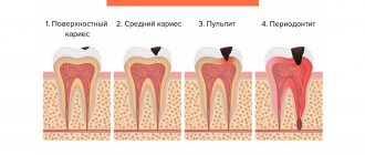
Stages of glaucoma
According to the severity of the process, 4 stages of glaucoma are distinguished:
- I initial stage - the early stage is characterized by periodic surges in intraocular pressure, causing sharp dilation of the pupils and headaches.
- Stage II of advanced glaucoma - advanced glaucoma is manifested by a significant narrowing of the visual field on the nasal side or the formation of a large arcuate scotoma. Increased intraocular pressure provokes pain in the affected eye; sometimes the enlargement of the eyeball is even visible visually. Visual acuity decreases. At this stage, it is most often impossible to do without surgery.
- Stage III of advanced glaucoma - at this stage, glaucoma is accompanied by a concentric narrowing of the visual field and the complete loss of its large areas, that is, a significant increase in the blind spot. Sharply increased intraocular pressure leads to other pathologies: retinal detachment, clouding of the lens, and the formation of hemorrhages. Drug therapy and diet for the third stage of glaucoma serve only as an aid; the main method of treatment in this case is surgery.
- IV terminal stage of glaucoma - complete loss of central vision and complete loss of objective vision. With terminal glaucoma, there is a serious increase in intraocular pressure, it is accompanied by severe headaches and eye pain, enlargement and clouding of the eyeball - buphthalmos or bull's eye.
Depending on age, glaucoma is divided into congenital (in children under 3 years of age), infantile (in children from 3 to 10 years of age), juvenile (in persons aged from 11 to 35 years of age) and glaucoma adults (in persons over 35 years of age). All forms are acquired, except congenital.
Who is suitable for laser eye surgery for glaucoma?
Indications for laser procedure for glaucoma:
- first and second stages of open-angle and closed-angle (acute) glaucoma;
- inability to reduce intraocular pressure and eliminate other symptoms of glaucoma using conservative therapy;
- allergic reactions to eye drops used to reduce pressure in the eye;
- when the patient cannot regularly take drops and tablets;
- as a prevention of damage to the second organ of vision, which does not have glaucoma.
The operation may be contraindicated for diabetic retinopathy, swelling and clouding of the cornea, and inflammatory processes in the eyeball. Also, the laser procedure is not used if the anterior chamber of the eye is very small.
The patient undergoes an examination before the operation, and in each specific case the decision to carry out the procedure is made by the doctor. He also chooses the type of laser surgery for glaucoma. There are two of them: trabeculoplasty and iridectomy.
Glaucoma treatment
Is glaucoma curable?
If there is a suspicion that a person has glaucoma, then it is possible to determine the stage of development of the disease during an examination by a specialist. The doctor will decide how to treat the patient based on the diagnostic data obtained. At the initial stage of development of the disease, it is possible to treat glaucoma without surgery, the so-called conservative method. Therapy is carried out using special eye drops that lower the pressure inside the eye, improve the outflow of fluid passing through the organ, or reduce its secretion.
A positive effect from such treatment is possible with the open-angle type of the disease, when there are no changes in the structure of the eyes. If a patient is diagnosed with angle-closure glaucoma, then medications are practically useless. And to eliminate problems, surgical treatment methods are already used. With their help, angle-closure glaucoma is curable both at the initial stage of the disease and at later stages.
Surgery for glaucoma
Surgery is indicated for open-angle glaucoma that responds poorly to prescribed medications. In case of an acute attack of angle-closure glaucoma, surgery is necessary if
if intraocular pressure cannot be normalized with drug treatment.
- Laser methods of treating glaucoma are considered the most modern and least traumatic operations:
1. Laser iridotomy is the removal of glaucoma without surgery. This type of treatment is highly effective, easily tolerated and safe. Removing glaucoma using a laser guarantees a complete cure for the disease. Laser iridotomy is performed without opening the eyeball and is a microsurgical intervention. The risk of complications is minimal.
2. Trabeculoplasty - the surface of the diaphragm is burned with a laser, which increases its tension and, as a result, permeability. As a result, more fluid swells from the anterior chamber, and the pressure decreases.
- Cryodestruction: the essence is similar to the previous method, but the effect is carried out not by a laser, but by cold, and the object is not the iris, but the sclera. It is exposed to cold, applying applications at several points at once. The operation is contraindicated for terminal stages of glaucoma, unsuccessful surgical interventions, or a history of pain. Cryodestruction is not as safe and more often causes complications. It is used if laser correction is contraindicated for a patient for some reason.
Prevention of glaucoma
Preventative measures will help reduce the damage that high blood pressure causes to the visual system, thereby preventing optic atrophy and blindness.
1. Do not overexert yourself - limit both physical and psycho-emotional stress. 2. Do not keep your head tilted - it is harmful for patients with glaucoma to engage in activities that require prolonged tilting of the head down. This applies to reading, drawing, drawing, knitting, embroidery and others. It is necessary to maintain a level head position when working at the computer or watching TV. 3. Set up the right lighting - it is dangerous for people with glaucoma to work in poor lighting. It is important to make it bright so as not to strain your eyes. 4. Give up bad habits, as smoking negatively affects the blood supply to the whole body. The transport of oxygen and nutrients to all elements of the eyeball is disrupted. 5. Choose loose clothing that does not interfere with blood circulation in the neck and head. 6. Avoid visual fatigue - it is important to take breaks while working at the computer, reading and watching movies. It is recommended to set aside 15-20 minutes every hour for rest. At this time, you need to really rest, and not change one strenuous activity to another. 7. Eat right - to prevent glaucoma, you need to include raw vegetables, fish, fruits in your diet, while reducing the amount of animal fats and sugar. 8. Consume moderate amounts of water and other drinks. You should not drink more than one glass of any liquid at a time. To be on the safe side, you can check your reaction to coffee or green tea: measure your blood pressure before and after. 9. Get proper rest, get a good night’s sleep, it is advisable not to stay up late, take a walk in the fresh air in the evening. You need to sleep on comfortable pillows. After waking up, it is recommended to do a warm-up without getting out of bed. 10. Do not refuse drug treatment. 11. Avoid sudden changes in lighting—changes in lighting intensity are a strong strain on the eyes, so before going to the cinema, for example, you need to use drops that prevent pupil dilation. 12. Constantly monitor your condition. Even with stable intraocular pressure, you should visit your doctor at least four times a year.
These measures will help avoid not only glaucoma, but also other diseases of the visual system. Prevention is recommended for everyone, since glaucoma can occur even in a completely healthy person. At Lensmaster you can undergo a free vision diagnostic by making an appointment through the website.
Intraocular fluid and its outflow pathways
Intraocular fluid (hereinafter referred to as IOF) plays a huge role in maintaining the level of intraocular pressure. It is one of the sources of nutrition for intraocular structures (lens, cornea, trabecular apparatus, vitreous body).
IG is produced by processes of the ciliary body located behind the iris and collects in the posterior chamber of the eye. Next, most of the liquid, washing the lens, flows through the pupil, enters the anterior chamber and passes through the ocular drainage system (trabecula and Schlemm's canal), which is located in the area of the angle of the anterior chamber. From the drainage system of the eye, the intraocular fluid enters the excretory collectors (graduates), and then into the superficial veins of the sclera.
About 85% of the intraocular fluid flows out this way, but there is another way of outflow, which takes about 15%.
IG may leave the eye, leaking through the stroma of the ciliary body and sclera into the veins of the choroid and sclera. This outflow pathway is called uveoscleral.
There is a certain balance between the production of intrauterine fluid and its outflow. When this balance is disturbed, the level of intraocular pressure changes, which is a prerequisite for the development of glaucoma.
Ophthalmohypertension of the eye
In recent years, a new diagnosis has appeared in ophthalmological practice: ocular hypertension. It is a manifestation of intraocular hypertension not associated with glaucoma. This term is a complex and very heterogeneous concept. Ocular hypertension can be essential, false or symptomatic.
In the false case, the increase in pressure is associated with the peculiarity of the eye’s reaction to tonometry or with the individual characteristics of the patient.
The cause of essential hypertension is an imbalance in the hydrodynamics of the eye associated with age-related changes. In particular, with age, the outflow of aqueous humor is often disrupted, while its secretion remains at a high level. Most often, this imbalance levels out over time. Symptomatic ocular hypertension is accompanied by a short-term or long-term increase in pressure. In this case, hypertension is only a symptom of another pathology (uveitis, glaucomatocyclic crisis, corticosteroid, toxic, diencephalic, endocrine hypertension). When the underlying disease is cured, intraocular hypertension also disappears. The course of this pathology is usually benign, that is, it does not lead to damage to the optic nerve. However, in some cases, ocular hypertension can develop into glaucoma, and therefore, any increase in intraocular pressure should be considered as the most important risk factor for the development of glaucoma.
Primary open angle glaucoma (POAG)
This disease is the most common type of glaucoma in adult patients. In this case, the level of intraocular pressure increases as a result of disruption of the outflow of aqueous humor. Causes include blockage of Schlemm's canal, degeneration of trabecular cells and intrascleral canals. Typically, this type of glaucoma has a gradual onset and progresses almost unnoticed by the patient. This is associated with late patients seeking medical help.
The complaints that the patient most often presents include a significant decrease in visual acuity, the appearance of rainbow circles near light sources, and blurred vision (temporary or permanent). All these symptoms are associated with increased intraocular pressure. Most often, changes in open-angle glaucoma can be detected on both sides, however, their severity is different.