The problem of osteoporosis existed in all periods of human development, but in the 20th-21st centuries the disease acquired the character of a “silent epidemic”. In modern medicine, osteoporosis is considered as a systemic disease, the main signs of which are structural changes in the microarchitecture of bone tissue and a decrease in bone mineral density (BMD). If a patient is diagnosed with osteoporosis, treatment is prescribed immediately. Without complex therapy, the disease is prone to steady progression and a decrease in BMD to critical levels, which is associated with a high risk of fractures.
Osteoporosis is not only a medical, but also a social problem. According to statistics, the disease ranks 4th in prevalence, second only to diabetes, cardiovascular diseases and cancer. Advanced osteoporosis is characterized by high mortality and disability.
Another important aspect is a significant decrease in the patient’s quality of life. In fact, doctors often assess the severity of the disease and the effectiveness of prescribed treatment methods only based on laboratory and instrumental data, forgetting about the patient’s well-being and complaints.
Signs and symptoms of osteoporosis
Osteoporosis is a disease that produces few symptoms in the early stages. Signs of pathology appear as bone tissue weakens:
- back pain that occurs with osteoporosis due to destruction of the vertebra;
- slouch;
- reduction in human height;
- fractures even with minor impact (femoral neck fractures are especially dangerous - in 20-25% of cases they lead to death in the first 6 months after injury, and in 40-45% - to disability);
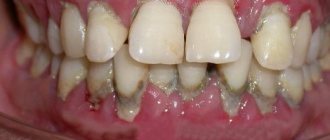
- loose gum tissue;
- pain in the joints, lower back;
- rapid fatigue due to physical exertion;
- discomfort when staying in one position for a long time;
- frequent bowel movements;
- thoracic kyphosis;
- heartburn;
- inability to take a full breath due to chest pain, feeling that there is not enough air;
- accelerated tooth decay;
- leg cramps.
You should consult a doctor for a preventative check if any of these symptoms occur, as well as in the following cases:
- treatment with corticosteroids for several months;
- the beginning of menopause;
- hip fracture in old age.
Early screening for osteoporosis is possible using a blood test, according to which we present the normal indicators according to the following criteria:
- total calcium – from 2.2 to 2.65 mmol/l;
- inorganic phosphorus – from 0.85 to 1.45 µmol/l;
- parathyroid hormone – from 9.5 to 75 pg/ml or from 0.7 to 5.6 pmol/l;
- DPID: for men (nmol DPID/mol creatinine) – from 2.3 to 5.4, for women – from 3 to 7.4;
- osteocalcin (ng/ml): for men - from 12 to 52, for women before menopause - from 6.5 to 42.3, for women after menopause - from 5.4 to 59.1.
Osteoporosis does not manifest itself until 20 to 30% of bone mass is lost. Therefore, after the age of 40, it is recommended to consult an endocrinologist for diagnostics once a year.
Forecast
With proper treatment and monitoring of the patient, the progression of bone loss and the risk of fractures can be slowed down. Fractures of the vertebrae and peripheral bones that have already occurred cannot, of course, be corrected. But even stopping the progression of the disease during osteotropic therapy is considered a success in treatment. If it is possible to achieve an increase in bone mass, then this has an even more positive effect on the course of the disease.
Since in the treatment of such a serious disease as osteoporosis we work more “for the future”, trying to stop the progression of the disease, the effect of treatment is not noticeable quickly.
It is important for the patient not to quit treatment halfway, but to constantly be in contact with the attending physician to discuss the details of treatment.
This is only possible with competent supervision (the so-called “management” of the patient by the primary attending physician with the involvement of advisory assistance from doctors of other specialties, if required by a specific clinical situation). The prognosis with this approach to therapy is most favorable in relation to the patient’s quality of life and treatment of the disease.
Types, forms and degrees of osteoporosis
This disease is classified according to several criteria. First of all, it is considered whether it is an independent (primary) disorder or a symptom of another disease (secondary).
Primary osteoporosis
This is juvenile, postmenopausal, idiopathic, senile osteoprosis.
- The juvenile type of the disease is typical for children and young people, is rare, and is most often caused by birth defects. The main manifestations are severe pain in the legs and back, curvature of the thoracic spine, and visible growth retardation. There is a tendency to compression fractures.
- The postmenopausal form is associated with accelerated bone loss in women 15 to 20 years after menstruation ceases. During this period, estrogens are produced in insufficient quantities and metabolism is disrupted. The disease can be acute or chronic and is accompanied by the occurrence of fractures of bones with a predominantly trabecular structure (vertebrae, distal parts of the radius) i Postnikova S.L. Features of postmenopausal osteoporosis / S.L. Postnikova // General Medicine. - 2004. - No. 4. - P. 41-45. .
- The idiopathic variety is more common in men, but in some cases it is diagnosed in women. The risk group by age is people 20-50 years old. The disease develops smoothly, primarily manifested by periodic pain in the spine, compression fractures are likely.
- Senile osteoporosis is associated with the aging of the body and occurs in both men and women after 70 years of age. Early symptoms are decreased vision, muscle weakness, and migraines. It manifests itself as loss of bone mass in both trabecular and cortical bone, which leads to femoral neck fractures i Postnikova S.L. Features of postmenopausal osteoporosis / S.L. Postnikova // General Medicine. - 2004. - No. 4. - P. 41-45. .
85% of cases of the disease relate to primary osteoporosis, mainly postmenopausal i Lesnyak O.M. Diagnosis, treatment and prevention of osteoporosis in general medical practice. Clinical recommendations / O.M. Lesnyak, N.V. Toroptseva // Russian family doctor. - 2014. - P. 4-17. .
Secondary osteoporosis
Secondary osteoporosis is a complication of various diseases (endocrine, inflammatory, hematological, gastroenterological) or drug therapy (for example, steroid).
Among the forms of secondary osteoporosis, the first place is occupied by glucocorticoid-induced osteoporosis (GIO), which develops in people of any age as a result of therapy with systemic glucocorticosteroids (SGCS).
In elderly patients, a decrease in bone density during long-term therapy with SGCS occurs 2-3 times faster than under physiological conditions. Osteoporotic fractures are reported in 30-50% of patients receiving long-term treatment with glucocorticosteroids. When taking glucocorticosteroids in a daily dose of >5 mc, the risk of fractures increases by 1.9 times compared to the general population, hip fractures by 2 times, vertebral fractures by almost 2.9 times i Baranova I.A. Glucocorticoid-induced osteoporosis / I.A. Baranova // Practical pulmonology. - 2008. - No. 1. - P. 3-9. .
Degrees of osteoporosis
- I (light). Bone density is slightly reduced, sometimes the patient experiences pain in the limbs or spine, and muscle tone decreases. Signs of low calcium levels in the body also appear: dry skin, hair loss, brittle nails.
- II (moderate). Structural changes in the bones are pronounced, the pain becomes constant, and due to damage to the spine, stooping appears. The pain syndrome intensifies with exercise, cramps appear in the calves, and disturbances in the functioning of the heart muscle.
- III (severe). Most of the bones are destroyed, the patient has poor posture, reduced height, and experiences constant severe back pain. Several parts of the spine are affected at once, increasing the risk of fracture of the femoral neck and collarbone.
- IV (very severe). On the x-ray, the bones are almost transparent, the vertebrae are “flattened”, and therefore the patient’s height is significantly reduced, the spinal canal is expanded, and the shape of the bones is changed. At this stage, the patient cannot care for himself.
By localization
The most severe form of the disease is damage to the spine. This increases the risk of getting a fracture and losing the ability to move. Based on localization, the following types of osteoporosis are distinguished:
- Cervical region. The length of the neck decreases, the angle of the head changes. Patients complain of dizziness, nausea, and muscle pain. There is a high risk of pinching the artery that supplies oxygen to the brain.
- Thoracic department. Posture changes noticeably, heart rate increases, and nails become brittle.
- Lumbar region. In this case, the lower back “sags” inward, which reduces the distance between the pelvis and ribs and causes an increase in the abdomen.
By joint damage
Osteoporosis can affect various joints:
- Hip. It is most often affected in old age and leads to disability due to the very low rate of bone tissue restoration. If combined with a spinal fracture, the patient becomes disabled.
- Knee. The cartilage wears out, movement becomes difficult, and severe pain occurs due to the fact that the bones are in contact with each other.
- Ankle. At the same time, it is difficult for a person not only to walk, but also to be at rest. My foot and lower leg hurt constantly.
Classification
Primary osteoporosis
This is the most common form of the disease. These include:
- postmenopausal osteoporosis
- senile (i.e. senile) osteoporosis
- juvenile (i.e. osteoporosis in adolescence)
- idiopathic osteoporosis (when no obvious cause of bone pain is found).
Secondary osteoporosis
It has many causes, when it is not the primary damage to bone tissue that occurs, but completely other diseases of the body, one of the manifestations of which is osteoporosis.
This group includes osteoporosis due to endocrine diseases, diseases of the gastrointestinal tract, kidney diseases, systemic diseases, blood diseases, and even taking many medications.
Diagnosis of osteoporosis
You can check the condition of the body and confirm or exclude the diagnosis of osteoporosis using laboratory tests and instrumental studies, of which the simplest and most informative is densitometry.
Lab tests:
- calcium in urine;
- clinical blood test;
- alkaline phosphatase (biochemistry indicator);
- TSH;
- markers of bone destruction;
- for men – testosterone.
Instrumental methods:
- radiography;
- densitometry;
- bone biopsy;
- bone scintigraphy;
- MRI.
How to check for osteoporosis using densitometry?
Ultrasound densitometry is a quick and painless diagnostic method. During the procedure, the speed of propagation of ultrasound waves through bone tissue is measured. Ultrasound travels faster through denser bones. The result of the study is recorded by a computer, the indicators are compared with the norm. The session lasts 2-3 minutes, and a conclusion is immediately issued.
To carry out this diagnosis, a Sonost-3000 densitometer is used - expert-level equipment that can detect loss of even 2-5% of bone mass.
Densitometry is recommended:
- nulliparous women;
- women over 45 years old;
- women who have given birth to 2 or more children;
- during early menopause;
- in case of menstrual irregularities;
- if you have bad habits;
- men over 50 years old;
- in case of deficiency of sex hormones.
You definitely need to undergo ultrasound densitometry if:
- Fractures often occur;
- there was a long course of glucocorticosteroids, diuretics, anticonvulsants and anticoagulants;
- diagnosed with hyperparathyroidism or other dysfunction of the parathyroid glands.
Also indicated are:
- change in posture;
- bone and muscle pain due to changing weather;
- pain in the lower back and chest with static load;
- senile stoop;
- night cramps in the legs;
- tooth decay;
- decreased growth;
- body weight deficiency;
- osteoporosis in close relatives;
- low testosterone levels in men;
- and etc.
Reasons for women
Osteoporosis is a disease that develops under the influence of many risk factors. There are primary and secondary osteoporosis. Primary osteoporosis develops with age. The reasons are considered to be:
- Complicated family history;
- Mature age;
- Short stature;
- Early menopause and late onset of menstruation;
- Instability of the menstrual cycle;
- Infertility;
- Breastfeeding for longer than one and a half years;
- History of bone fractures;
- Taking hormonal medications;
- Low physical activity;
- Pathology of the endocrine system (thyrotoxicosis, hyperparathyroidism, type 1 diabetes mellitus);
- Excessive drinking and smoking.
Secondary osteoporosis can be caused by the following diseases:
- Itsenko-Cushing's disease and syndrome;
- Thyrotoxicosis;
- Rheumatoid arthritis;
- Diabetes mellitus type 1;
- Conditions after gastric resection,
- Chronic renal failure;
- Chronic liver diseases.
There is a high risk of developing osteoporosis in people taking medications whose side effect is weight loss (immunosuppressants, glucocorticosteroids, anticonvulsants, gonadotropin-releasing hormone antagonists, aluminum-containing antacids).
Smoking, alcohol abuse, caffeine, excess or insufficient physical activity are factors that can cause osteoporosis. The likelihood of developing osteoporosis is higher in people who eat insufficient amounts of calcium, vitamin D, a lot of meat, or cannot tolerate dairy products. Generally recognized factors that increase the risk of developing osteoporosis are the presence of osteoporosis in parents, sisters and brothers, early menopause, and a sedentary lifestyle. Make an appointment
Osteoporosis therapy and clinical recommendations
First of all, you need to know which doctor treats osteoporosis: since the causes of the disease can be different, different specialists can treat it - a rheumatologist, an orthopedic traumatologist, an endocrinologist.
Effective treatment of osteoporosis (both early and after 50 years) may include physiotherapeutic procedures, medication, lifestyle and diet adjustments, and physical exercise.
Drug therapy
- Taking vitamin D and calcium supplements. Consumption of these elements in the right dosages leads to a rapid increase in bone mineral density and a decrease in the incidence of fractures.
- Bisphosphonates. Treatment with these drugs reduces the risk of fractures by 30-50% and increases bone density.
- Hormone therapy (replacement). It is carried out for the prevention and treatment of the postmenopausal form of the disease. Treatment leads to the cessation of bone thinning, the prevention of fractures, and the elimination of urogenital and autonomic complications of menopause.
- Calcitonins. They inhibit bone tissue resorption and have a pronounced analgesic effect.
- Ossein-hydroxyapatite complex. Normalizes calcium homeostasis, improves bone metabolism, stimulates bone formation, restores the balance between the processes of bone formation and resorption.
Non-drug therapy
- Wearing a corset (orthoses)
Indicated for back pain and compression fractures of the spine. The corset should be worn constantly or intermittently, always taking it off at night.
- Physical education, walking, aerobic exercise.
Regular walks in the fresh air are beneficial. Loads should not be excessive; it is necessary to exclude power sports and those that involve the likelihood of mechanical impacts (for example, playing with a ball).
Diagnostic methods
In order to make an accurate diagnosis, you need to contact an orthopedic traumatologist. Diagnosis of osteoporosis involves a combination of several methods:
- taking an anamnesis, visual examination - during the process, the specialist finds out the duration of existence of the existing symptomatic picture, as well as characteristic anamnestic signs;
- densitometry – makes it possible to fully assess bone density;
- X-ray examination - despite the low information content, the method is widely used in diagnostics and allows one to determine existing signs of the development of the disease;
- laboratory tests - a set of tests that involve determining the level of essential vitamins and minerals.
Complications from osteoporosis
The main complication is injuries to the musculoskeletal system, especially compression fractures of the spine and microfractures due to sudden compression of the joints. In this case, a large load is not necessary; you can simply trip and fall, resulting in a fracture.
, compression of the spinal cord and nerve endings may occur . Because of this, loss of sensitivity in various parts of the body, as well as paralysis and disability, is possible.
Fractures are especially dangerous in old age - only 9% of people (according to statistics for the Russian Federation) return to normal life after this.
Osteoporosis can also have the following consequences:
- impediment to growth, which is especially important for a child or adolescent;
- decrease in height - approximately 2-4 cm per year;
- poor posture – “hump” in the thoracic region (thoracic kyphosis);
- disruption of internal organs due to incorrect posture.
general description
The most common fracture sites for osteoporosis are the vertebrae, femoral neck, and radius bone in the wrist area.
These are dangerous areas. Vertebral fractures can damage the spinal nerves and even the spinal cord. A hip fracture confines an elderly patient to a bed, and this forced immobility entails many problems: pneumonia, thrombosis, bedsores, etc. A quarter of patients with a hip fracture are cured, a quarter dies and half remain disabled. Osteoporotic fractures are characterized by the fact that they do not require trauma to occur. Often, it is enough to lift a heavy bag from the ground, fall from your own height or from a sofa, or you may not notice the moment of injury at all.
The incidence of osteoporosis increases sharply after age 50. Women suffer from it 3 times more often than men.
Sources
- Baranova I.A. Glucocorticoid-induced osteoporosis / I.A. Baranova // Practical pulmonology. - 2008. - No. 1. - P. 3-9.
- Baranova I. Caution: osteoporosis! / I. Baranova // Asthma and allergies. - 2004. - No. 4. - P. 18-19.
- Zulkarneev R.A. Prevention and treatment of osteoporosis / R.A. Zulkarneev, R.R. Zulkarneev // Kazan Medical Journal. - 2003. - T. 84. - No. 3. - P. 230-232.
- Lesnyak O.M. Diagnosis, treatment and prevention of osteoporosis in general medical practice. Clinical recommendations / O.M. Lesnyak, N.V. Toroptseva // Russian family doctor. - 2014. - P. 4-17.
- Postnikova S.L. Features of postmenopausal osteoporosis / S.L. Postnikova // General Medicine. - 2004. - No. 4. - P. 41-45.
- Rodionova I.V. Systemic osteoporosis and osteoporosis of the lower jaw / I.V. Rodionova [and others] // Nurse. - 2015. - No. 5. - P. 32-34.
Non-steroidal anti-inflammatory drugs
Movalis
For osteoporosis, the medication is used in tablets and solution for injection. Injections are required only for the first 2-3 days during the exacerbation period, after which the patient is transferred to enteral treatment. The solution is injected deep into the muscle in an amount of 7.5-15 mg of the active ingredient. Injections are given strictly once a day.
The drug Movalis in the form of a solution for intramuscular administration
The tablets are taken at an individually selected time, strictly during meals, since the active substance can adversely affect the gastrointestinal tract. The dose is 7.5 mg also once a day. If necessary, the amount of Movalis is increased to 15 mg and reduced after achieving an adequate result.
Revmoxicam
The medication is also available in the form of injections and oral tablets. Treatment involves intramuscular administration of 7.5-15 mg of Revmoxicam in the morning. The drug is administered strictly once a day and no more than three days in a row. After an acute attack of osteoporosis is relieved, tablets are taken with meals at a dose of 7.5-15 mg. The tablets are taken daily in an individually selected course.
The drug Revmoxicam in tablets
Diclofenac
A classic anti-inflammatory, taken during an exacerbation of the disease. Take the medication in the form of tablets at a dose of 75 mg of Diclofenac. For severe pain, treatment can be prescribed with injections, which are given intramuscularly. The dosage is also 75 mg daily. The duration of treatment is always negotiated separately for each patient.
Anti-inflammatory drug Diclofenac
Attention! Nonsteroidal anti-inflammatory drugs are prescribed to patients with diffuse osteoporosis at any stage of development.
Porosity of bone tissue is the main sign of osteoporosis on x-ray
Drugs against bone destruction
Osteochin
The drug has a positive effect on bone tissue, producing new cells and stopping bone destruction. At the same time, there is a positive effect on joint tissue and intervertebral discs. The duration of taking Osteochin is at least six months; for diffuse type osteoporosis, the drug is usually prescribed for a course of at least two to three years. The dose of the medication is 200 mg three times a day after meals.
Osteochin has a positive effect on bone tissue, producing new cells
Miacalcic
The drug is released in the form of a nasal spray. For osteoporosis, you need to take 200 IU/day. To get the maximum effect, it is recommended to take individually selected doses of calcium and vitamin D at the same time as Miacalcic. The duration of the course is long, determined and reviewed by a doctor every six months.

