Hemolytic disease (HDN) - symptoms and treatment
The development of hemolytic disease is possible only through contact between the blood of the mother and the fetus. During pregnancy, thanks to the placenta, fetal red blood cells enter the mother's body in small quantities, insufficient for the production of antibodies. At the time of delivery, due to abortion, miscarriage or complicated pregnancies, red blood cells enter the mother's bloodstream in large quantities, which causes the production of class M antibodies (IgM). These antibodies are formed almost immediately after contact with Rh-positive fetal blood. They provide temporary immunity from any foreign substances, but IgM are not able to penetrate the placenta to the baby.
Antibodies of class M are then transformed into antibodies of class G (IgG). They are produced 3 months after contact with Rh-positive red blood cells, provide long-term immunity for several years and are able to pass through the placenta into the fetal blood. This explains the fact that during the first pregnancy, these immune particles are not dangerous for the fetus, because during a normal pregnancy, the child’s blood mixes with the mother’s blood only in the last months of pregnancy or after childbirth, when IgG has not yet been developed.
During the first pregnancy, only recognition of the fetal red blood cells occurs, i.e., a primary immune response, which is also called “irritation” of the mother’s immune system. The term “sensitization” is also used for this process, and when applied to Rh conflict - “Rh sensitization”. The primary immune response is not dangerous to the fetus.
As a rule, a conflict regarding the Rh factor develops during a second pregnancy. This is due to the fact that by the time of the next conception, class G antibodies are already present in the mother’s body, so they begin to attack the red blood cells of the fetus already in the early stages. In this regard, the likelihood of developing this disease, as well as its severity, increases with each subsequent pregnancy. The disease occurs in 63% of children from women with sensitization.
It is important to understand that in the case of an abortion during the first pregnancy, regardless of the method used, the likelihood of sensitization (antibody production) in women with a negative Rh factor increases significantly. At the same time, the risk of infertility increases.
Maternal antibodies destroy fetal red blood cells in the liver and spleen, which disrupts the functioning of these organs. With a large amount of antibodies, damage to red blood cells occurs inside the vessels. In response to the death of red blood cells, the liver, spleen and bone marrow begin to produce reticulocytes (red blood cell precursor cells), which leads to their increase. This explains the development of symptoms of anemia and hepatosplenomegaly (enlarged liver and spleen) [7].
The breakdown product of red blood cells is indirect bilirubin, a bile pigment. Bilirubin is a toxic enzyme that damages the tissues of the brain, liver, lungs, kidneys, etc. A critical increase in the level of indirect bilirubin leads to irreversible damage to brain structures - bilirubin encephalopathy (kernicterus).
Factors in the development of kernicterus are prematurity, infections, hypoxia (lack of oxygen in the fetus), metabolic disorders (low or increased blood glucose levels), hemorrhages, taking certain medications (sulfonamides, salicylates, furosemide, diazepam, etc.) and alcohol consumption [2].
What is the reason?
If the mother has Rh negative blood and the child has Rh positive blood, then Rh incompatibility occurs. Because of this, the mother’s immune system can identify the fetus’s red blood cells as potentially dangerous, foreign, and begin to produce antibodies against the Rh factor located on them. Having attached to the child’s red blood cells, antibodies destroy them. Moreover, this process begins during the period of intrauterine development of the fetus and continues after the birth of the child. If the fetus has Rh-negative blood, and the mother has Rh-positive blood, then this situation does not arise.
Why does jaundice last?
In some cases, jaundice may be more severe: with higher levels of free bilirubin and/or lasting up to 3-4 months. There are several reasons for this:
- A reduced volume of breast milk or formula, and nutritional deficiencies immediately after birth somewhat worsen and prolong the course of jaundice.
- Fully breastfeeding in the first months of life can prolong the period of jaundice and increase bilirubin levels. This condition is called breast milk jaundice.
The cause of “breast milk jaundice” is still not clear. You can talk about such jaundice if it:
- delayed longer than 1-2 months of life;
- bilirubin level remains above 170-180 µmol/l;
- there is a tendency to a slow decrease in bilirubin;
- temporary withdrawal of breastfeeding leads to a sharp decrease in blood bilirubin.
Both conditions are safe for the baby and do not require treatment. In case of breastfeeding jaundice, in rare cases, with very high blood bilirubin levels, the doctor may recommend a temporary refusal of breast milk. However, in the vast majority of cases this is not required. Neither Russian nor international clinical recommendations recommend weaning such a baby.
Is it necessary to treat physiological jaundice?
Physiological jaundice in newborns does not require treatment with medications, but you should adhere to some recommendations:
- A nursing mother needs to put her baby to her breast more often and not take long breaks between feedings. This promotes normal bowel movements in the child, and most of the bilirubin is excreted from the body in feces;
- bottle-fed babies also require frequent feeding with small portions, with mandatory supplementation with water.
How does HDN occur?
Features of the course of HDN are:
- earlier (as early as 1 day) onset of jaundice;
- the ability to achieve higher (sometimes critical) bilirubin numbers;
- high intensity of skin coloring;
- the presence of other symptoms - swelling, enlargement of the liver and spleen, lethargy, refusal to eat;
- possible addition of neurological symptoms;
- presence of anemia in a blood test;
- positive Coombs test.
HDN can be either mild or quite severe. As a rule, jaundice is easier when there is blood group incompatibility, and more severe when there is incompatibility according to the Rh factor.
What is physiological jaundice of newborns?
Up to 90% of the total number of jaundices are physiological. This means that they do not require medication correction, do not harm the child’s health and go away on their own over time.
Features of physiological jaundice are:
- the appearance of signs of jaundice no earlier than 2-3 days of life. Earlier onset of symptoms often has an unfavorable course;
- increasing manifestations by the end of the first week of life;
- the duration of the course is no more than 2-3 weeks. If jaundice persists, it makes sense to look for the cause;
- low intensity of skin coloring. Physiological jaundice is characterized by a change in skin color on the face and torso, as well as staining of the sclera. In this case, the baby’s arms and legs usually remain pink;
- no other symptoms other than skin coloring;
- no change in the color of urine and feces;
- no changes in the general blood test;
- in a biochemical blood test, the level of blood bilirubin increases due to indirect. At the same time, the level of liver enzymes (ALT, AST) remains normal;
- The blood bilirubin level never reaches critical values sufficient for the development of neurological disorders.
Why do newborns develop jaundice so often?
The frequent occurrence of jaundice in newborns is due to the following reasons:
- the newborn has a large number of red blood cells in the blood, which helps prevent the development of hypoxia;
- The structure of hemoglobin in a fetus differs from the structure of hemoglobin in an already born baby and an adult. Fetal hemoglobin is called fetal hemoglobin. It has a greater affinity for oxygen and less resistance to changes in environmental conditions. Immediately after birth, there is an intensive breakdown of fetal hemoglobin and its replacement with “ordinary” hemoglobin. This sharply increases the amount of free hemoglobin, and then bilirubin in the blood;
- characteristic of a newborn, the immaturity of liver enzymes involved in the conversion of free bilirubin into bound bilirubin;
- lack of intestinal microflora in the newborn, which also takes part in the removal of bilirubin from the body.
Due to these features, normal bilirubin in a newborn is several times higher than in adults. In children who, during intrauterine development or childbirth, encountered certain traumatic factors (prematurity, fetal hypoxia, intrauterine infection, bleeding), the risk of jaundice increases.
What is the treatment for TTH?
Most often, phototherapy is used to treat tension-type headache: the newborn is placed for a while under a lamp emitting light of a certain wavelength. As a rule, phototherapy is well tolerated by babies, but in some cases it can cause dry skin and mucous membranes. Therefore, frequent breastfeeding and additional fluid administration are recommended for children undergoing phototherapy. The duration of phototherapy depends on the level of bilirubin in the blood.
Less commonly, HDN is treated with medications, red blood cells, or intravenous immunoglobulin preparations. In exceptional situations, a replacement blood transfusion is performed. These manipulations are carried out only in a hospital setting.
In some cases, with mild HDN, treatment is not required.
Why does newborns' skin turn yellow?
The most numerous blood cells are erythrocytes. Their main function is to transport oxygen from the lungs to tissues and organs through the bloodstream. The protein that is part of the red blood cell and holds the oxygen molecule is called hemoglobin.
The average lifespan of red blood cells is 3-4 months. The process of disintegration of old blood cells and the formation of new ones occurs in the body constantly and continuously. As a result of the breakdown of red blood cells, hemoglobin is released from the cells and is transformed into bilirubin.
Free (indirect) bilirubin enters the liver cells through the bloodstream. There, with the help of special enzymes, it is converted into so-called bound (direct) bilirubin. Through the system of bile ducts, as part of bile, it enters the intestines and partially back into the blood, and is then excreted from the body along with feces and urine, coloring them a characteristic yellow color. Failure at any of the above stages causes the accumulation of excess bilirubin in the blood.
Since bilirubin is highly soluble in adipose tissue, its excess quickly and easily accumulates in the subcutaneous fat, causing staining of the skin and mucous membranes. For the same reason, a pronounced excess of bilirubin can accumulate in the tissues of the nervous system, causing symptoms of its damage.
How not to miss the symptoms of hemolytic disease
While the mother is pregnant, signs of blood incompatibility do not manifest themselves in any way in either the mother or the fetus. And after birth, HDN clinically manifests itself in different ways, depending on what form it takes: anemic, icteric and edematous. There are also cases of a combination of these forms. Let's look at them separately.
1. Anemic form. It is considered the easiest. Its manifestations are pallor of the skin, neurological disorders, for example, too much sleep, lethargy, apathy, poor appetite, and a sluggish sucking reflex. In addition, there are signs of enlargement of the spleen and liver, observed over time.
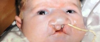
2. Jaundice form. The most common form. It is diagnosed in almost 90% of cases. With this form, jaundice is the most important symptom. Literally in the first hours of life, the skin and mucous membranes acquire a yellow tint, and enlargement of the liver and spleen is possible. The severity of the jaundice form is determined by the distribution throughout the body and the intensity of jaundice. This is determined visually using the Cramer scale. There are five degrees in total, with the first affecting the face and neck, and with the fifth the whole body. The intensity of jaundice depends on the level of bilirubin, which gives the skin its yellow color. A critical level of this enzyme can affect the neurons of the brain, its structures, and cause a serious and dangerous complication, bilirubin encephalopathy.
3. Edema form (“hydrops fetalis”). This is the most severe form, most often diagnosed in utero. The icteric coloration of the membranes, amniotic fluid, and umbilical cord did not go unnoticed by doctors. From the moment of birth, the child has swelling throughout the body - subcutaneous, abdominal, chest. The condition of the newborn is serious. Children diagnosed with this particular form of the disease require intensive treatment, including blood transfusions.
Immediately after birth, it is important for children, especially those at risk, to determine their blood type . Children who do not have the same blood type or Rh group as their mother should be examined by a doctor several times during the first day of life.
Mom herself may notice jaundice, as well as excessive pallor of the mucous membranes and skin. In this case, you must immediately inform your doctor.
HDN should not be confused with other diseases of newborns:
- hereditary hemolytic or posthemorrhagic anemia;
- non-immune hydrops fetalis;
- various infections, etc.
Only a doctor can make an accurate diagnosis; do not try to make a diagnosis yourself or downplay its importance.